. 2000 May 16;249(1-2):53-65.
doi: 10.1016/s0378-1119(00)00136-0.

GP73, a novel Golgi-localized protein upregulated by viral infection

Affiliations

GP73, a novel Golgi-localized protein upregulated by viral infection

R D Kladney et al. Gene. .

Abstract

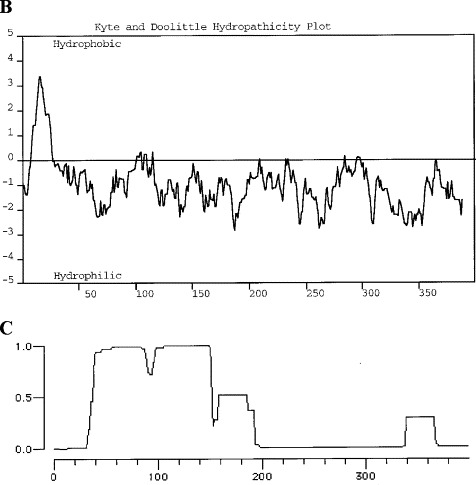
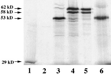
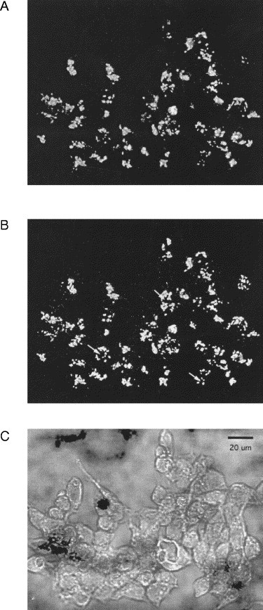
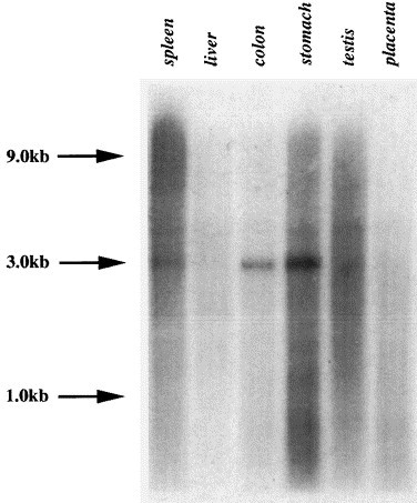
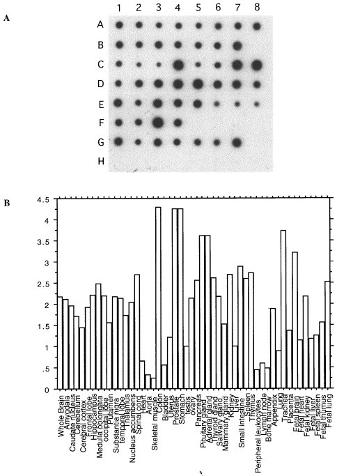
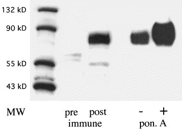
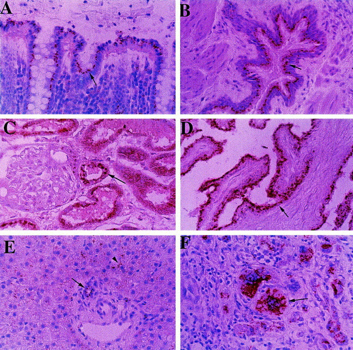
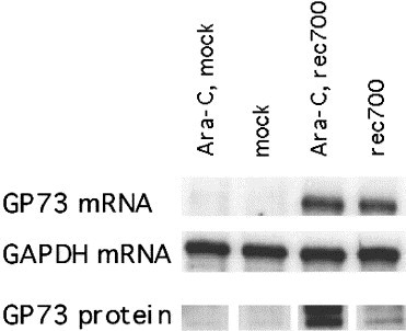
We report the isolation and characterization of GP73, a novel 73kDa human Golgi protein. The GP73 cDNA was cloned by differential screening of a cDNA library derived from the liver of a patient with adult giant-cell hepatitis (GCH), a rare form of hepatitis with presumed viral etiology. In vitro transcription-translation studies indicate that GP73 is an integral membrane protein, and immunolocalization experiments using epitope-tagged GP73 demonstrate that the protein is localized to the Golgi apparatus. Northern blot analysis of RNA from multiple human tissues reveals a single GP73 mRNA transcript with a size of approximately 3.0kb. Immunohistochemical studies using rabbit polyclonal antisera directed against recombinant GP73 demonstrate that the protein is preferentially expressed by epithelial cells in many human tissues. In normal livers, GP73 is consistently present in biliary epithelial cells, whereas hepatocytes show little or no signal. In contrast, livers of patients with GCH display strong GP73 immunoreactivity in multinucleated hepatocytes. GP73 mRNA and protein are expressed in highly differentiated HepG2 hepatoma cells after infection with adenovirus in vitro. We conclude that GP73 represents a novel, epithelial cell-specific integral membrane Golgi protein that can be upregulated in response to viral infection.


Figures

Fig. 1
Fig. 1
(A) Nucleotide and deduced amino acid sequence of GP73 (GenBank accession number AF236056). Two potential starting methionines are underlined. The presumptive transmembrane domain is underlined. Several O- or N-linked glycosylation consensus sites are marked by a caret (^). The stop codon is marked by asterisks. (B) Hydropathicity analysis of GP73. The hydropathicity analysis was carried out using the algorithm by Kyte and Doolittle. A hydrophobic stretch is present in the N-terminal portion of the protein, corresponding to a predicted membrane-spanning domain. The remainder of the protein is predicted to be hydrophilic. (C) Coiled coil analysis of GP73. The analysis was performed according to the algorithm by Lupas. The graph plots the likelihood for a given amino acid to be part of a coiled coil domain.
Fig. 1
Fig. 1
(A) Nucleotide and deduced amino acid sequence of GP73 (GenBank accession number AF236056). Two potential starting methionines are underlined. The presumptive transmembrane domain is underlined. Several O- or N-linked glycosylation consensus sites are marked by a caret (^). The stop codon is marked by asterisks. (B) Hydropathicity analysis of GP73. The hydropathicity analysis was carried out using the algorithm by Kyte and Doolittle. A hydrophobic stretch is present in the N-terminal portion of the protein, corresponding to a predicted membrane-spanning domain. The remainder of the protein is predicted to be hydrophilic. (C) Coiled coil analysis of GP73. The analysis was performed according to the algorithm by Lupas. The graph plots the likelihood for a given amino acid to be part of a coiled coil domain.
Fig. 2
Fig. 2
In vitro transcription–translation of GP73. 35S-labeled GP73 or β-lactamase proteins were transcribed and translated in a reticulocyte in vitro system in the absence or presence of canine microsomes. The reaction products were separated by SDS–PAGE, and visualized by autoradiography. Molecular sizes are marked by arrows. Lane 1: control reaction using β-lactamase cDNA. Lane 2: alkaline extract of material contained in lane 1. Lane 3: transcription–translation of GP73 cDNA in the absence of microsomes. Lane 4: transcription–translation of GP73 in the presence of microsomes. Lane 5: alkaline extract (pH 11.5) of material contained in lane 4. Lane 6: PNGase F treatment of solubilized microsomes contained in lane 4.
3
3
Subcellular localization of GP73 protein. 293 cells were stably transfected with an ecdysone-inducible expression vector containing the full-length GP73 ORF and an in-frame V5 epitope. Cells were induced with 5 μM ponasterone A for 16 h, and analyzed by indirect immunofluorescence (63× objective, water immersion, numerical aperture 1.25) using antibodies directed epitope-tagged GP73 and against the Golgi marker protein giantin. (A) GP73 immunoreactivity, (B) giantin immunoreactivity, (C) transmission microscopy of cells stained in (A) and (B). The nuclei of selected cells are marked by arrows in (B) to demonstrate the perinuclear localization of the GP73 and giantin signals. The 20 μm bar in (C) indicates the magnification.
Fig. 4
Fig. 4
Northern blot analysis of GP73 expression in human tissues. Poly(A+) mRNA (2 μg/lane) from normal human tissues was separated by northern blotting, and probed with a 32P-labelled GP73 cRNA fragment as described in Section 2.4. The positions of the 1.0, 3.0, and 9.0 kb molecular weight markers are indicated by arrows.
Fig. 5
Fig. 5
mRNA dot blot analysis of GP73 expression in multiple human tissues. (A) RNA dot blots: mRNA from 50 normal human tissues was spotted onto a nitrocellulose membrane, and probed with a cDNA probe directed against GP73 as described in Section 2. The samples are arranged as follows: A1–8 (whole brain, amygdala, caudate nucleus, cerebellum, cerebral cortex, frontal lobe, hippocampus, medulla oblongata); B1–7 (occipital lobe, putamen, substantia nigra, temporal lobe, thalamus, nucleus acumbens, spinal cord); C1–8 (heart, aorta, skeletal muscle, colon, bladder, uterus, prostate, stomach); D1–8 (testis, ovary, pancreas, pituitary gland, adrenal gland, thyroid gland, salivary gland, mammary gland); E1–8 (kidney, liver, small intestine, spleen, thymus, peripheral leukocyte, lymph node, bone marrow); F1–4 (appendix, lung, trachea, placenta); G1–7 (fetal brain, fetal heart, fetal kidney, fetal liver, fetal spleen, fetal thymus, fetal lung). H1–7 contain control samples that are not expected to yield hybridization signals (100 ng yeast total RNA, 100 ng yeast tRNA, 100 ng Escherichia coli rRNA, 100 ng E. coli DNA, 100 ng Poly r(A), 100 ng human Cot1DNA, 100 ng human DNA, 500 ng human DNA). (B) Quantification of the results obtained in (A). The autoradiogram shown in (A) was evaluated by densitometry, as described in Section 2. GP73 signals (corrected for the corresponding ubiquitin signals) are expressed relative to the signal obtained in normal liver.
Fig. 6
Fig. 6
Western blot analysis of GP73 expression in HepSK-1 and GP73-transfected 293 cells. Cellular proteins were extracted from cultured HepSK-1 and GP73-transfected 293 cells, separated by SDS–PAGE (15 μg/lane), transferred to a PVDF membrane, and probed with a polyclonal rabbit antibody (dilution 1:5000) raised against GP73 (aa 41–400). The positions of molecular weight (MW) markers are indicated. Pre-, post-immune: lysates of HepSK-1 cells were probed with prei-mmune sera or post-immune sera from week 8 after injection respectively. PonA: 293 cells were incubated in the absence (−) or presence (+) of 5 μM ponasterone A for 16 h, and cellular lysates were probed with post-immune sera. Signals were detected using an HRP-conjugated secondary antibody (1:1500), and visualized by ECL. The immune sera specifically recognize a broad protein band of approximately 73 kDa (GP73). GP73 expression is strongly induced after ponasterone A induction in 293 cells.
Fig. 7
Fig. 7
Expression of GP73 protein in normal human tissues. Tissue- and cell-specific GP73 expression was studied in normal human autopsy tissues and in the liver of a patient with GCH by indirect immunohistochemistry, as noted in Section 2. GP73 immunoreactivity is represented by the red signal, with a blue background provided by the hematoxylin counterstain. (A) Colon, (B) lung, (C) kidney, (D) prostate, (E) normal liver, (F) liver with GCH (original magnification ×400). GP73 is predominantly expressed in cells of the epithelial lineage (indicated by arrows), including colonic enterocytes (A), bronchial epithelial cells (B), proximal and distal tubular epithelial cells (C), and prostate epithelial cells (D). In normal liver, GP73 is expressed in biliary epithelial cells (arrow) and in occasional hepatocytes (arrowhead) (E). In GCH, strong staining is present in heptocyte-derived multinucleated syncitia (arrow).
Fig. 8
Fig. 8
Expression of GP73 mRNA and protein in adenovirus-infected HepG2 cells. HepG2 cells were infected with rec700 adenovirus or mock-infected. Aliquots of cells were infected in the presence of Ara C (20 μg/ml) to inhibit viral DNA replication and the expression of late viral genes. GP73 mRNA was measured by ribonuclease protection assay as described in Section 2.4. GAPDH mRNA levels were measured to document that comparable amounts of mRNA (20 μg) had been loaded in each lane. GP73 protein levels were measured by western blotting on 15 μg aliquots of cellular proteins, as described in Section 2.5.

References

    1. Alconada A., Bauer U., Sodeik B., Hoflack B. Intracellular traffic of herpes simplex virus glycoprotein E: characterization of the sorting signals required for its trans-Golgi network localization. J. Virol. 1999;73:377–387. - PMC - PubMed
    1. Altschul S.F., Gish W., Miller W., Myers E.W., Lipman D.J. J. Mol. Biol. 1990;215:403–410. - PubMed
    1. Ausubel F.M., Brent R., Kingston R.E., Moore D.D., Seidman J.G., Smith J.A., Struhl K. Current Protocols in Molecular Biology. Wiley; New York: 1987.
    1. Berger E.G. The Golgi apparatus: from discovery to contemporary studies. In: Berger E.G., Roth J., editors. The Golgi Apparatus. Birkhäuser; 1997. pp. 1–36.
    1. Blobel G., Dobberstein B. Transfer of proteins across membranes. II. Reconstitution of functional rough microsomes from heterologous components. J. Cell Biol. 1975;67:852–862. - PMC - PubMed

MeSH terms

Associated data