

Molecular basis of functional voltage-gated K+ channel diversity in the mammalian myocardium
- PMID: 10835033
- PMCID: PMC2269952
- DOI: 10.1111/j.1469-7793.2000.t01-1-00285.x
Molecular basis of functional voltage-gated K+ channel diversity in the mammalian myocardium
Abstract
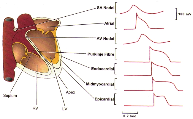
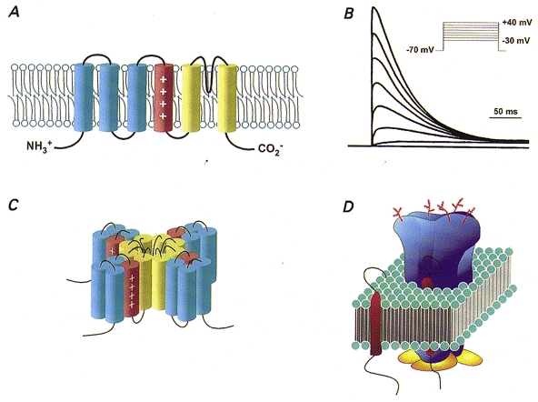
In the mammalian heart, Ca2+-independent, depolarization-activated potassium (K+) currents contribute importantly to shaping the waveforms of action potentials, and several distinct types of voltage-gated K+ currents that subserve this role have been characterized. In most cardiac cells, transient outward currents, Ito,f and/or Ito,s, and several components of delayed reactivation, including IKr, IKs, IKur and IK,slow, are expressed. Nevertheless, there are species, as well as cell-type and regional, differences in the expression patterns of these currents, and these differences are manifested as variations in action potential waveforms. A large number of voltage-gated K+ channel pore-forming (alpha) and accessory (beta, minK, MiRP) subunits have been cloned from or shown to be expressed in heart, and a variety of experimental approaches are being exploited in vitro and in vivo to define the relationship(s) between these subunits and functional voltage-gated cardiac K+ channels. Considerable progress has been made in defining these relationships recently, and it is now clear that distinct molecular entities underlie the various electrophysiologically distinct repolarizing K+ currents (i.e. Ito,f, Ito,s, IKr, IKs, IKur, IK,slow, etc.) in myocyardial cells.
Figures
References
-
- Abbott GW, Sesti F, Splawski I, Buck ME, Lehmann MH, Timothy KW, Keating MT, Goldstein SA. MiRP1 forms IKr potassium channels with HERG and is associated with cardiac arrhythmia. Cell. 1999;97:175–187. - PubMed
-
- Accili EA, Kiehn J, Wible BA, Brown AM. Interactions among inactivating and noninactivating Kv-beta subunits, and Kv-alpha-1.2, produce potassium currents with intermediate inactivation. Journal of Biological Chemistry. 1997;272:28232–28236. - PubMed
-
- Antzelevitch C, Sicouri S, Lukas A, Nesterenko VV, Liu D-W, Didiego JM. Regional differences in the electrophysiology of ventricular cells: physiological implications. In: Zipes DP, Jalife J, editors. Cardiac Electrophysiology: From Cell to Bedside. Philadelphia PA, USA: W. B. Saunders Co.; 1994. pp. 228–245.
-
- Anumonwo J M B, Freeman LC, Kwok WM, Kass RS. Delayed rectification in single cells isolated from guinea pig sinoatrial node. American Journal of Physiology. 1992;262:H921–925. - PubMed
Publication types
MeSH terms
Substances
LinkOut - more resources
Full Text Sources
Other Literature Sources
Molecular Biology Databases
Miscellaneous