. 2000 Jun 1;19(11):2435-43.
doi: 10.1093/emboj/19.11.2435.

RAP1 controls rhoptry targeting of RAP2 in the malaria parasite Plasmodium falciparum

Affiliations

RAP1 controls rhoptry targeting of RAP2 in the malaria parasite Plasmodium falciparum

D L Baldi et al. EMBO J. .

Abstract

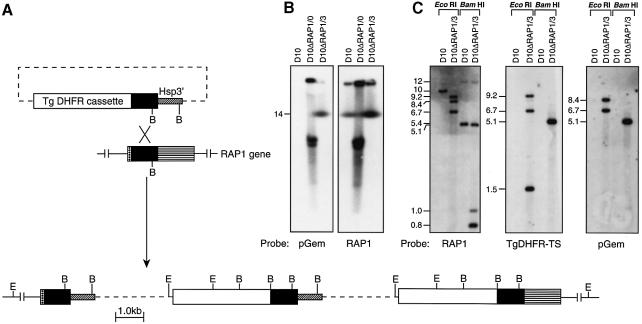
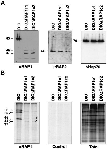
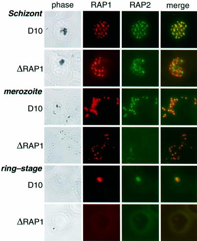
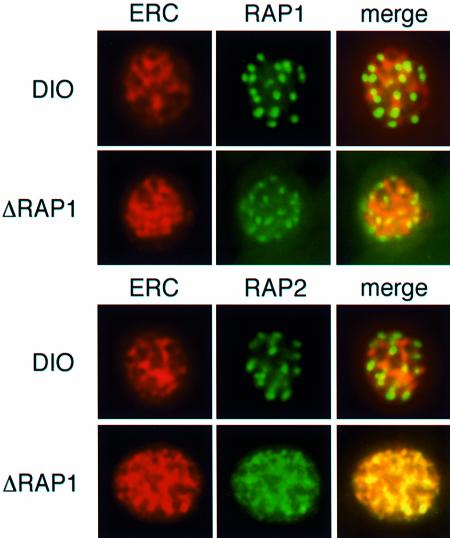
Rhoptry associated protein 1 (RAP1) and 2 (RAP2), together with a poorly described third protein RAP3, form the low molecular weight complex within the rhoptries of Plasmodium falciparum. These proteins are thought to play a role in erythrocyte invasion by the extracellular merozoite and are important vaccine candidates. We used gene-targeting technology in P.falciparum blood-stage parasites to disrupt the RAP1 gene, producing parasites that express severely truncated forms of RAP1. Immunoprecipitation experiments suggest that truncated RAP1 species did not complex with RAP2 and RAP3. Consistent with this were the distinct subcellular localizations of RAP1 and 2 in disrupted RAP1 parasites, where RAP2 does not traffic to the rhoptries but is instead located in a compartment that appears related to the lumen of the endoplasmic reticulum. These results suggest that RAP1 is required to localize RAP2 to the rhoptries, supporting the hypothesis that rhoptry biogenesis is dependent in part on the secretory pathway in the parasite. The observation that apparently host-protective merozoite antigens are not essential for efficient erythrocyte invasion has important implications for vaccine design.


Figures

None
Fig. 1. Disruption of the RAP1 gene. (A) Integration of plasmid pHC1ΔRAP1 by single-site homologous recombination produces a ‘pseudodiploid’, in which the upstream copy of RAP1 is truncated, while the downstream copy lacks a promoter element (Crabb and Cowman, 1996; Wu et al., 1996; Crabb et al., 1997a,b). Two copies of the plasmid have been integrated as shown. E, EcoRI; B, BamHI. (B) Hybridization of a pulsed-field gel electrophoresis blot, containing separated P.falciparum chromosomes (Corcoran et al., 1986), with pGEM plasmid sequences alone (left) reveals multiple transgenic sequences in D10ΔRAP1/0, corresponding to episomal DNA (Crabb and Cowman, 1996; Crabb et al., 1997a). After cycling cultures with/without pyrimethamine, stable parasite line D10ΔRAP1/3 exhibits a single band that co-migrates with chromosome 14. The upper band on both blots corresponds to DNA remaining in the wells. Hybridization of an identical blot with labelled RAP1 (right) confirms that the pGEM sequences are present on the same chromosome as RAP1. (C) Digestion of genomic DNA with EcoRI or BamHI confirms that the RAP1 gene has been disrupted in D10ΔRAP1/3, yielding the expected restriction pattern.
None
Fig. 2. RAP1 and RAP2 expression and association in wild type and ΔRAP1 mutants. (A) Purified schizonts of the D10 parental parasites and two clonal mutants (ΔRAP1c1 and ΔRAP1c2) were separated by SDS–PAGE, immunoblotted and probed with antibodies specific for RAP1 (monoclonal 7H8/50) (Schofield et al., 1986; Howard et al., 1998b), RAP2 (monoclonal IC3/94) or Hsp70 (polyclonal antiserum). The D10ΔRAP1 mutants produced no wild-type RAP1 protein (left) and only low levels of two truncated RAP1 proteins (∼46 and 37 kDa). The mutants produced reduced levels of wild-type RAP2 (centre), but normal levels of Hsp70 (right). (B) Purified trophozoites were metabolically labelled for 6 h with [35S]methionine and immunoprecipitated with monoclonal antibody 7H8/50 (left panel); immunoprecipitation with a negative control monoclonal antibody is shown (centre panel), total labelled protein is also shown (right panel). RAP2 and RAP3 proteins co-immunoprecipitated with RAP1 in wild-type D10 parasites, but not in the ΔRAP1 mutants.
None
Fig. 3. RAP1 specifies rhoptry localization of RAP2. Individual rows show D10 and D10ΔRAP1c1 schizonts, merozoites and ring-stage parasites, as labelled. Each row shows phase-contrast images, staining with polyclonal rabbit anti-RAP1 (red), staining with monoclonal anti-RAP2 (green), and merged RAP1 + RAP2 staining (yellow indicates co-localization) for the same parasite. Top panel: D10 schizont showing punctate rhoptry localization of both RAP1 and RAP2. Second panel: the truncated RAP1 protein in D10ΔRAP1c1 schizonts (see Figure 2) still localizes to the rhoptries, but RAP2 exhibits diffuse non-rhoptry fluorescence. Third panel: RAP1 and RAP2 co-localize in D10 merozoites. Fourth panel: RAP1 exhibits similar (presumably rhoptry) focal staining in D10ΔRAP1c1 parasites, but RAP2 is not detectable. Fifth panel: in wild-type parasites, both RAP1 and RAP2 proteins are carried into ring-stage parasites after infection. Sixth panel: neither RAP1 nor RAP2 are carried into the erythrocyte during merozoite infection. Ring-stage parasites were identified by staining with DAPI (not shown).
None
Fig. 4. RAP2 is located in the ER lumen of ΔRAP1 parasites. Individual rows show mature schizonts of D10 and ΔRAP1 parasites (D10ΔRAP1c1) as labelled. Each row shows staining with polyclonal rabbit anti-PfERC (red) and anti-RAP1 (green) or staining with monoclonal anti-RAP2 (green). Merged images are PfERC + RAP1 or RAP2 (yellow indicates co-localization) for the same parasite. Top panel: PfERC and RAP1 do not co-localize in D10 schizonts. Second panel: PfERC and RAP1 do not co-localize in ΔRAP1 schizonts. Third panel: PfERC and RAP2 do not co-localize in D10 schizonts. Fourth panel: PfERC and RAP2 co-localize in ΔRAP1 schizonts.
None
Fig. 5. Comparison of the growth rates of the parental D10 line and the RAP1 mutants. Synchronized parasites of D10 and D10ΔRAP1c1 were plated at 1% parasitaemia. Smears were taken every 8 h and parasitaemia counted for 132 h. Closed circles correspond to D10 and open circles to D10ΔRAP1c1. The arrows indicate when the parasites were subcultured to 2%.

References

    1. Aikawa M., Miller,L.H., Johnson,J. and Rabbege,J. (1978) Erythrocyte entry by malarial parasites. A moving junction between erythrocyte and parasite. J. Cell Biol., 77, 72–82. - PMC - PubMed
    1. Anders E.M., Hartley,C.A. and Jackson,D.C. (1990) Bovine and mouse serum B inhibitors of influenza A viruses are mannose-binding lectins. Proc. Natl Acad. Sci. USA, 87, 4485–4489. - PMC - PubMed
    1. Andres D.A., Rhodes,J.D., Meisel,R.L. and Dixon,J.E. (1991) Characterisation of the carboxyl-terminal sequences responsible for protein retention in the endoplasmic reticulum. J. Biol. Chem., 266, 14277–14288. - PubMed
    1. Bushell G.R., Ingram,L.T., Fardoulys,C.A. and Cooper,J.A. (1988) An antigenic complex in the rhoptries of Plasmodium falciparum. Mol. Biochem. Parasitol., 28, 105–112. - PubMed
    1. Camus D. and Hadley,T.J. (1985) A Plasmodium falciparum antigen that binds to host erythrocytes and merozoites. Science, 230, 553–556. - PubMed

Publication types

Substances