Comparative Study
. 2000 Jun 1;19(11):2452-64.
doi: 10.1093/emboj/19.11.2452.

Structural basis for recognition of the translocated intimin receptor (Tir) by intimin from enteropathogenic Escherichia coli

Affiliations
Comparative Study

Structural basis for recognition of the translocated intimin receptor (Tir) by intimin from enteropathogenic Escherichia coli

M Batchelor et al. EMBO J. .

Abstract

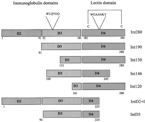
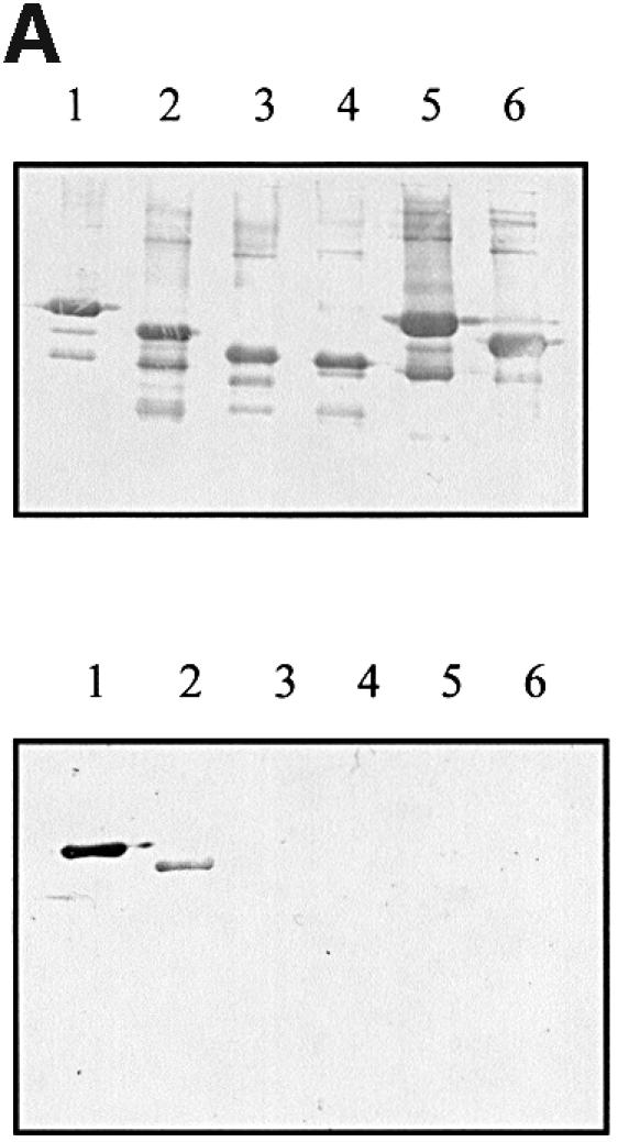
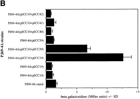
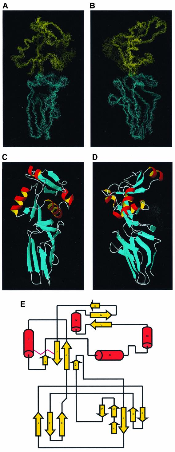
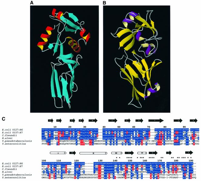
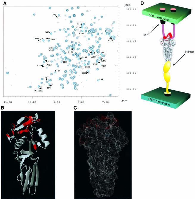
Intimin is a bacterial adhesion molecule involved in intimate attachment of enteropathogenic and enterohaemorrhagic Escherichia coli to mammalian host cells. Intimin targets the translocated intimin receptor (Tir), which is exported by the bacteria and integrated into the host cell plasma membrane. In this study we localized the Tir-binding region of intimin to the C-terminal 190 amino acids (Int190). We have also determined the region's high-resolution solution structure, which comprises an immunoglobulin domain that is intimately coupled to a novel C-type lectin domain. This fragment, which is necessary and sufficient for Tir interaction, defines a new super domain in intimin that exhibits striking structural similarity to the integrin-binding domain of the Yersinia invasin and C-type lectin families. The extracellular portion of intimin comprises an articulated rod of immunoglobulin domains extending from the bacterium surface, conveying a highly accessible 'adhesive tip' to the target cell. The interpretation of NMR-titration and mutagenesis data has enabled us to identify, for the first time, the binding site for Tir, which is located at the extremity of the Int190 moiety.


Figures

None
Fig. 1. Schematic representation of the overlapping Int280-derived polypeptides. The two IgSF-like domains (D2 and D3), the C-type lectin-like domain (D4) and the conserved motifs in Int280 are shown at the top. The position of W150 within Int190 is indicated. Numbers on both sides of the fragments mark the first and last amino acids of each fragment within the Int280 domain.
None
Fig. 2. (A) Detection of Int–Tir interactions using gel overlays. Western blots of MBP–Int derivatives were reacted with a rabbit MBP antiserum (top) or overlayed with Tir-M (bottom). Similar levels of MBP–Int280 (lane 1), MBP–Int190 (lane 2), MBP–Int150 (lane 3), MBP–Int146 (lane 4), MBP–IntD3&D4 (lane 5) and MBP–IntD3 (lane 6); fusion proteins were detected with polyclonal antiserum (top), while Tir-M only bound to MBP–Int280 (bottom, lane 1) and MBP–Int190 (bottom, lane 2). (B) Detection of Int–Tir interactions using the yeast two-hybrid system. β-galactosidase assays showing a 14-fold increase in enzymatic activity in strains co-expressing the whole Tir polypeptide and Int280 and a 7.5-fold increase when Int190 was co-expressed with Tir compared with the parent, cured and the other single and double transformants.
None
Fig. 3. (A) Cα traces representing the superimposition of the 15 refined Int188 structures. (B) Cα traces representing the superimposition of the 15 refined Int188 structures. The orientations of (A) and (B) are related by a 90° rotation. (C) Schematic representation of Int188 for the orientation displayed in (A). (D) Schematic representation of Int188 domains for the orientation displayed in (B). (E) A ‘flattened’ illustration highlighting the topology of Int188. Helices are represented as open tubes and β-strands as arrows.
None
Fig. 4. The structural comparison of Int188 topology with D4/D5 from Invasin (Hamburger et al., 1999). (A) Schematic representation of Int188. Helices are shown as red tubes and strands as blue arrows. (B) Schematic representation of D4/D5 from invasin. Helices are shown as purple tubes, strands as yellow arrows. (C) Sequence alignment and topology for Int188 and invasin. The approximate location of secondary structure elements is also indicated; helices are delineated as open tubes and β-strands as black arrows. The amino acids positions are shown for Int190 from EPEC O127:H6 and correspond to residues 750–939 in full-length intimin. The residues highlighted in blue and red represent identity and conservation with the EPEC O127:H6 sequence, respectively. Intimins from E.coli O127:H6, E.coli O26:H- and E.coli O157:H7 have been immunologically categorized into types α, β and γ, respectively. The two invasin sequences, Y.pseudotuberculosis and Yersinia enterocolitica, are aligned on the basis of the structural superimposition provided by DALI. Asterisks represent amide resonances that are significantly perturbed upon the addition of Tir55 peptide.
None
Fig. 5. (A) The 1H-15N HSQC NMR spectrum for Int188 (blue) with the identical region of the 1H-15N HSQC NMR spectrum for Int188 with Tir55 peptide overlaid (black). Peaks corresponding to amides that are affected upon addition of Tir55 are labelled with their residue number (for consistency these are numbered according to Int190). (B) Schematic representation of the structure of Int188. Residues shown in red indicate chemical shift/line-width perturbation in the presence of Tir55, namely Y140, K142, I147, I148, S149, W150, T154, Q156, D157, A158, V162, A163, S164, T165, K170, Q171, N176, I177, S180, E181, N183, A184, Y185, T187 and V189. These data are also summarized in Figure 3A. The data illustrate a possible binding site for Tir55, which is composed of highly concerted patch of residues in D4. For clarity Int188 is rotated by 180° with respect to Figure 3A. (C) A dot representation of the solvent-accessible surface for Int188. The residues that show chemical shift/line-width perturbation in the presence of Tir55 are shown in red. The data illustrate a possible binding site for Tir55, which is composed of a highly concerted patch of residues in D4. (D) Schematic representation of the quaternary structure of the entire extracellular region of intimin in complex with Tir peptide. IGSF domains D1 and D2 are shown in yellow. The Tir-binding fragment of intimin (i.e. Int190, D3 and D4) is illustrated by the space filling representation shown in (B). Tir is shown schematically: the two predicted transmembrane helices are shown as tubes and the intimin-binding peptide, which is manually docked onto the binding site, as a red ribbon.
None
Fig. 6. FAS reaction (A, D, G), intimin staining (B, E, H) and overlaid images (C, F and I) of infected HEp-2 cells. CVD206(pCVD438) (A–C) showed both FAS and intimin staining. No staining was observed using strain CVD206 alone (D, E) although adhering bacteria could be observed by phase contrast (F). Substitution of W899 in strain CVD206(pICC54) resulted in FAS negative staining although surface intimin expression was not affected (G–I).
None
Fig. 7. (A) Detection of Int190–Tir interactions using gel overlays. Western blots of MBP–Int mutants were reacted with a rabbit MBP antiserum or overlayed with Tir-M. Similar levels of MBP–Int190 (lane 1) and MBP–Int190W150A (lane 2) fusion proteins were detected with polyclonal antiserum, while Tir-M only bound to MBP–Int190 (lane 3). (B) Detection of Int–Tir interactions using the yeast two hybrid system. β-galactosidase assays showing a 7-fold increase in enzymatic activity in strains co-expressing the whole Tir polypeptide and Int190 compared with Tir–Int190A150, Tir–Int280A240 and single plasmid transformants.

References

    1. Adu-Bobie J., Frankel,G., Bain,C., Goncaleves,A.G., Trabulsi,L.R., Douce,G., Knutton,S. and Dougan,G. (1998) Detection of intimin α, β, γ and δ, four intimin derivatives expressed by attaching and effacing microbial pathogens. J. Clin. Microbiol., 36, 662–668. - PMC - PubMed
    1. Bax A., Clore,G.M. and Gronenborn,A.M.J. (1990) 1H-1H correlation via isotropic mixing of 13C magnetization, a new three-dimensional approach for assigning 1H and 13C spectra of 13C enriched proteins. Magn. Res., 88, 425–431.
    1. Boyington J.G., Riaz,A.N., Patamawam,A., Coligan,J.E., Brook,A.G. and Sun,P.D. (1999) Structure of the CD94 reveals a novel C-type lectin fold: implications for the NK cell-associated CD94/NKG2 receptors. Immunity, 10, 75–82. - PubMed
    1. Brünger A.T. (1993) X_PLOR Manual. Version 3.1. Yale University Press, New Haven, CT.
    1. Cornilescu G., Delaglio,F. and Bax,A. (1999) Protein backbone angle restraints from searching a database for chemical shift and sequence homology. J. Biomol. NMR, 13, 289–302. - PubMed

Publication types

MeSH terms

Associated data