. 2000 Jun 1;19(11):2465-74.
doi: 10.1093/emboj/19.11.2465.

Definitive hematopoietic stem cells first develop within the major arterial regions of the mouse embryo

Affiliations

Definitive hematopoietic stem cells first develop within the major arterial regions of the mouse embryo

M F de Bruijn et al. EMBO J. .

Abstract

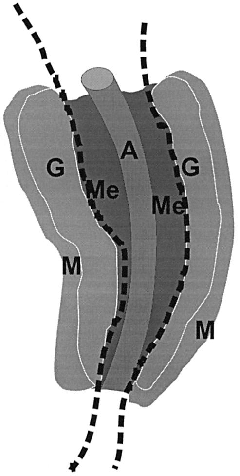
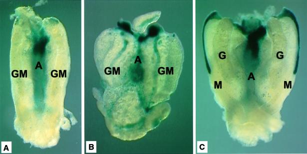
The aorta-gonad-mesonephros (AGM) region is a potent hematopoietic site within the mammalian embryo body, and the first place from which hematopoietic stem cells (HSCs) emerge. Within the complex embryonic vascular, excretory and reproductive tissues of the AGM region, the precise location of HSC development is unknown. To determine where HSCs develop, we subdissected the AGM into aorta and urogenital ridge segments and transplanted the cells into irradiated adult recipients. We demonstrate that HSCs first appear in the dorsal aorta area. Furthermore, we show that vitelline and umbilical arteries contain high frequencies of HSCs coincident with HSC appearance in the AGM. While later in development and after organ explant culture we find HSCs in the urogenital ridges, our results strongly suggest that the major arteries of the embryo are the most important sites from which definitive HSCs first emerge.


Figures

None
Fig. 1. Schematic representation of subdissected regions of the AGM. E11 and E12 AGM tissue is dissected along the dotted lines to separate the dorsal aorta and surrounding mesenchyme from the urogenital ridges, the latter of which contain the developing gonads and pro/mesonephroi. A, dorsal aorta; Me, mesenchyme; G, gonad; M, mesonephros.
None
Fig. 2. Multilineage repopulation analysis of transplant recipients engrafted with aorta- or UGR-derived HSCs. Southern blot analysis was performed on products obtained from donor-marker specific PCRs. PCR reactions were performed on DNA isolated from hematopoietic tissues and sorted hematopoietic cells from representative transplant recipients receiving cells from (A) E11 aorta, (B) E12 aorta and (C) E12 UGR. At >4 months post-transplantation, transplant recipients were killed and hematopoietic tissues isolated. Lymphoid, myeloid and erythroid lineage cells were sorted and DNAs examined for the presence of the donor cell genetic marker (human β-globin). Percentage contribution was controlled by mixes of the donor-transgene marker with unmarked DNA (100, 10, 1 and 0%). Donor-marker contribution was determined by PhosphorImager quantitation of specific 32P hybridizing probe. The donor signal was normalized to the hybridization signal for the myogenin PCR product. Percentage donor marker contributions are indicated below each lane. Pb, peripheral blood; Th, thymus; Ln, lymph nodes; S, spleen; B, splenic B lymphocytes; T, splenic T lymphocytes; E, bone marrow erythroid precursors; M, bone marrow myeloid precursors; L, bone marrow lymphoid precursors; Bm, bone marrow.
None
Fig. 3. Multilineage repopulation analysis of transplant recipients engrafted with cultured aorta- or UGR-derived HSCs. DNAs were isolated from hematopoietic tissue and sorted hematopoietic cells from representative transplant recipients receiving cells from (A) E11 cultured aorta and (B) E11 cultured UGRs. At >4 months post-transplantation, transplant recipients were killed and hematopoietic tissues isolated. Lymphoid, myeloid and erythroid lineage cells were sorted and DNAs examined for the presence of the donor cell genetic marker (human β-globin) as described in Figure 2.
None
Fig. 4. Cbfa2-LacZ expression in subregions of the AGM. X-gal staining was performed on (A) E11 AGM directly after dissection, (B) E11 AGM after 3 days in organ explant culture and (C) E12 AGM directly after dissection. Abundant blue staining is observed in the area containing the dorsal aorta and mesenchyme in all three tissues. However, the UGRs stained brightly only at E12 or after explant culture of E11 AGMs. The stripe of expression in the anterior mesonephros at E12 is from the Mullerian duct. Whether HSC activity is located in the strongly X-gal-positive Mullerian ducts or elsewhere in the mesonephroi remains to be determined.
None
Fig. 5. Multilineage repopulation analysis of a transplant recipient engrafted with E10 vitelline and umbilical arteries. At >4–7 months post-transplantation, transplant recipients that received vitelline and umbilical arteries were killed and hematopoietic tissues isolated. The results from a representative multilineage analysis are shown. Lymphoid, myeloid and erythroid lineage cells were sorted and DNAs examined for the presence of the donor cell genetic marker (human β-globin) as described in Figure 2.

References

    1. Baldwin H.S. et al. (1994) Platelet endothelial cell adhesion molecule-1 (PECAM-1/CD31): alternatively spliced, functionally distinct isoforms expressed during mammalian cardiovascular development. Development, 120, 2539–2553. - PubMed
    1. Bertwistle D., Walmsley,M.E., Read,E.M., Pizzey,J.A. and Patient,R.K. (1996) GATA factors and the origins of adult and embryonic blood in Xenopus: responses to retinoic acid. Mech. Dev., 57, 199–214. - PubMed
    1. Brown L., Rodaway,A., Schilling,T., Jowett,T., Ingham,P., Patient,R. and Sharrocks,A. (2000) Insights into early vasculogenesis revealed by expression of the ETS-domain transcription factor FLi-1 in wild-type and mutant zebrafish embryos. Mech. Dev., 90, 237–252. - PubMed
    1. Carmeliet P. et al. (1996) Abnormal blood vessel development and lethality in embryos lacking a single VEGF allele. Nature, 380, 435–439. - PubMed
    1. Chiquoine A. (1954) The identification, origin and migration of the primordial germ cells in the mouse embryo. Anat. Rec., 118, 135–146. - PubMed

Publication types

MeSH terms