. 2000 Jun 1;19(11):2483-91.
doi: 10.1093/emboj/19.11.2483.

TREK-1 is a heat-activated background K(+) channel

Affiliations

TREK-1 is a heat-activated background K(+) channel

F Maingret et al. EMBO J. .

Abstract

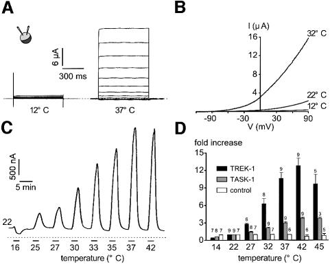
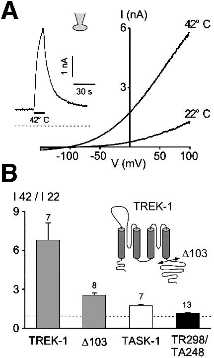
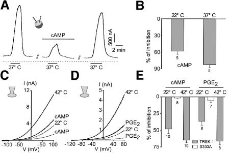
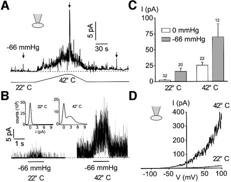
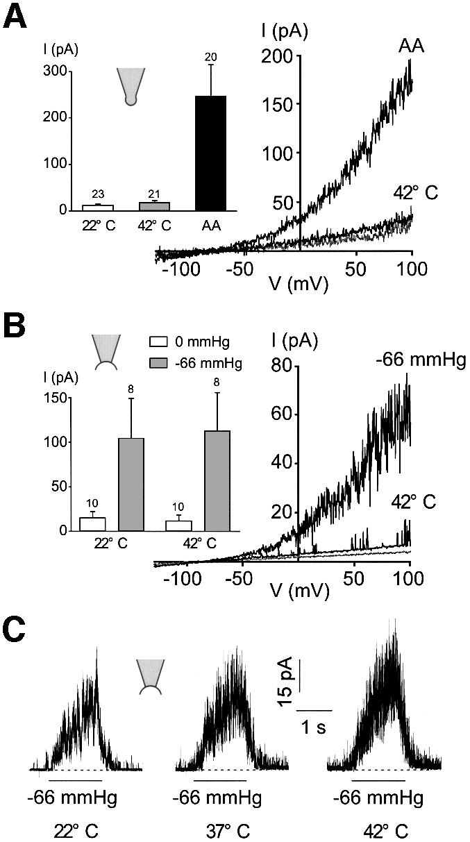
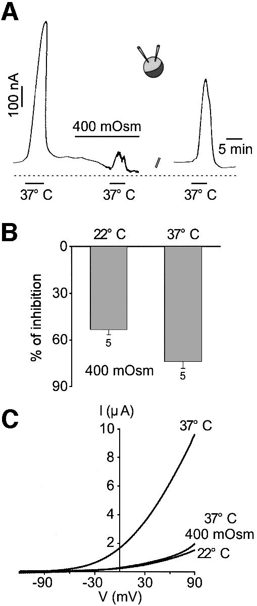
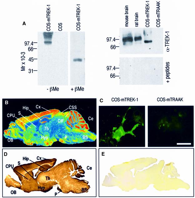
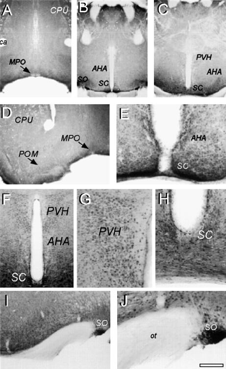
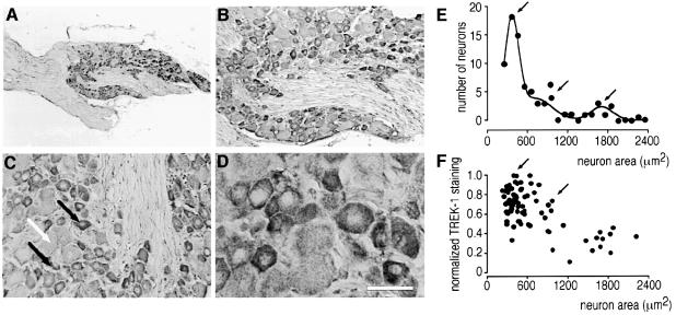
Peripheral and central thermoreceptors are involved in sensing ambient and body temperature, respectively. Specialized cold and warm receptors are present in dorsal root ganglion sensory fibres as well as in the anterior/preoptic hypothalamus. The two-pore domain mechano-gated K(+) channel TREK-1 is highly expressed within these areas. Moreover, TREK-1 is opened gradually and reversibly by heat. A 10 degrees C rise enhances TREK-1 current amplitude by approximately 7-fold. Prostaglandin E2 and cAMP, which are strong sensitizers of peripheral and central thermoreceptors, reverse the thermal opening of TREK-1 via protein kinase A-mediated phosphorylation of Ser333. Expression of TREK-1 in peripheral sensory neurons as well as in central hypothalamic neurons makes this K(+) channel an ideal candidate as a physiological thermoreceptor.


Figures

None
Fig. 1. TREK-1 is a temperature-sensitive K+ channel in Xenopus oocyte. (A) The two-microelectrode voltage-clamp technique was used to voltage-clamp oocytes expressing TREK-1. I–V curves of an oocyte expressing TREK-1 maintained at 12 and 37°C. The holding potential is –80 mV and increment voltage steps of 20 mV are applied every 5 s from –130 to 90 mV. (B) Voltage ramps of 800 ms duration applied from a holding potential of –80 mV are recorded at 12, 22 and 32°C in a TREK-1-expressing oocyte. (C) A TREK-1-expressing oocyte is voltage-clamped at a holding potential of –20 mV. The zero current is indicated by a dotted line. Cooling from 22 to 16°C inhibits TREK-1 basal activity while a gradual increase in temperature up to 42°C reversibly stimulates TREK-1 current amplitude. (D) Stimulation of current amplitude (fold increase It/I22°C) measured at –20 mV in control, TREK-1- and TASK-1-expressing oocytes. Note that at temperatures >42°C, TREK-1 is decreased irreversibly. At 22°C, current amplitude measured at –20 mV is 76 ± 3 nA (n = 7), 318 ± 45 nA (n = 9) and 1483 ± 197 nA (n = 9) for control, TREK-1- and TASK-1-expressing oocytes, respectively.
None
Fig. 2. TREK-1 is a heat-activated K+ channel in COS cells. (A) Voltage ramps of 800 ms duration are applied from a holding potential of –80 mV. The cell is bathed with an external medium containing 5 mM K+. Currents are recorded at 22 and 42°C, as indicated. The inset illustrates TREK-1 current induced by a temperature jump from 22 to 42°C (indicated by a horizontal bar) at a holding potential of 0 mV in physiological K+ conditions. Zero current is indicated by a dotted line. (B) Temperature sensitivity expressed as the ratio of current amplitude I42°C/I22°C measured at 0 mV of TREK-1, a C-terminally deleted TREK-1 mutant (Δ103), TASK-1 and a chimera containing the core of TREK-1 and the C-terminus of TASK-1 (TR298/TA248). Numbers of experiments are indicated. The cartoon illustrates the C-terminal deletion.
None
Fig. 3. cAMP and PGE2 reverse thermal stimulation of TREK-1. (A) Effect of 0.5 mM CPT-cAMP on TREK-1 current evoked by a temperature jump from 22 to 37°C in Xenopus oocytes. The current is measured at a holding potential of –20 mV. cAMP is applied for 5 min and is washed out for 13 min. (B) Summary of the effects of 0.5 mM CPT-cAMP (percentage inhibition) on TREK-1 current amplitude measured in oocytes at a holding potential of –20 mV and recorded at 22 and 37°C. (C) Voltage ramps of 800 ms duration are applied from a holding potential of –80 mV in a COS cell expressing TREK-1. Currents are measured at 22 and 42°C in the absence and presence of 0.5 mM CPT-cAMP. cAMP is superfused for 2 min. (D) Voltage ramps of 800 ms duration are applied from a holding potential of –80 mV in a COS cell expressing TREK-1. Currents are measured at 22 and 42°C in the absence and presence of 5 µM PGE2 superfused for 2 min. (E) Summary of the experiments performed in COS cells (percentage inhibition) in the presence of 0.5 mM cAMP or 5 µM PGE2 at 22 and 42°C. The mutant Ser333 is not sensitive to both 0.5 mM cAMP and 5 µM PGE2 at 22°C. At 22°C, current amplitude measured at 0 mV is 14.3 ± 3.9 and 98.0 ± 18.5 pA/pF for TREK-1 and TREK-1Ser333 mutant, respectively. At 37°C, current amplitude is 43.7 ± 7.5 and 168.6 ± 28.3 pA/pF for TREK-1 and TREK-1Ser333 mutant, respectively.
None
Fig. 4. Heat opens TREK-1 in the cell-attached patch configuration. (A) Cell-attached patch recording at 0 mV in a COS cell expressing TREK-1. Channel activity is recorded at atmospheric pressure and during a stretch of –66 mmHg at 22 and 42°C. (B) Channel activity elicited by a stretch of –66 mmHg is largely potentiated at 42°C. The same experiment as in (A). Zero current is indicated by a dotted line. The inset shows Gaussian fits of the amplitude histograms at 22 and 42°C of channel activity recorded at atmospheric pressure (50 bins per histogram). Recordings of 3.5 s duration were filtered at 5 kHz and sampled at 50 kHz. (C) Summary of the experiments (mean current, I) performed at atmospheric pressure and during a stretch of –66 mmHg recorded at 22 and 42°C at 0 mV in cell-attached patches of COS cells expressing TREK-1. (D) Voltage ramps of 800 ms duration from a holding potential of –80 mV in a cell-attached patch from a COS cell expressing TREK-1 recorded at 22 and 42°C.
None
Fig. 5. Heat activation of TREK-1 is lost upon excision. (A) Thermal activation of TREK-1 at 42°C is lost in an outside-out patch, while addition of 10 µM AA at 22°C in the bath solution opens TREK-1 channels. Voltage ramps of 800 ms duration are applied from a holding potential of –80 mV. The inset illustrates the summary of the outside-out patch experiments performed at 22 and 42°C and in the presence of 10 µM AA at 22°C. The patches are held at a holding potential of 0 mV and channel activity is represented as the mean current I. (B) In inside-out patches: heat activation is lost, while a stretch of –66 mmHg opens TREK-1 channels. Voltage ramps of 800 ms duration are applied from a holding potential of –80 mV. The inset illustrates TREK-1 channel activity (mean current I) in inside-out patches measured at 0 mV and recorded at 22 and 42°C at atmospheric pressure and during a stretch of –66 mmHg. (C) A stretch of –66 mmHg elicits TREK-1 opening in an inside-out patch held at 0 mV and is not sensitive to temperature as indicated.
None
Fig. 6. Hyperosmolarity reverses thermal activation of TREK-1. (A) TREK-1 current is recorded at –20 mV with the two-microelectrode voltage-clamp technique in a Xenopus oocyte. A temperature jump from 22 to 37°C reversibly opens TREK-1. Increasing the osmolarity up to 400 mOsm with mannitol reversibly depresses the opening of TREK-1 by heat. Mannitol is washed out for 20 min. (B) Summary of the experiments illustrating the effect of 400 mOsm hyperosmolarity with mannitol on TREK-1 current amplitude recorded at 22 and 37°C and measured at –20 mV. (C) Voltage ramps of 800 ms duration are applied from a holding potential of –80 mV at 22 and 37°C. A 400 mOsm hyperosmolarity with mannitol reverses TREK-1 current stimulation by heat.
None
Fig. 7. Characterization of TREK-1 antibodies by western blotting and immunocytochemistry. (A) Left: total proteins of COS cells transfected with mTREK-1 cDNA were separated on a 10% polyacrylamide gel (5 µg/lane) under non-reducing (–βMe) or reducing conditions (+βMe). Western blots were incubated with affinity-purified α-TREK-1 (1:2000) polyclonal rabbit antibodies. Mock-transfected COS cells were used as a negative control (empty expression pCD8 vector). Antigen–antibody complexes were visualized by using an enhanced chemiluminescence method (Super Signal, Pierce). Right: synaptic membranes from mouse and rat brain, and COS cells expressing mTREK-1 or mTRAAK. A 30 µg aliquot of brain synaptic membranes or 5 µg of total proteins of COS cells were loaded per lane. Blots were incubated with α-TREK-1 (1:2000) alone (top) or the α-TREK-1 (1:2000) pre-incubated with a mixture of the two GST fusion proteins (bottom). (B) Digitized autoradiograph illustrating TREK-1 mRNA distribution as observed by in situ hybridization on mouse sagittal brain sections using a specific 49mer oligonucleotide complementary to mouse TREK-1. The same in situ panel has been published previously by Fink et al. (1996) as Figure 3B. The description of the in situ hybridization in this previous work was not correct as it stated that only a very low level of expression of TREK-1 was detected in the hypothalamus. This in situ hybridization shows on the contrary a very strong expression of TREK-1 in the hypothalamic area. (C) Immunocytochemistry experiments on COS cells expressing mTREK-1 (left) or mTRAAK-transfected (right). Immunocomplexes were revealed by fluorescence microscopy. Scale bar, 20 µm. (D) Immunohistochemistry with α-TREK-1 antibody on a sagittal mouse brain section. Immunostaining was visualized using the peroxidase–DAB technique. (E) Signals were blocked by pre-absorption of the α-TREK-1 antibody with an excess of the antigenic fusion proteins. BS, brainstem; Ce, cerebellum; CIF, inferior colliculus; CPU, caudate–putamen; CSS, superior colliculus; Cx, neocortex; Hip, hippocampus; Hyp, hypothalamus; OB, olfactory bulb; P, pontine nuclei; S, septum; Th, thalamus.
None
Fig. 8. Immunolocalization of TREK-1 in the mouse preoptic and anterior hypothalamus. Immunohistochemistry was performed on mouse brain coronal sections at the level of PO/AH (A–J). TREK-1 immunostaining was visualized using the peroxidase–DAB technique as described in Materials and methods. (AC) Low-power microphotographs at brain levels 0.4, –0.7 and –0.9. (D–J) Medium- and high-power microphotographs of distinct areas showing (D) MPO/POM, (E and H) SC, (F and G) PVH and AHA, and (I and J) SO. AHA, anterior hypothalamic area; ca, anterior commissure; CPU, caudate–putamen; MPO, medial preoptic area; ot, optic tract; POM, magnocellular preoptic area; PVH, paraventricular nucleus; SC, suprachiasmatic nucleus; SO, supraoptic nucleus.
None
Fig. 9. Immunolocalization of TREK-1 in mouse DRG neurons. TREK-1 immunostaining was visualized using the peroxidase–DAB technique as described in Materials and methods. (AD) TREK-1 immunostaining within DRG at increasing magnification. Note in (C) the high labelling in small and medium sized (black arrows) and low labelling in large (white arrow) sensory neurons. Scale bar, 50 µm. (E) Number of neurons as a function of size (in µm2). The histogram was fitted with the sum of Gaussian functions. Bin size was 50 µm2. (F) Normalized TREK-1 expression as a function of the size of the neurons. Small and medium sized neurons express high levels of TREK-1 (as indicated by arrows).

References

    1. Boulant J.A. (1998a) Cellular mechanisms of temperature sensitivity in hypothalamic neurons. Prog. Brain Res., 115, 3–8. - PubMed
    1. Boulant J.A. (1998b) Hypothalamic neurons. Mechanisms of sensitivity to temperature. Ann. N Y Acad. Sci., 856, 108–115. - PubMed
    1. Braun H.A., Bade,H. and Hensel,H. (1980) Static and dynamic discharge patterns of bursting cold fibers related to hypothetical receptor mechanisms. Pflugers Arch., 386, 1–9. - PubMed
    1. Buckler K.J. (1997) A novel oxygen-sensitive potassium current in rat carotid body type I cells. J. Physiol., 498, 649–662. - PMC - PubMed
    1. Caterina M.J. and Julius,D. (1999) Sense and specificity: a molecular identity for nociceptors. Curr. Opin. Neurobiol., 9, 525–530. - PubMed

Publication types

MeSH terms