. 2000 Jun 1;14(11):1343-52.

Notch signaling is essential for vascular morphogenesis in mice

Affiliations

Notch signaling is essential for vascular morphogenesis in mice

L T Krebs et al. Genes Dev. .

Abstract

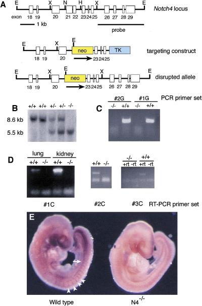
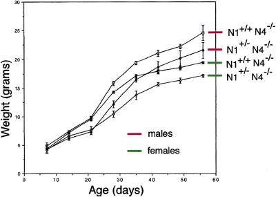
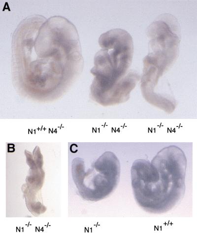
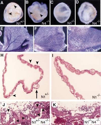
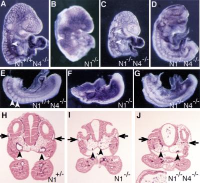
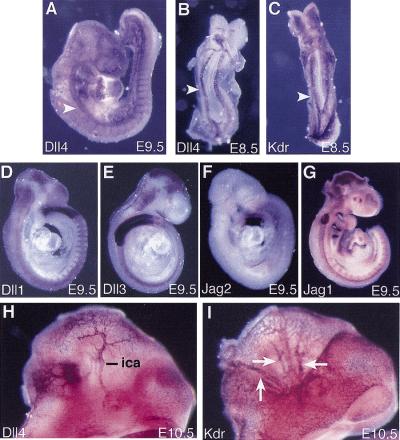
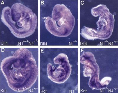
The Notch gene family encodes large transmembrane receptors that are components of an evolutionarily conserved intercellular signaling mechanism. To assess the role of the Notch4 gene, we generated Notch4-deficient mice by gene targeting. Embryos homozygous for this mutation developed normally, and homozygous mutant adults were viable and fertile. However, the Notch4 mutation displayed genetic interactions with a targeted mutation of the related Notch1 gene. Embryos homozygous for mutations of both the Notch4 and Notch1 genes often displayed a more severe phenotype than Notch1 homozygous mutant embryos. Both Notch1 mutant and Notch1/Notch4 double mutant embryos displayed severe defects in angiogenic vascular remodeling. Analysis of the expression patterns of genes encoding ligands for Notch family receptors indicated that only the Dll4 gene is expressed in a pattern consistent with that expected for a gene encoding a ligand for the Notch1 and Notch4 receptors in the early embryonic vasculature. These results reveal an essential role for the Notch signaling pathway in regulating embryonic vascular morphogenesis and remodeling, and indicate that whereas the Notch4 gene is not essential during embryonic development, the Notch4 and Notch1 genes have partially overlapping roles during embryogenesis in mice.


Figures

Figure 1
Figure 1
Targeted disruption of the Notch4 gene. (A) Targeting scheme. (Top) The genomic organization of a portion of the Notch4 gene. Exons are indicated by white boxes. (Middle) The structure of the targeting vector. The deleted exons 21 and 22 encode amino acids 1249–1434 in the extracellular domain of the Notch4 protein. (Bottom) The predicted structure of the Notch4 locus following homologous recombination of the targeting vector. (E) EcoRI; (H) HindIII; (N) NcoI; (X) XbaI. (B) DNA isolated from embryos of the intercross of Notch4+/− heterozygous mice was digested with EcoRI, blotted, and hybridized with the indicated probe. Sizes of hybridizing fragments are indicated. Genotypes of progeny are indicated at the top of the lane. (C) Genomic PCR analysis with primers located in region deleted in the Notch4d1 mutant allele. Genotypes are indicated at top. (D) RT–PCR analysis. RT–PCR primer sets are indicated at the bottom of each panel. Primer set #1C flanks the deleted region; primers sets #2C and #3C are located at the 3′ end of the Notch4 cDNA. Genotypes are indicated at the top of the lane. (+rt) Plus reverse transcriptase; (−rt) without reverse transcriptase. (E) Whole mount in situ hybridization of a wild-type and a Notch4−/− embryo with an antisense Notch4 riboprobe encoding the intracellular domain of the Notch4 protein. Notch4 expression in the wild-type embryo is observed in intersomitic blood vessels (arrowheads) and the dorsal aorta (arrow). No Notch4 expression is observed in the mutant embryo.
Figure 2
Figure 2
Genetic interactions of Notch1 and Notch4 mutations. Growth curve of Notch1+/− Notch4−/− (♦,⋄) vs. Notch1+/+ Notch4−/− (▪, □) littermates. The weights of Notch1+/− Notch4−/− and Notch1+/+ Notch4−/− mutant animals are plotted against age. Males (red) and females (green) are plotted separately. Data presented are from four mice in each group. Error bars indicate the s.d..
Figure 3
Figure 3
Synergistic effects in Notch1−/− Notch4−/− double mutant embryos. (A) Two Notch1−/− Notch4−/− double homozygous mutant embryos and littermate control isolated at E9.5. (B) Severely affected Notch1−/− Notch4−/− double homozygous mutant embryo. The double homozygous mutant embryos in both A and B have not completed embryonic turning and have open neural tubes. (C) A typical Notch1−/− embryo and control littermate at E9.5. The Notch1−/− mutant embryo has completed turning and has a closed neural tube.
Figure 4
Figure 4
Yolk sac and placental defects in mutant embryos. (A–D) Morphology of embryos in their yolk sacs at E9.5. Large vitelline blood vessels (arrowheads) are observed in the Notch1+/− and Notch4−/− yolk sacs, but not in the Notch1−/− and Notch1−/− Notch4−/− mutants. (E–G) PECAM-1-stained yolk sacs. The yolk sacs of the Notch1−/− and Notch1−/− Notch4−/− mutant embryos are at the primitive vascular plexus stage and have not undergone vascular remodeling to form the large and small blood vessels of the mature yolk sac. (H,I) Histological sections of PECAM-1-stained yolk sacs. The Notch1+/− yolk sac (H) has differentiated both small capillaries (arrowheads) and large vitelline collecting vessels (arrow). The Notch1−/− yolk sac (I) exhibits a disorganized, confluent vascular plexus. (J,K) Histological sections of placentas at E9.5. In the Notch1+/− Notch4−/− control embryo (J), embryonic blood vessels containing nucleated erythrocytes (arrowheads) have invaded the labyrinthine layer of the placenta. In the Notch1−/− Notch4−/− mutant embryo (K), embryonic blood vessels are present at the edge of the placenta but have not invaded the labyrinthine layer.
Figure 5
Figure 5
Defects in vascular remodeling in Notch1−/− and Notch1−/− Notch4−/− mutant embryos. (A–G) PECAM-1-stained whole mount embryos. (A–D) Defective morphogenesis of the main trunk of the anterior cardinal vein (arrowhead) in Notch1−/− (B) and Notch1−/− Notch4−/− mutant embryos (C,D). The double mutant embryo in D is more severely affected than the embryo in C. (E–G) In the Notch1+/− Notch4−/− control embryo (E), intersomitic vessels (arrowheads) differentiate, whereas these vessels are not observed in the Notch1−/− (F) and Notch1−/− Notch4−/− (G) mutant embryos. (H–J) Histological sections of PECAM-1-stained embryos at the level of the otic vesicle. In the N1+/− control embryo (H), both the dorsal aortae (arrowheads) and the anterior cardinal veins (arrows) have open lumens and normal morphology. In the Notch1−/− Notch4−/− mutant embryo (J), endothelial cells have differentiated but both the dorsal aortae and the anterior cardinal veins have an abnormal, collapsed morphology. In the less-severely affected Notch1−/− mutant embryo (I), the anterior cardinal veins still have an open lumen but the dorsal aortae are collapsed.
Figure 6
Figure 6
Expression pattern of the Dll4 gene suggests that it encodes the ligand for the Notch1 and Notch4 receptors in the embryonic vasculature. Whole-mount in situ hybridization with the indicated probes of embryos isolated at E8.5 (B,C), E9.5 (A,D–G), and E10.5 (H,I). Dll4 expression is observed in the dorsal aorta (arrowheads) and, at E9.5, the intersomitic vessels (A). The other genes encoding Notch ligands do not exhibit obvious expression in the vasculature at E9.5 (D–G). At E10.5, Dll4 expression is observed in the internal carotid artery (ica) (H), but is not observed in branches of the primary head vein (arrows in I), which express Kdr.
Figure 7
Figure 7
Dll4 and Kdr expression in mutant embryos. Embryos of the indicated genotypes were hybridized with riboprobes for Dll4 (A–C) and Kdr (D–F). Note the more severe dysmorphogenesis of the Notch1−/− Notch4−/− double mutant embryos (C,F) compared with the Notch1−/− single mutant embryos (B,E). Dll4 expression was not downregulated in either the Notch1−/− or Notch1−/− Notch4−/− mutant embryos. However, the expression patterns of both Dll4 and Kdr reveal disruptions in vascular morphogenesis in the mutant embryos.

References

    1. Adams RH, Wilkinson GA, Weiss C, Diella F, Gale NW, Deutsch U, Risau W, Klein R. Roles of ephrinB ligands and EphB receptors in cardiovascular development: Demarcation of arterial/venous domains, vascular morphogenesis, and sprouting angiogenesis. Genes & Dev. 1999;13:295–306. - PMC - PubMed
    1. Artavanis-Tsakonas S, Rand MD, Lake RJ. Notch signaling: Cell fate control and signal integration in development. Science. 1999;284:770–776. - PubMed
    1. Baldwin HS, Shen HM, Yan HC, De Lisser HM, Chung A, Mickanin C, Trask T, Kirschbaum NE, Newman PJ, Albeda SM, Buck CA. Platelet endothelial cell adhesion molecule-1 (PECAM-1/CD31): Alternatively spliced, functionally distinct isoforms expressed during mammalian cardiovascular development. Development. 1994;120:2539–2553. - PubMed
    1. Carmeliet P, Collen D. Role of vascular endothelial growth factor and vascular endothelial growth factor receptors in vascular development. Curr Top Microbiol Immunol. 1999;237:133–158. - PubMed
    1. Chan Y-M, Jan YN. Roles for proteolysis and trafficking in Notch maturation and signal transduction. Cell. 1998;94:423–426. - PubMed

Publication types

MeSH terms

LinkOut - more resources