. 2000 Jun 1;14(11):1364-76.

Tissue- and stage-specific modulation of Wingless signaling by the segment polarity gene lines

Affiliations

Tissue- and stage-specific modulation of Wingless signaling by the segment polarity gene lines

V Hatini et al. Genes Dev. .

Abstract

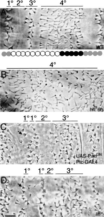
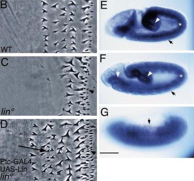
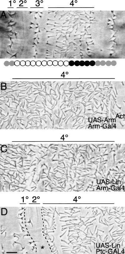
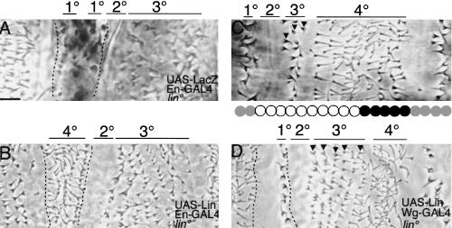
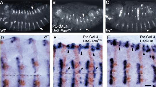
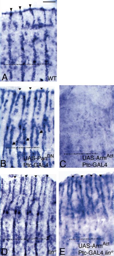
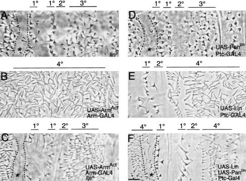
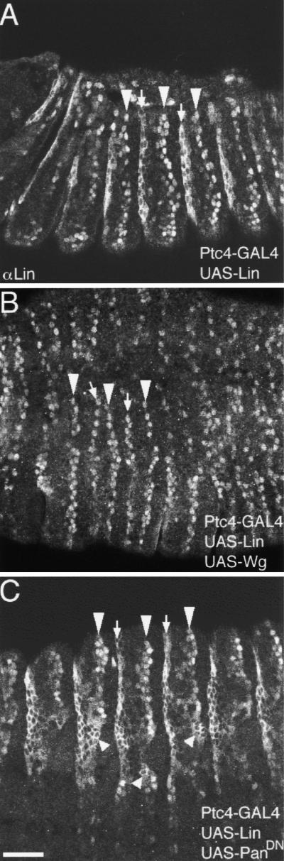
Wnt signaling controls a variety of developmental programs but the mechanisms by which the same signal leads to distinct outputs remain unclear. To address this question, we identified stage-specific modulators of Wingless (Wg) signaling in the Drosophila embryonic epidermis. We show that lines (lin) is essential for Wg-dependent patterning in dorsal epidermis. lin encodes a novel protein that acts cell-autonomously, downstream or in parallel to Armadillo (Arm) and upstream of Wg-dependent target genes. Lin can accumulate in nuclei of cells signaled by Wg, suggesting that signaling promotes entry of Lin into the nucleus, where it cooperates with Arm and Pangolin. Thus, a stage-specific modulator is used to mediate Wg signaling activity in dorsal patterning. Hedgehog (Hh) controls half of the parasegmental pattern dorsally and antagonizes Wg function to do so. Lin can accumulate in the cytoplasm of cells signaled by Hh, suggesting that Hh antagonizes Wg function by prohibiting Lin from entering the nucleus.


Figures

Figure 1
Figure 1
Contribution of Hh, Wg, and Lin to dorsal pattern. (A) Wild-type (WT) dorsal cuticle pattern. (1°, 2°, 3°, 4°). Four distinct cell types visible across the parasegment. We previously mapped En/Hh (gray circles) and Wg (black circles) expression domains that underlie this cuticle pattern (Heemskerk and DiNardo 1994; Bokor and DiNardo 1996). (B) hhts, inactivated at 6 hr AEL. 1°–3° fates are replaced by 4° fates. (C) Ptc–GAL4; UAS–PanDN. When Wg signaling is blocked, the 4° fates are lost and excess 3° fates develop. In addition, an ectopic 1° cell row develops (arrow). (D) lin. 4° fates are missing, and excess 3° fates develop. In addition, an ectopic 1° cell row develops (arrow). Bar, 10 μm in all panels.
Figure 2
Figure 2
Rescue of lin mutants and lin mRNA expression. (A) Smallest deficiency [Df(2R) P15; broken line] that uncovers lin and the position of the lin gene. (B) Wild-type ventral denticle belt composed of six denticle rows. (C) lin. Anteriormost three denticle rows are replaced with smooth cuticle, and denticle row 4 appears large like row 5. Excess denticles are produced posterior to the denticle belt (arrowhead), a phenotype similar to that obtained when late Wg function is attenuated (Bejsovec and Martinez-Arias 1991). (D) lin Ptc–GAL4; UAS–Lin. Anterior denticle rows are restored, and excess row 2/3 denticles are produced (arrow). Note that excess denticles posterior to the belt (arrowhead) are not restored to smooth cuticle. (E–G) RNA in situ. Low levels of lin are ubiquitously expressed in epidermis (closed arrows), with higher levels in hindgut (open arrowhead) and foregut (open arrow) and in mesoderm (asterisk). Bar, 5 μm in B–D; 100 μm in E–G.
Figure 2
Figure 2
Rescue of lin mutants and lin mRNA expression. (A) Smallest deficiency [Df(2R) P15; broken line] that uncovers lin and the position of the lin gene. (B) Wild-type ventral denticle belt composed of six denticle rows. (C) lin. Anteriormost three denticle rows are replaced with smooth cuticle, and denticle row 4 appears large like row 5. Excess denticles are produced posterior to the denticle belt (arrowhead), a phenotype similar to that obtained when late Wg function is attenuated (Bejsovec and Martinez-Arias 1991). (D) lin Ptc–GAL4; UAS–Lin. Anterior denticle rows are restored, and excess row 2/3 denticles are produced (arrow). Note that excess denticles posterior to the belt (arrowhead) are not restored to smooth cuticle. (E–G) RNA in situ. Low levels of lin are ubiquitously expressed in epidermis (closed arrows), with higher levels in hindgut (open arrowhead) and foregut (open arrow) and in mesoderm (asterisk). Bar, 5 μm in B–D; 100 μm in E–G.
Figure 3
Figure 3
Wg and Lin are sufficient for specifying the 4° cell fate. (A) Wild type. (B) UAS–ArmS10; Arm–GAL4. Activation of Wg signaling broadly shows replacement of 1°–3° fates by excess 4° fates. (C) UAS–Lin Arm–GAL4. Ubiquitous overexpression of Lin also leads to replacement of 1°–3° cell fates with the 4° fates. (D) Ptc–GAL4; UAS–Lin. Occasionally, overexpression of Lin is not sufficient to specify 4° fate in place of 2° (asterisk). Bar, 10 μm in all panels.
Figure 4
Figure 4
lin acts cell-autonomously. (A) lin En–GAL4; UAS–LacZ. Activity stain reveals the En domain in lin mutants (enclosed by broken lines), which includes ectopic and normal 1° cell types and two to three rows of smooth cuticle in between. (B) Expression of UAS–Lin with En–GAL4 in lin mutants transforms all cells in the En domain into 4° cells. (C) Wild type. Two to three rows differentiate as 3° cells (arrowheads). (D) Expression of UAS–Lin with Wg–GAL4 in lin mutants restores 4° fate to five to six cells rows where Wg is expressed, but not more anteriorly. (Arrowheads) Five rows that still differentiate as 3° cells. Note that the ectopic 1° cell type is restored to smooth cuticle. Bar, 10 μm in all panels.
Figure 5
Figure 5
Lin and Wg similarly control wg gene expression. (A) Wild-type, stage 13. Wg protein is expressed in stripes in both dorsal (open arrowhead) and ventral (open arrow) epidermis. (B) In embryos blocked for late Wg signaling, Wg expression is lost ventrally and is fading dorsally. (C) lin. Wg expression is maintained ventrally, but lost dorsally. In both B and C, Wg protein is missing from most dorsal cells, but expression is up-regulated in a few individual cells. (D) Wild type, Wg RNA (blue), En protein (brown). wg is expressed anterior to the En domain. (E) In embryos overexpressing activated Arm, an ectopic wg stripe is induced posterior to the En domain ventrally (arrows) and dorsally (arrowheads). (F) In embryos overexpressing Lin, an ectopic stripe of wg is induced posterior to the En domain, but only dorsally (arrowhead). Bar, 50 μm in A–C; 10 μm in D–F.
Figure 6
Figure 6
Lin and Wg similarly control ve gene expression. (A) Wild type, Ve RNA in situ. ve is expressed in one epidermal stripe extending from the dorsal edge (arrowheads) to the ventral midline (broken line). ve is expressed posteriorly adjacent to the En domain (Gritzan et al. 1999). Also note ve expression in some mesodermal and nerve cells underlying the epidermis. (B) Blockage of Wg signaling activity by expression of dominant-negative Pan with the Ptc–GAL4 driver leads to an ectopic stripe of ve that extends from the dorsal edge (arrowheads) to the ventral midline (arrows). The ectopic ve stripe is anteriorly adjacent to the En domain (Gritzan et al. 1999). (C) Reciprocally, activation of the Wg pathway by expression of activated Arm with the Ptc–GAL4 driver leads to repression of ve expression. (D) In lin mutants, an ectopic stripe of ve is found dorsally (arrowheads), but not ventrally. (E) Activation of Wg signaling by expression of activated Arm, with the Ptc–GAL4, in lin mutants represses ve expression ventrally but not dorsally. Bar, 20 μm in all panels.
Figure 7
Figure 7
Lin is essential for Wg signaling and can promote Wg-dependent readouts when Wg signaling is deficient (A) lin. (B) Activation of Wg signaling results in global specification of 4° cell types. (C) However, activation of Wg signaling in lin mutants, by expression of activated Arm with Arm–GAL4, does not lead to global specification of 4° cell types. Note that the ectopic 1° row is preceded by a region of differentiating smooth cuticle (asterisk), as in A. (D) Pattern in embryos blocked for late Wg signaling. (E) Ptc–GAL4; UAS–Lin. Excess rows of 4° cells. (F) Overexpression of Lin in embryos deficient in Wg signaling leads to differentiation of the 4° cell type, in place of the 3°, in cells flanking the En domain. Note that the ectopic 1° cell row is not rescued. However, anterior to this region, smooth cuticle is replaced with the 4° (asterisk). Bar, 10 μm in all panels.
Figure 8
Figure 8
Differential subcellular localization of Lin. (A) Ptc–GAL4; UAS–Lin. Anterior to the En domain, Lin localizes to nuclei (open arrowheads), whereas posterior to the En domain, Lin localizes to the cytoplasm (open arrows). (B) Coexpression of Lin and Wg with the Ptc–GAL4 driver leads to the localization of Lin in nuclei both anterior and posterior to the En domain. (C) Coexpression of Lin and dominant-negative Pan with the Ptc–GAL4 driver leads to the localization of Lin to the cytoplasm anterior to the En domain in lateral epidermis (small open arrowheads). Dorsally, the differential localization of Lin is maintained. Bar, 50 μm in all panels.

References

    1. Akiyoshi S, Inoue H, Hanai J, Kusanagi K, Nemoto N, Miyazono K, Kawabata M. c-Ski acts as a transcriptional co-repressor in transforming growth factor-beta signaling through interaction with smads. J Biol Chem. 1999;274:35269–35277. - PubMed
    1. Alexandre C, Lecoutois M, Vincent J-P. Wingless and Hedgehog pattern Drosophila denticle belts by regulating the production of short-range signals. Development. 1999;126:5689–5698. - PubMed
    1. Baker NE. Molecular cloning of sequences from wingless a segment polarity gene in Drosophila: The spatial distribution of a transcript in embryos. EMBO J. 1987;6:1765–1773. - PMC - PubMed
    1. ————— Embryonic and imaginal requirements for wg, a segment polarity gene in Drosophila. Dev Biol. 1988;125:96–108. - PubMed
    1. Behrens J, von Kries JP, Kuhl M, Bruhn L, Wedlich D, Grosschedl R, Birchmeier W. Functional interaction of β-catenin with the transcription factor LEF-1. Nature. 1996;382:638–642. - PubMed

Publication types

MeSH terms