. 2000 Jun 1;14(11):1390-9.

N-myc can functionally replace c-myc in murine development, cellular growth, and differentiation

Affiliations

N-myc can functionally replace c-myc in murine development, cellular growth, and differentiation

B A Malynn et al. Genes Dev. .

Abstract

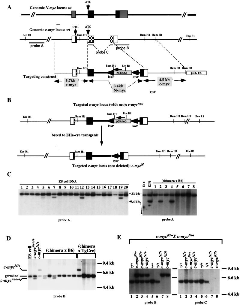
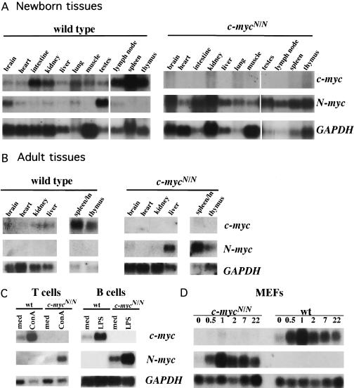
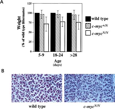
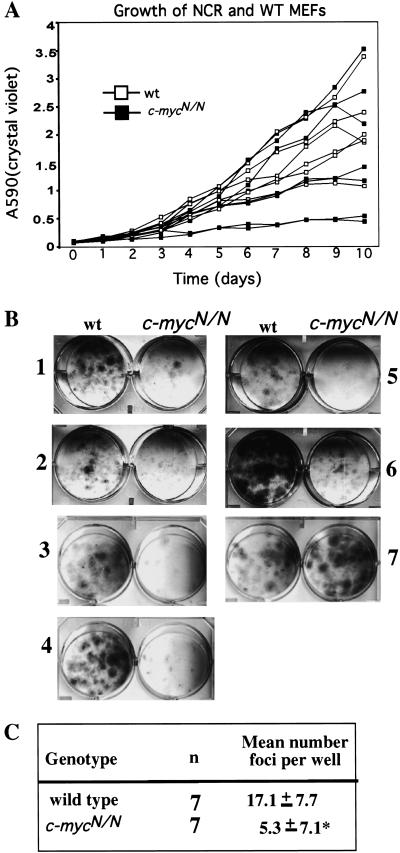
Members of the myc family of cellular oncogenes have been implicated as transcriptional regulators in pathways that govern cellular proliferation and death. In addition, N-myc and c-myc are essential for completion of murine embryonic development. However, the basis for the evolutionary conservation of myc gene family has remained unclear. To elucidate this issue, we have generated mice in which the endogenous c-myc coding sequences have been replaced with N-myc coding sequences. Strikingly, mice homozygous for this replacement mutation can survive into adulthood and reproduce. Moreover, when expressed from the c-myc locus, N-myc is similarly regulated and functionally complementary to c-myc in the context of various cellular growth and differentiation processes. Therefore, the myc gene family must have evolved, to a large extent, to facilitate differential patterns of expression.


Figures

Figure 1
Figure 1
Targeted replacement of c-myc coding sequences with N-myc coding sequences. (A) Schematic diagrams of the intron–exon structures of the genomic N-myc and c-myc loci and the N- into c-myc replacement construct. The majority of the coding sequences of N-myc (black boxes) and c-myc (hatched boxes) are contained within exons 2 and 3. The 5′ and 3′ untranslated regions are represented as shaded boxes for N-myc and open boxes for c-myc. In the targeting construct, called pNCR11, N-myc exons 2 and 3 and the intervening intron are flanked by 5′ and 3′ c-myc homology arms. PGKneo, surrounded by loxP sites, was inserted within the N-myc intron. (B) Schematic diagrams of the targeted c-myc loci before and after Cre-mediated recombination. pNCR11 was transfected into embryonic stem cells and screened for homologous recombination into the c-myc locus. A schematic of the structure of the targeted locus, called c-mycneo, is shown. Mice derived from the targeted ES cells were bred to transgenic mice carrying the EIIa–cre transgene and offspring were screened for transmission of the Cre-mediated recombination, resulting in deletion of the neo gene. A schematic of the structure of the targeted locus, called c-mycN, is shown. (C) Southern blot analysis for germ-line transmission of the c-mycneo targeted locus. Targeting of ES cells was screened by digestion of DNA with EcoRI and hybridization to probe A (left, lanes 5,7,12,19). Appropriate targeting was confirmed by digestion with multiple restriction enzymes and probes (data not shown). Targeted ES cells were injected into blastocysts and chimeric mice were bred either to C57BL/6 or 129Sv/Ev mice. Offspring were tested for germ-line transmission by digesting tail DNA samples with EcoRI and hybridizing to probe A (right, lanes 2,4; data not shown). (D) Southern blot analysis for Cre-mediated deletion of the neo gene to produce c-mycN/N mice. c-mycneo-transmitting chimera were bred to EIIa–cre transgenic mice. Tail-DNA samples were analyzed by digestion with BamHI and Southern blots were hybridized to probe B. The germ line BamHI fragment hybridizing to probe B is ∼6.0 kb; the c-mycneo allele results in a ∼5.5-kb fragment, because of inserted BamHI site in the PGKneo sequence. After Cre-mediated deletion of the PGKneo gene, the elimination of this BamHI site results in a probe B-hybridizing fragment of ∼7.0 kb. (E) Mating of heterozygous c-mycN/+ mice yielded homozygous mice that survived in the absence of a c-myc gene. Tail DNA from 10-day-old offspring of a c-mycN/+ heterozygous mating was digested with BamHI and Southern blots were hybridized to probe B (left). The diagnosed genotypes are designated above each lane and included wild type (c-myc+/+), heterozygous (c-mycN/+), and homozygous (c-mycN/N) (left). The Southern blot was stripped and hybridized to probe C. DNA from c-mycN/N mice failed to hybridize to this probe, which is composed of both coding exons of c-myc DNA, proving that these mice are unable to produce c-myc-encoded transcripts (right).
Figure 2
Figure 2
Expression of c-mycN allele. (A,B) Expression of N-myc and c-myc transcripts in wild-type and c-mycN/N mice. Replicate northern blots of total RNA, extracted from tissues of adult (B) or newborn (A) wild-type and homozygous knock-in mice, were hybridized to probes specific for the c-myc or N-myc coding exons 2 and 3. Blots were subsequently hybridized to GAPDH to control for RNA loading. (C) Mitogen-stimulated up-regulation of N-myc and c-myc genes in lymphocytes. B or T lymphocytes, enriched from spleen or lymph nodes of wild-type or c-mycN/N mice, were incubated in vitro with or without stimulation with LPS or ConA, respectively. RNA was extracted, electrophoresed, and replicate northern blots were hybridized as above. (D) Serum-stimulated up-regulation of N-myc and c-myc genes in MEFs. Subconfluent MEFS were cultured in low (0.5%) serum for 48 hr. Media with 10% serum was added at time 0 and cells were harvested for RNA extraction at various times thereafter and electrophoresed. Replicate Northern blots were hybridized as above.
Figure 3
Figure 3
Potential differences between c-mycN/N and wild-type mice. (A) The average weight of c-mycN/N mice. Mice from individual litters were weighed at various times after birth. For each litter, the weights of heterozygous or homozygous mutant mice were calculated as a percentage of the average weight of wild-type littermates. The mean (±s.d.) percentage of heterozygous and homozygous mutant mice was calculated. The mean weight of c-mycN/N mice was significantly different from that of wild-type or heterozygous mice (P < 0.001). (B) Histological analysis of c-mycN/N mice. Sections of skeletal muscle from wild-type (left) or homozygous c-mycN/N (right) day 1 newborns are shown. The appearance of muscular dystrophy periodic and observed in 1/2 of the homozygotes.
Figure 4
Figure 4
Development and proliferation of c-mycN/N lymphocytes. (A) Flow cytometric analysis of lymphocytes from wild-type and homozygous c-mycN/N mice. Single cell populations isolated from thymus (top left), spleen (right), and bone marrow (bottom left) from wild-type or homozygous c-mycN/N mice were stained with a panel of antibodies that identify various subpopulations of T- or B-lineage lymphocytes, as indicated. Live cells were gated based on forward and side-scatter profile. The percentage of gated cells in particular quadrants or boxes are shown. Similar percentages of cell subpopulations were observed for homozygous c-mycN/N and wild-type mice. Absolute numbers of cells in the various fractions were also calculated. No significant differences were found (data not shown). (B,C,D,E) In vitro proliferation of wild-type and homozygous c-mycN/N lymphocytes. T (B,C) and B lymphocytes (D,E) were enriched from lymph node and spleen, respectively, of homozygous c-mycN/N or wild-type littermates and cultured either with medium or various doses of ConA ± IL-2 (B), anti-CD3 ± IL-2 (C), LPS (D), or anti-Ig ± IL-4 (E). The incorporation of 3H-thymidine was measured and indicated as counts per minute.
Figure 5
Figure 5
Growth potential of wild type and homozygous c-mycN/N mouse embryonic fibroblasts. (A) P3 MEFS were plated at 5 × 104 cells per well in replicate wells and cultured for the indicated number of days. Triplicate wells were fixed and stained with crystal violet. Adsorption at OD540 was measured for each time point for individual MEF lines and directly correlates with cell numbers (Kamijo et al. 1997). Each symbol represents the mean of triplicate readings of triplicate wells. The standard deviation was <1%. (B,C) P3 MEFS were plated at 3500 cells per well in triplicate, cultured for 12 days, and fixed and stained with crystal violet. A representative plate is shown. The number of foci per well was determined, the mean (±s.d.) was calculated for each cell line. The mean number of foci per well from c-mycN/N MEFs was significantly less than that of wild-type MEFs (*P < 0.01).

References

    1. Amati B, Brooks MW, Levy N, Littlewood TD, Evan GI, Land H. Oncogenic activity of the c-Myc protein requires dimerization with Max. Cell. 1993;72:233–245. - PubMed
    1. Auffray C, Rougeon F. Purification of mouse immunoglobulin heavy-chain messenger RNAs from total myeloma tumor RNA. Eur J Biochem. 1980;107:303–314. - PubMed
    1. Barrett J, Birrer MJ, Kato GJ, Dosaka-Akita H, Dang CV. Activation domains of L-Myc and c-Myc determine their transforming potencies in rat embryo cells. Mol Cell Biol. 1992;12:3130–3137. - PMC - PubMed
    1. Bush A, Mateyak M, Dugan K, Obaya A, Adachi S, Sedivy J, Cole M. c-myc null cells misregulate cad and gadd45 but not other proposed c-Myc targets. Genes & Dev. 1998;12:3797–3802. - PMC - PubMed
    1. Charron J, Malynn BA, Fisher P, Stewart V, Jeannotte L, Goff SP, Robertson EJ, Alt FW. Embryonic lethality in mice homozygous for a targeted disruption of the N-myc gene. Genes & Dev. 1992;6:2248–2257. - PubMed

Publication types

MeSH terms

LinkOut - more resources