

Impaired emotional declarative memory following unilateral amygdala damage
- PMID: 10837507
- PMCID: PMC311327
- DOI: 10.1101/lm.7.3.180
Impaired emotional declarative memory following unilateral amygdala damage
Abstract
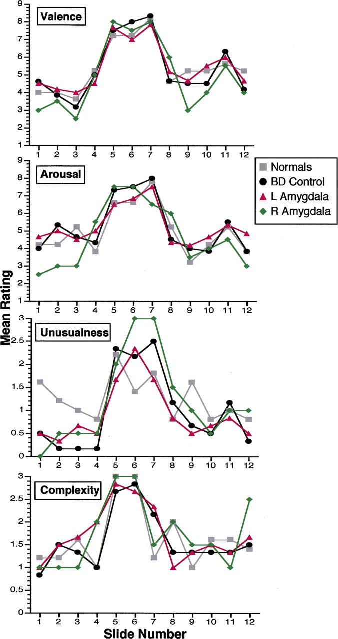
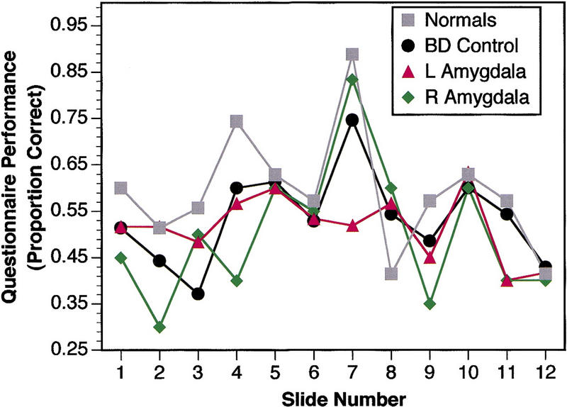
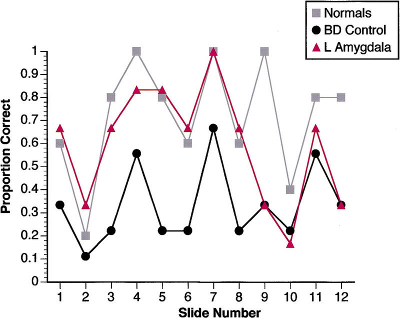
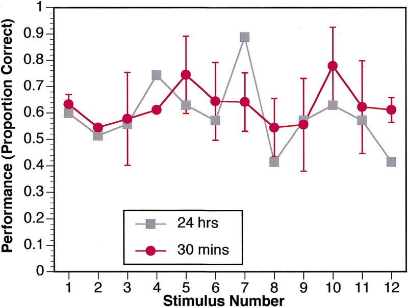
Case studies of patients with bilateral amygdala damage and functional imaging studies of normal individuals have demonstrated that the amygdala plays a critical role in encoding emotionally arousing stimuli into long-term declarative memory. However, several issues remain poorly understood: the separate roles of left and right amygdala, the time course over which the amygdala participates in memory consolidation, and the type of knowledge structures it helps consolidate. We investigated these questions in eight subjects with unilateral amygdala damage, using several different measures. For comparison, our main task used stimuli identical to those used previously to investigate emotional declarative memory in patients with bilateral amygdala damage. Contrasts with both brain-damaged and normal control groups showed that subjects with left amygdala damage were impaired in their memory for emotional stimuli, despite entirely normal memory for neutral stimuli (because of a number of caveats, the findings from subjects with right amygdala damage were less clear). Follow-up experiments suggested that the normal facilitation of memory for emotional stimuli may develop over an extended time course (>30 min), consistent with prior findings, and that the specific impairment we report may depend in part on the lexical nature of the task used (written questionnaire). We stress the complex and temporally extended nature of memory consolidation and suggest that the amygdala may influence specific components of this process.
Figures

References
-
- Adolphs R, Cahill L, Schul R, Babinsky R. Impaired declarative memory for emotional material following bilateral amygdala damage in humans. Learn & Mem. 1997;4:291–300. - PubMed
-
- Babinsky R, Calabrese P, Durwen HF, Markowitsch HJ, Brechtelsbauer D. The possible contribution of the amygdala to memory. Behav Neurol. 1993;6:167–170. - PubMed
-
- Berlyne DE, Borsa DM, Craw MA, Gelman RS, Mandell EE. Effects of stimulus complexity and induced arousal on paired-associate learning. J Verbal Learn Verbal Behav. 1965;4:291–299.
-
- Bianchin M, Mello e Souza T, Medina JH, Izquierdo I. The amygdala is involved in the modulation of long-term memory, but not in working or short-term memory. Neurobiol Learn Mem. 1999;71:127–131. - PubMed
-
- Bradley MM, Greenwald MK, Petry MC, Lang PJ. Remembering pictures: Pleasure and arousal in memory. J Exp Psychol Learn Mem & Cog. 1992;18:379–390. - PubMed
Publication types
MeSH terms
LinkOut - more resources
Full Text Sources
Medical