. 2000 Jun 5;191(11):1895-903.
doi: 10.1084/jem.191.11.1895.

In vivo identification of glycolipid antigen-specific T cells using fluorescent CD1d tetramers

Affiliations

In vivo identification of glycolipid antigen-specific T cells using fluorescent CD1d tetramers

K Benlagha et al. J Exp Med. .

Abstract

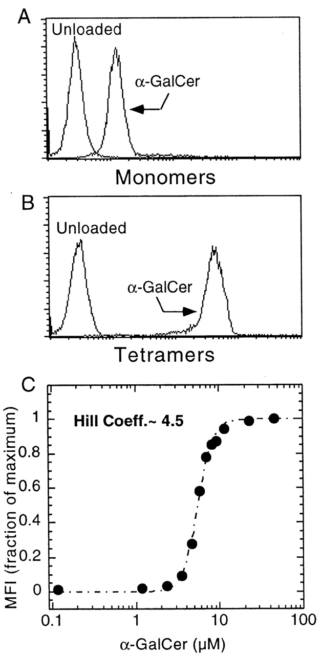
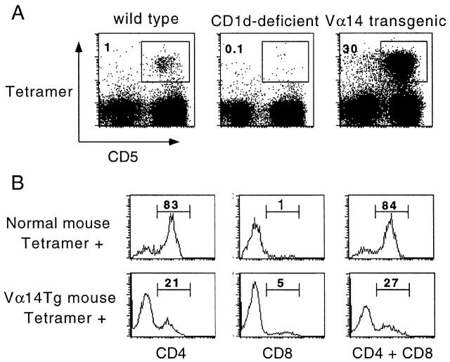
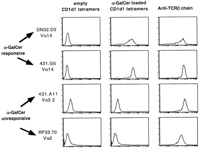
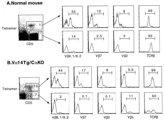
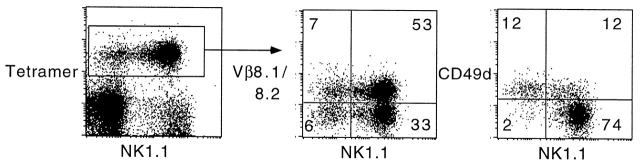
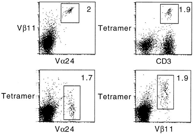
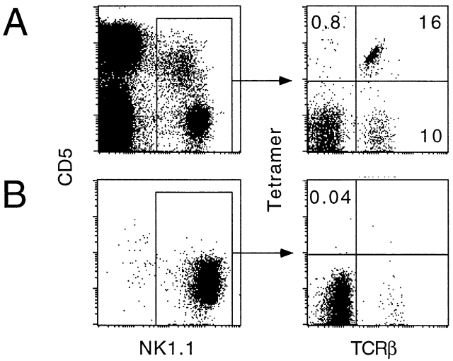
The CD1 family of major histocompatibility complex (MHC)-like molecules specializes in presenting lipid and glycolipid antigens to alpha/beta T lymphocytes, but little is known about the size of the CD1-restricted T cell population or the frequency of T lymphocytes specific for a given glycolipid antigen. Here, we report the generation and use of mouse CD1d1-glycolipid tetramers to visualize CD1d-restricted T cells. In contrast with previous BIAcore-based estimates of very short half-lives for CD1d-glycolipid complexes, we found that the dissociation rate of several different CD1d-glycolipid complexes was very slow. Fluorescent tetramers of mouse CD1d1 complexed with alpha-galactosylceramide (alphaGalCer), the antigen recognized by mouse Valpha14-Jalpha281/Vbeta8 and human Valpha24-JalphaQ/Vbeta11 natural killer T (NKT) cell T cell receptors (TCRs), allowed us for the first time to accurately describe, based on TCR specificity, the entire population of NKT cells in vivo and to identify a previously unrecognized population of NK1.1-negative "NKT" cells, which expressed a different pattern of integrins. In contrast, natural killer (NK) cells failed to bind the tetramers either empty or loaded with alphaGalCer, suggesting the absence of a CD1d-specific, antigen-nonspecific NK receptor. Mouse CD1d1-alphaGalCer tetramers also stained human NKT cells, indicating that they will be useful for probing a range of mouse and human conditions such as insulin-dependent diabetes mellitus, tumor rejection, and infectious diseases where NKT cells play an important role.


Figures

Figure 1
Figure 1
Long life span of CD1d1–glycolipid complexes. (A) Plate-bound CD1d1 (2.5 μg/ml) was loaded with 0.1 or 0.3 μM αGalCer for 2 h, washed, and chased for different periods of time before adding the Vα14-Jα281 NKT cell hybridoma DN32D3 and measuring IL-2 release in the supernatant (mean ± SD). (B) Plate-bound CD1d1 was first incubated with 30 μg/ml of LAM or GM1 for 6 h, washed, and chased for different periods of time. 0.1 μM αGalCer was added for 2 h and washed before incubation with DN32D3. (C) Microwells were coated overnight with 2.5 μg/ml CD1d1 or BSA or 0.1μM αGalCer, washed, then pulsed for 2 h with 0.1 μM αGalCer as indicated, and washed again before adding the CD1d-restricted Vα14 hybridomas DN32D3 or a control CD1d-restricted non-Vα14 hybridoma, 431.A11 (Vα3.2-Jα8/Vβ8), and measuring IL-2 release (mean ± SD). 431A11 and DN32D3 produced similar amounts of IL-2 when stimulated with CD1d1-expressing thymocytes.
Figure 1
Figure 1
Long life span of CD1d1–glycolipid complexes. (A) Plate-bound CD1d1 (2.5 μg/ml) was loaded with 0.1 or 0.3 μM αGalCer for 2 h, washed, and chased for different periods of time before adding the Vα14-Jα281 NKT cell hybridoma DN32D3 and measuring IL-2 release in the supernatant (mean ± SD). (B) Plate-bound CD1d1 was first incubated with 30 μg/ml of LAM or GM1 for 6 h, washed, and chased for different periods of time. 0.1 μM αGalCer was added for 2 h and washed before incubation with DN32D3. (C) Microwells were coated overnight with 2.5 μg/ml CD1d1 or BSA or 0.1μM αGalCer, washed, then pulsed for 2 h with 0.1 μM αGalCer as indicated, and washed again before adding the CD1d-restricted Vα14 hybridomas DN32D3 or a control CD1d-restricted non-Vα14 hybridoma, 431.A11 (Vα3.2-Jα8/Vβ8), and measuring IL-2 release (mean ± SD). 431A11 and DN32D3 produced similar amounts of IL-2 when stimulated with CD1d1-expressing thymocytes.
Figure 2
Figure 2
Binding of CD1d1–αGalCer complexes to a Vα14-Jα281/Vβ8 NKT cell hybridoma. (A) Biotinylated monomers, revealed with streptavidin-PE. (B) Streptavidin-PE tetramers. (C) Binding curve of CD1d1 tetramers loaded at different concentrations of αGalCer; binding was measured by flow cytometry analysis (mean fluorescence intensity, MFI). Hill Coeff., Hill coefficient (see Materials and Methods).
Figure 4
Figure 4
CD1d1–αGalCer tetramers stained CD1d1-restricted Vα14-Jα281 NKT cells in vivo. (A) Spleen cells from wild-type, CD1d-deficient, or Vα14-Jα281 TCR α chain transgenic mice in a C57BL/6 background were stained with CD1d1–αGalCer tetramers-PE, CD5-Cychrome, and anti–I-Ab–FITC. Dot plots were gated on MHC class II splenocytes. (B) Splenocytes from normal mice and Vα14-Jα281 TCR α chain transgenics were stained with tetramer-Cychrome, CD5-APC, and anti-CD4 or CD8-FITC. Histograms display the frequency of CD4+ and/or CD8+ cells among gated CD5+ tetramer+ splenocytes.
Figure 3
Figure 3
CD1d1–αGalCer tetramers specifically stained Vα14-Jα281 NKT cell hybridomas. “Empty” or αGalCer-loaded tetramers and anti–TCR-β mAb were used to stain two canonical Vα14-expressing, αGalCer-responsive hybridomas (DN32D3 and 431G5), one CD1d1-restricted αGalCer-unresponsive Vα3.2 hybridoma (431A11), and one Kb-restricted Vα2 hybridoma (RF33.70).
Figure 5
Figure 5
CD1d1–αGalCer tetramer+ cells exhibit intermediate levels of surface TCR and a bias in Vβ usage. Splenocytes from normal mice (top) and Vα14-Jα281 TCR α chain transgenics in a C57BL/6.TCR Cα2/− background (bottom) were stained with tetramer-Cychrome, CD5-APC, and anti-Vβ–FITC. Histograms display the frequency of Vβ1 cells among CD5+ tetramer+ and CD5+ tetramer splenocytes gated as indicated.
Figure 6
Figure 6
A subset of CD1d1–αGalCer tetramer+ cells are NK1.1 and express upregulated levels of CD49d. Liver lymphocytes from normal C57BL/6 mice were stained with tetramer-Cychrome, NK1.1-PE, and anti-Vβ–FITC or CD49d-FITC. Note that most tetramer+ NK1.1+ cells were CD49d whereas tetramer+ NK1.1 cells were CD49d+. Both subsets exhibited the same bias in Vβ8 (middle) and Vβ7 and Vβ2 (not shown). Similar results were found for splenocytes and in Vα14-Jα281 TCR α chain transgenic mice.
Figure 7
Figure 7
Mouse CD1d1–αGalCer tetramers stain human Vα24/Vβ11 NKT cells. Human PBLs were stimulated with 0.1 μM αGalCer in vitro and stained after 7 d in culture. In this experiment, CD1d1-mediated αGalCer presentation to NKT cells is provided by the PBLs themselves. After centrifugation through Ficoll, the cells were preincubated with anti-TCR antibodies for 1 h before adding tetramer-PE. Note that anti-Vα24 completely blocks tetramer binding.
Figure 8
Figure 8
CD1d1–αGalCer tetramers do not stain NK cells. Fresh splenocytes (A) or spleen-derived A-LAK cells (B) were stained with CD5-APC, NK1.1-PE, tetramer-Cychrome, and TCR-β–FITC. Less than 1% of NK cells (α/β sTCRNK1.1+) were tetramer+.

References

    1. Calabi F., Jarvis J.M., Martin L.H., Milstein C. Two classes of CD1 genes. Eur. J. Immunol. 1989;19:285–292. - PubMed
    1. Porcelli S.A., Modlin R.L. The CD1 systemantigen-presenting molecules for T cell recognition of lipids and glycolipids. Annu. Rev. Immunol. 1999;17:297–329. - PubMed
    1. Kawano T., Cui J., Koezuka Y., Toura I., Kaneko Y., Motoki K., Ueno H., Nakagawa R., Sato H., Kondo E. CD1d-restricted and TCR-mediated activation of valpha14 NKT cells by glycosylceramides. Science. 1997;278:1626–1629. - PubMed
    1. Brossay L., Chioda M., Burdin N., Koezuka Y., Casorati G., Dellabona P., Kronenberg M. CD1d-mediated recognition of an alpha-galactosylceramide by natural killer T cells is highly conserved through mammalian evolution. J. Exp. Med. 1998;188:1521–1528. - PMC - PubMed
    1. Spada F.M., Koezuka Y., Porcelli S.A. CD1d-restricted recognition of synthetic glycolipid antigens by human natural killer T cells. J. Exp. Med. 1998;188:1529–1534. - PMC - PubMed

Publication types