. 2000 Jun 5;191(11):1945-56.
doi: 10.1084/jem.191.11.1945.

Enhanced growth of primary tumors in cancer-prone mice after immunization against the mutant region of an inherited oncoprotein

Affiliations

Enhanced growth of primary tumors in cancer-prone mice after immunization against the mutant region of an inherited oncoprotein

C T Siegel et al. J Exp Med. .

Abstract

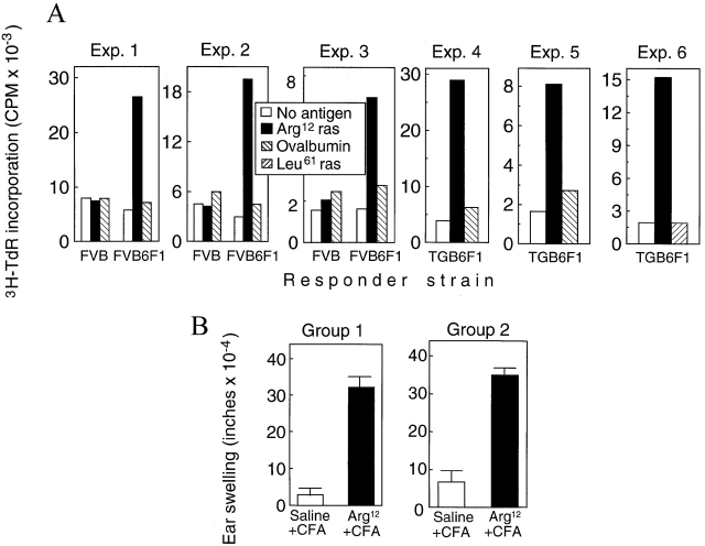
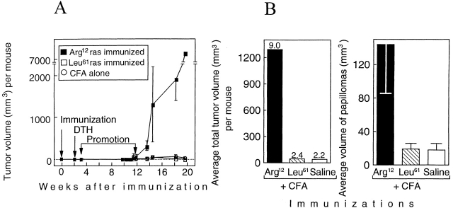
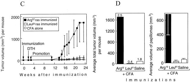
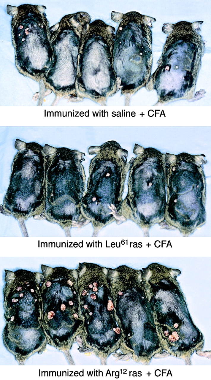
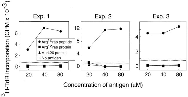
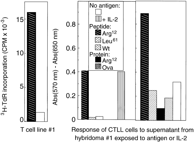
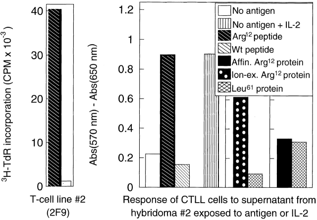
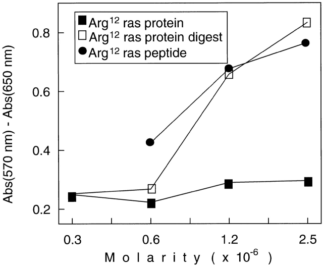
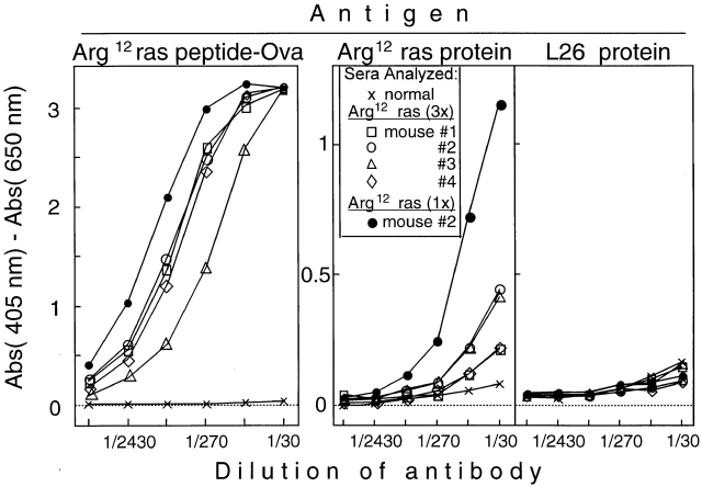
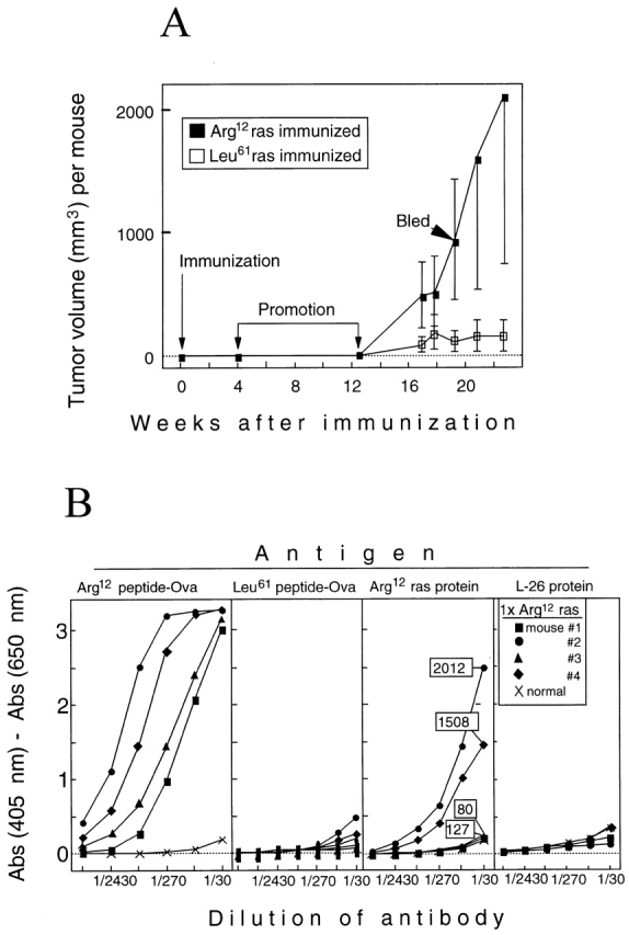
One major objective of tumor immunologists is to prevent cancer development in individuals at high risk. (TG.AC x C57BL/6)F1 mice serve as a model for testing the feasibility of this objective. The mice carry in the germline a mutant ras oncogene that has an arginine at codon 12 instead of glycine present in the wild-type, and after physical (wounding) or chemical promotion, these mice have a high probability for developing papillomas that progress to cancer. Furthermore, F1 mice immunized with Arg(12) mutant ras peptide in complete Freund's adjuvant (CFA) develop T cells within 10 d that proliferate in vitro on stimulation with the Arg(12) mutant ras peptide. Within 14 d, these mice have delayed-type hypersensitivity to the peptide. Immunization with CFA alone or with a different Arg(12) mutant ras peptide in CFA induced neither response. To determine the effect of immunization on development of tumors, mice immunized 3 wk earlier were painted on the back with phorbol 12-myristate 13-acetate every 3 d for 8 wk. The time of appearance and the number of papillomas were about the same in immunized and control mice, but the tumors grew faster and became much larger in the mice immunized with the Arg(12) mutant ras peptide. Thus, the immunization failed to protect against growth of papillomas. The peptide-induced CD4(+) T cells preferentially recognized the peptide but not the native mutant ras protein. On the other hand, mice immunized with Arg(12) mutant ras peptide and bearing papillomas had serum antibodies that did bind native mutant ras protein. Together, these studies indicate that active immunization of cancer-prone individuals may result in immune responses that fail to eradicate mutant oncogene-expressing tumor cells, but rather induce a remarkable enhancement of tumor growth.


Figures

Figure 1
Figure 1
FVB6F1 mice and cancer-prone TGB6F1 mice carrying a mutant ras transgene mount a specific DTH response when immunized against the mutant ras peptide, and lymphocytes from these mice mount a specific proliferative response to the mutant peptide. (A) The proliferative response of lymphocytes from immunized FVB, FVB6F1, and TGB6F1 mice was analyzed by harvesting draining LNCs 7–10 d after immunization and pulsing them with 3H-TdR after 2 d of culture with or without antigen, as indicated. Three representative experiments using cells taken from a single or a pool of animals are shown. (B) DTH reactions were assayed in TGB6F1 9-wk -old (Group 1)and 13-wk-old (Group 2) mice that were naive or had been immunized with the Arg12 mutant ras peptide in CFA or with saline in CFA. 14 d after the immunization, mice were challenged with Arg12 mutant ras peptide in one ear and saline in the other. The ear swelling was measured 24 h later. Bars represent SEM of groups consisting of 10 mice.
Figure 2
Figure 2
Immunization against a mutant region of the ras oncoprotein results in enhanced growth of primary tumors in mice carrying the mutant ras gene in the germline. (A) Kinetics of tumor development in relation to immunization, DTH analysis, and promotion. Chemical promotion with PMA once every 3 d (20 treatments) was begun 3 wk after and ended 11.7 wk after immunization. Five out of five Arg12 mutant ras–immunized mice, five out of five Leu61 mutant ras–immunized mice, and three out of five mice immunized with CFA developed two or more papillomas. (B) The average total volume of tumors per mouse and the average volume of papillomas within each group of mice. Tumor volumes in B were measured 2.5 wk after the end of PMA treatment. The numbers on top of the bars at left represent the average number of papillomas per mouse within that group. Five mice per group were used. (C and D) The design of the experiment and results were similar, except the experiment was done ∼1 yr later with 10 mice per group. Tumor volumes in D were measured 12.1 wk after the end of the PMA treatment. 8/10 Arg12 mutant ras–immunized mice, 1/10 Leu61 ras–immunized mice, and 3/10 mice immunized with CFA alone developed two or more papillomas. The data in Fig. 2A–D remain virtually unchanged when all mice that developed no or only one papilloma after promotion are excluded as potential “nonresponders” (references 12, 13; described in Materials and Methods).
Figure 2
Figure 2
Immunization against a mutant region of the ras oncoprotein results in enhanced growth of primary tumors in mice carrying the mutant ras gene in the germline. (A) Kinetics of tumor development in relation to immunization, DTH analysis, and promotion. Chemical promotion with PMA once every 3 d (20 treatments) was begun 3 wk after and ended 11.7 wk after immunization. Five out of five Arg12 mutant ras–immunized mice, five out of five Leu61 mutant ras–immunized mice, and three out of five mice immunized with CFA developed two or more papillomas. (B) The average total volume of tumors per mouse and the average volume of papillomas within each group of mice. Tumor volumes in B were measured 2.5 wk after the end of PMA treatment. The numbers on top of the bars at left represent the average number of papillomas per mouse within that group. Five mice per group were used. (C and D) The design of the experiment and results were similar, except the experiment was done ∼1 yr later with 10 mice per group. Tumor volumes in D were measured 12.1 wk after the end of the PMA treatment. 8/10 Arg12 mutant ras–immunized mice, 1/10 Leu61 ras–immunized mice, and 3/10 mice immunized with CFA alone developed two or more papillomas. The data in Fig. 2A–D remain virtually unchanged when all mice that developed no or only one papilloma after promotion are excluded as potential “nonresponders” (references 12, 13; described in Materials and Methods).
Figure 3
Figure 3
Enhanced tumor development in TGB6F1 mice with the Arg12 mutant ras peptide. Five mice per group were immunized with the Arg12 mutant ras peptide in CFA, the Leu61 mutant ras peptide in CFA, or with saline in CFA. Pictures were taken 1.5 wk after the end of promotion (mice anesthetized for photography). Although the difference between the specifically immunized and the control groups is apparent, the bottom panel shows that papillomas developed more slowly in one of the five Arg12 mutant ras peptide–immunized mice, and some of the papillomas in the Arg12 mutant ras of the other four similarly immunized group remained small. The results shown here and in Fig. 2A and Fig. B are derived from the same experiment; pictures shown here were taken 1 wk earlier than results shown for the same mice in Fig. 2 B.
Figure 4
Figure 4
Photomicrograph illustrating the appearance of a papilloma on the back skin of an Arg12 mutant ras–immunized mouse at time of sacrifice. The lower magnification (original magnification: ×20) shows the keratinization of the papilloma. At the higher magnification (original magnifications: ×100 and ×200), the dysplastic epithelial changes and the neutrophilic inflammatory infiltrate at the epidermal–dermal interphase become evident. These inflammatory infiltrates do not contribute significantly to the tumor volume and are similarly found in papillomas of control mice (not shown). Hematoxylin and eosin staining.
Figure 5
Figure 5
Arg12 mutant ras peptide is preferentially recognized by T cells from mice immunized with the Arg12 mutant ras peptide or by T hybridoma cells derived from these T cells. Top: proliferative response of LNCs in vitro from mice immunized with Arg12 mutant ras peptide, and with CFA 9 d earlier. Three representative experiments using cells taken from a single or pool of animals are shown. Mutant L26 (MutL26) protein is used as a control. Middle: proliferation of an anti-Arg12 mutant ras T cell line when cultured with the Arg12 mutant ras peptide (left). T cell hybridoma derived from this T cell line remains Arg12 mutant ras peptide specific, but does not respond to the Arg12 mutant ras protein (right). Cells from the T cell line or T cell hybridoma were cultured with antigen, and spleen cells were provided as APCs. The antigen-specific stimulation of the T cells caused increased 3H-TdR incorporation, whereas the antigen-specific stimulation of the T cell hybridoma caused IL-2 release, which was analyzed using growth stimulation of the IL-2–dependent CTLL cells as determined by a colorimetric assay (described in Materials and Methods). Bottom: same as top, except a different T cell line 2F9 8 was used. This T cell line 2F9 is also specific for the Arg12 mutant ras peptide, and the T cell hybridoma derived from this T cell line again only recognizes the mutant peptide and not the intact protein. Affinity-purified (Affin.) Arg12 mutant ras protein was prepared from a recombinant GST fusion protein as described. The ion exchange (Ion-ex.) matrix–purified Arg12 and Leu61 mutant ras proteins were a gift from Dr. R.G. Fenton. The average of duplicate samples are shown. Wt, wild-type.
Figure 5
Figure 5
Arg12 mutant ras peptide is preferentially recognized by T cells from mice immunized with the Arg12 mutant ras peptide or by T hybridoma cells derived from these T cells. Top: proliferative response of LNCs in vitro from mice immunized with Arg12 mutant ras peptide, and with CFA 9 d earlier. Three representative experiments using cells taken from a single or pool of animals are shown. Mutant L26 (MutL26) protein is used as a control. Middle: proliferation of an anti-Arg12 mutant ras T cell line when cultured with the Arg12 mutant ras peptide (left). T cell hybridoma derived from this T cell line remains Arg12 mutant ras peptide specific, but does not respond to the Arg12 mutant ras protein (right). Cells from the T cell line or T cell hybridoma were cultured with antigen, and spleen cells were provided as APCs. The antigen-specific stimulation of the T cells caused increased 3H-TdR incorporation, whereas the antigen-specific stimulation of the T cell hybridoma caused IL-2 release, which was analyzed using growth stimulation of the IL-2–dependent CTLL cells as determined by a colorimetric assay (described in Materials and Methods). Bottom: same as top, except a different T cell line 2F9 8 was used. This T cell line 2F9 is also specific for the Arg12 mutant ras peptide, and the T cell hybridoma derived from this T cell line again only recognizes the mutant peptide and not the intact protein. Affinity-purified (Affin.) Arg12 mutant ras protein was prepared from a recombinant GST fusion protein as described. The ion exchange (Ion-ex.) matrix–purified Arg12 and Leu61 mutant ras proteins were a gift from Dr. R.G. Fenton. The average of duplicate samples are shown. Wt, wild-type.
Figure 6
Figure 6
The mutant Arg12 ras peptide and digests of the Arg12 mutant ras protein but not the intact protein stimulate Arg12 mutant ras peptide–specific T hybridoma cells. Equimolar concentrations of either the Arg12 mutant ras peptide, the undigested protein, or protein digested by exposure to the bacterial endoprotease Glu-C were tested for antigen-specific stimulation of hybridoma R12-H2 derived from the 2F9 T cell line. Antigen recognition by the hybridoma cells resulted in IL-2 release, which was analyzed using growth stimulation of the IL-2–dependent CTLL cells in a colorimetric assay. Proteins were prepared and digested as described in Materials and Methods.
Figure 7
Figure 7
TGB6F1 mice immunized repeatedly with the Arg12 mutant ras peptide produce Arg12 mutant ras peptide–specific antibodies, and some of these antibodies also recognize the intact protein specifically. Mice were immunized intraperitoneally with 100 μg of the Arg12 or Leu61 mutant ras peptide in CFA and boosted twice at 2-wk intervals with the peptide in IFA. 2 wk after the last boost, sera from immunized mice were tested for antibodies specific for mutant ras peptide or protein. Sera of all four mice have antibody that binds mutant ras peptide and also the mutant ras protein, but the antibodies did not bind a control protein. For comparison, the serum of mouse no. 2 (•), which was immunized only once (1x) with the Arg12 mutant ras peptide in CFA and then promoted with PMA followed by development of papillomas, was included. This serum is identical to that of mouse no. 2 (•) shown in the middle panel. 3x, three times.
Figure 8
Figure 8
TGB6F1 mice immunized only once but then promoted with PMA can produce antibody specific for Arg12 mutant ras peptide, and antibody titers correlate directly with tumor burden. (A) Experimental design and kinetics of tumor growth. TGB6F1 mice were immunized with 150 μg of either Arg12 or Leu61 mutant ras peptide in CFA (four mice per group) and then promoted with PMA as described in Materials and Methods. Kinetics of tumor development and time of serum sampling in this group is indicated. (B.) Sera of all of the four TGB6F1 mice singly (1x) immunized with the Arg12 mutant ras peptide and then promoted with PMA have antibody that binds Arg12 mutant ras peptide and not the Leu61 mutant ras peptide, but only antibodies in sera of mice nos. 2 and 4 also bind mutant ras protein. These antibodies were Arg12 mutant ras protein specific because they did not bind to the L26 control protein. Protein binding antibody titers correlated with the host tumor burden. Numbers in the rectangles represent total tumor volume in mm3 for the indicated mouse at the time the serum was taken. Sera from Leu61 mutant peptide–immunized mice do not have antibodies that bind either ras peptide (data not shown).

References

    1. Fearon E.R. Human cancer syndromesclues to the origin and nature of cancer. Science. 1997;278:1043–1050. - PubMed
    1. Leder A., Kuo A., Cardiff R.D., Sinn E., Leder P. v-Ha-ras transgene abrogates the initiation step in mouse skin tumorigenesiseffects of phorbol esters and retinoic acid. Proc. Natl. Acad. Sci. USA. 1990;87:9178–9182. - PMC - PubMed
    1. Barbacid M. ras genes. Annu. Rev. Biochem. 1987;56:779–827. - PubMed
    1. Germolec D.R., Spalding J., Yu H.S., Chen G.S., Simeonova P.P., Humble M.C., Bruccoleri A., Boorman G.A., Foley J.F., Yoshida T., Luster M.I. Arsenic enhancement of skin neoplasia by chronic stimulation of growth factors. Am. J. Pathol. 1998;153:1775–1785. - PMC - PubMed
    1. Cannon R.E., Spalding J.W., Trempus C.S., Szczesniak C.J., Virgil K.M., Humble M.C., Tennant R.W. Kinetics of wound-induced v-Ha-ras transgene expression and papilloma development in transgenic Tg.AC mice. Mol. Carcinog. 1997;20:108–114. - PubMed

Publication types