Review
. 2000 Jun;64(2):412-34.
doi: 10.1128/MMBR.64.2.412-434.2000.

Type I restriction systems: sophisticated molecular machines (a legacy of Bertani and Weigle)

Affiliations
Review

Type I restriction systems: sophisticated molecular machines (a legacy of Bertani and Weigle)

N E Murray. Microbiol Mol Biol Rev. 2000 Jun.

Abstract

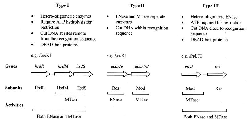
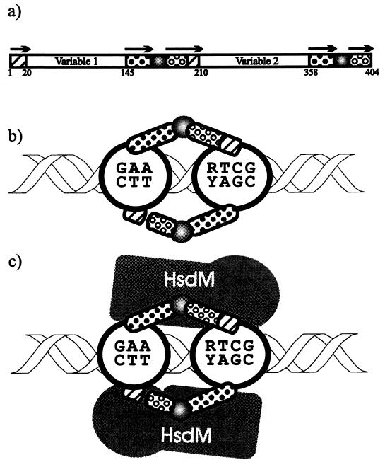
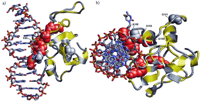
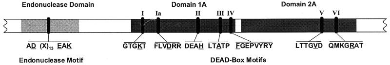
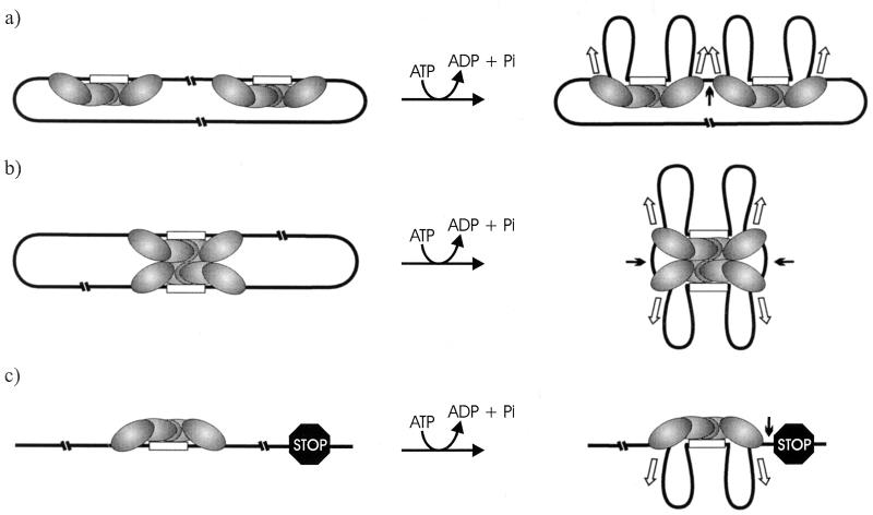
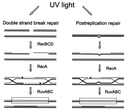
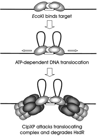
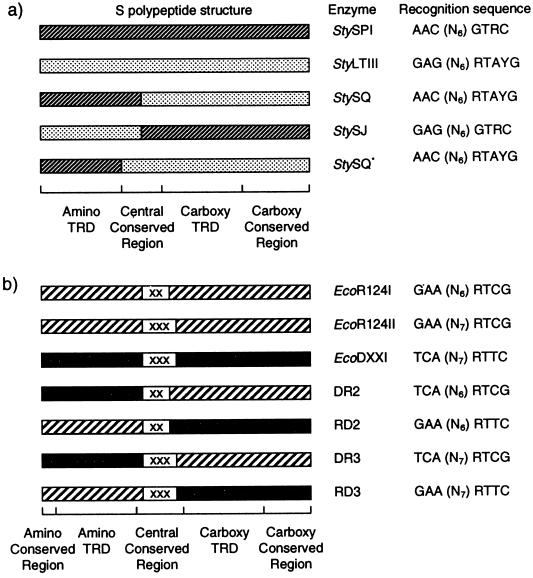
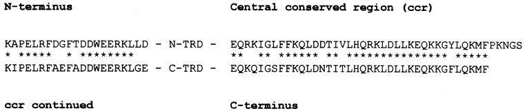
Restriction enzymes are well known as reagents widely used by molecular biologists for genetic manipulation and analysis, but these reagents represent only one class (type II) of a wider range of enzymes that recognize specific nucleotide sequences in DNA molecules and detect the provenance of the DNA on the basis of specific modifications to their target sequence. Type I restriction and modification (R-M) systems are complex; a single multifunctional enzyme can respond to the modification state of its target sequence with the alternative activities of modification or restriction. In the absence of DNA modification, a type I R-M enzyme behaves like a molecular motor, translocating vast stretches of DNA towards itself before eventually breaking the DNA molecule. These sophisticated enzymes are the focus of this review, which will emphasize those aspects that give insights into more general problems of molecular and microbial biology. Current molecular experiments explore target recognition, intramolecular communication, and enzyme activities, including DNA translocation. Type I R-M systems are notable for their ability to evolve new specificities, even in laboratory cultures. This observation raises the important question of how bacteria protect their chromosomes from destruction by newly acquired restriction specifities. Recent experiments demonstrate proteolytic mechanisms by which cells avoid DNA breakage by a type I R-M system whenever their chromosomal DNA acquires unmodified target sequences. Finally, the review will reflect the present impact of genomic sequences on a field that has previously derived information almost exclusively from the analysis of bacteria commonly studied in the laboratory.


Figures

FIG. 1
FIG. 1
Distinguishing characteristics and organization of the genetic determinants and subunits of the different types of R-M systems. ENase, restriction endonuclease; Mtase, methyltransferase. Modified with permission from a figure in reference .
FIG. 2
FIG. 2
Specificity polypeptide of EcoR124I. (a) Organization indicating the two variable regions (TRDs) and the regions conserved in sequence for all members of one family. Repeats of similar sequence (below the arrows) are identified by their conserved marking. The number of the amino acid residue at the beginning and end of each region is given below the diagram. (b) Model of Kneale (86), in which the repeated sequences form linkers joining the TRDs in a rotationally symmetrical configuration. The nucleotide sequences identified by each TRD are shown. (c) Consequent model of the methyltransferase, in which the two HsdM subunits bind to the linker region to generate an enzyme with pseudodyad symmetry.
FIG. 3
FIG. 3
Experimental support for the symmetrical arrangement of the TRDs (large open circles or arrows) within specificity subunits. (a) Predicted arrangement of two truncated subunits of EcoDXXI and the consequent recognition of a hyphenated symmetrical target sequence. (b) The HsdS subunits of StySKI (type IB) and EcoR124I (type IC). The IB family of enzymes have a long N-terminal conserved sequence, while the IC family has a long C-terminal conserved tail. The amino-terminal TRD of StySKI and the carboxy-terminal TRD of EcoR124I (indicated by arrows) have 36% amino acid identity. The DNA targets recognized are indicated. The complement of the EcoR124I 3′ target sequence (CGAY) is a degenerate version of the StySKI 5′ target sequence (CGAT), consistent with a symmetrical organization of two similar TRDs. (c) Circularly permuted variants of the specificity gene of EcoAI (type IB) can retain activity. The normal organization of regions within an HsdS polypeptide is given in the first diagram, followed by an active but circularly permuted variant. The black segments identify the well-conserved repeats.
FIG. 4
FIG. 4
Model of the amino acid segment comprising residues 43 to 157 of the amino terminus of EcoKI interacting with DNA as proposed by Sturrock and Dryden (166). (a) Side view. (b) Bird's eye view. Residues in red are substitutions that cause the loss of restriction and modification; those in yellow have no detectable effect on activity. Residue 141, in gray, was previously thought to lead to a loss of both activities but is now known to impair rather than inactivate. The DNA structure is from the complex of M · HhaI with its DNA target sequence (85) and therefore shows an extrahelical cytosine rather than adenine. The figure is modified with permission from that published in O'Neill et al. (131).
FIG. 5
FIG. 5
Domains and motifs of HsdR of EcoKI. The N- and C-terminal regions are omitted, since their roles are not known. The two domains that include the DEAD-box motifs correlate with IA and 2A, as determined for the structures of DNA helicases (36). Substitutions for the underlined amino acids confer a restriction-deficient phenotype.
FIG. 6
FIG. 6
ATP-dependent DNA translocation. (a) Model of Studier and Bandyopadhyay (165). EcoKI bound to adjacent target sequences translocates DNA towards itself. Collision blocks translocation and stimulates the nicking of each DNA strand. Two domains of HsdR flanking the DNA are indicated. (b) In this variant, the EcoKI complexes have dimerized prior to translocation (51). (c) When translocation is impeded by some other protein or structure, endonuclease activity is stimulated (77).
FIG. 7
FIG. 7
Generation of unmodified target sequences following UV irradiation. Methylated strands of DNA are shown as thick lines, and unmethylated strands are shown as thin lines. Homologous recombination, involved in the repair of double-strand breaks or postreplicative repair, can generate regions of unmethylated, double-stranded DNA via annealing of two unmethylated strands (regions within boxes). In addition, the SOS mutagenesis pathway leads to new (unmodified) target sequences as the result of base changes.
FIG. 8
FIG. 8
Model for the mechanism of ClpXP-dependent RA (111). When a type IA or IB R-M complex binds to an unmodified target sequence, translocation is initiated and ClpXP recognizes the HsdR subunit of the complex. ClpXP degrades HsdR, preventing further translocation and endonuclease activity.
FIG. 9
FIG. 9
Evolution of type I R-M systems with new specificities. (a) Recombination between hsdS genes produces hybrid genes and chimeric S polypeptides. StySPI and StyLTIII are naturally occurring type I R-M systems (see Table 2). StySQ and StySJ have hybrid hsdS genes (55, 56). The regions originating from StySPI are hatched, and those originating from StyLTIII are stippled. Reassortment of the target recognition domains (TRDs) accordingly gave rise to recombinant recognition sequences (56, 127). Site-directed mutagenesis of the central conserved region of the StySQ hsdS gene produced StySQ*, comprising only the amino-terminal variable region from StySPI and the remainder from StyLTIII. The StySQ* target sequence confirms that the amino-terminal variable region is in fact a TRD responsible for recognition of the trinucleotide component of the sequence (33). (b) Sequence specificity may also be altered by changing the length of the nonspecific spacer of the target sequence. The S polypeptides of EcoR124I and EcoR124II differ only in the number of times a short amino acid motif (X = TAEL) is repeated within their central conserved regions (142), resulting in extension of the spacer in the target sequence from six nucleotides (N6) in EcoR124I in N7 in EcoR124II. The recognition sequence of EcoDXXI also contains a nonspecific spacer of seven nucleotides, corresponding to three TAEL repeats in its S polypeptide (64). Chimeric S polypeptides recognize the predicted target sequences (64). Modified from a figure in Barcus and Murray (10) with permission.
FIG. 10
FIG. 10
Repeated amino acid sequences in an HsdS polypeptide of L. lactis. The sequence (153) has been aligned to emphasize the repeats flanking the predicted target recognition domains.

References

    1. Abadjieva A, Patel J, Webb M, Zinkevich V, Firman K. A deletion mutant of the type IC restriction endonuclease EcoR1241 expressing a novel DNA specificity. Nucleic Acids Res. 1993;21:4435–4443. - PMC - PubMed
    1. Allen D J, Makhov A, Grilley M, Taylor J, Thresher R, Modrich P, Griffith J D. MutS mediates heteroduplex loop formation by a translocation mechanism. EMBO J. 1997;16:4467–4476. - PMC - PubMed
    1. Alm R A, Ling L S, Moir D T, King B L, Brown E D, Doig P C, Smith D R, Noonan B, Guild B C, deJonge B L, Carmel G, Tummino P J, Caruso A, Uria-Nickelsen M, Mills D M, Ives C, Gibson R, Merberg D, Mills S D, Jiang Q, Taylor D E, Vovis G F, Trust T J. Genomic-sequence comparison of two unrelated isolates of the human gastric pathogen Helicobacter pylori. Nature. 1999;397:176–180. - PubMed
    1. Althorpe N J, Chilley P M, Thomas A T, Brammar W J, Wilkins B M. Transient transcriptional activation of the Incl1 plasmid anti-restriction gene (ardA) and SOS inhibition gene (psiB) early in conjugating recipient bacteria. Mol Microbiol. 1999;31:133–142. - PubMed
    1. Arber W. Host-controlled modification of bacteriophage. Annu Rev Microbiol. 1965;19:365–378. - PubMed

Publication types

MeSH terms

Substances

LinkOut - more resources