. 2000 Jun 20;97(13):7266-71.
doi: 10.1073/pnas.130189697.

Human centromere protein A (CENP-A) can replace histone H3 in nucleosome reconstitution in vitro

Affiliations

Human centromere protein A (CENP-A) can replace histone H3 in nucleosome reconstitution in vitro

K Yoda et al. Proc Natl Acad Sci U S A. .

Abstract

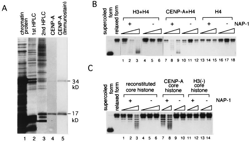
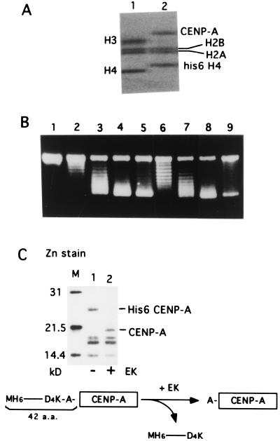
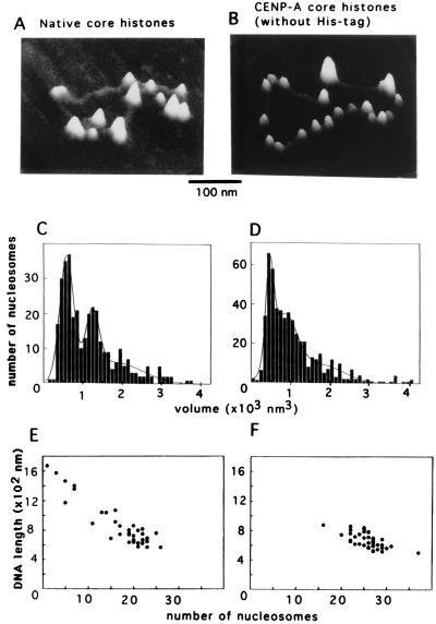
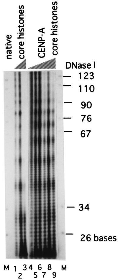
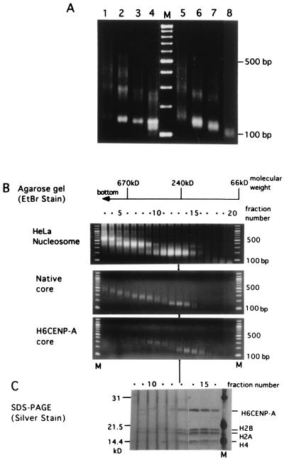
Centromere protein A (CENP-A) is a variant of histone H3 with more than 60% sequence identity at the C-terminal histone fold domain. CENP-A specifically locates to active centromeres of animal chromosomes and therefore is believed to be a component of the specialized centromeric nucleosomes on which the kinetochores are assembled. Here we report that CENP-A, highly purified from HeLa cells, can indeed replace histone H3 in a nucleosome reconstitution system mediated by nucleosome assembly protein-1 (NAP-1). The structure of the nucleosomes reconstituted with recombinant CENP-A, histones H2A, H2B, and H4, and closed circular DNAs had the following properties. By atomic force microscopy, "beads on a string" images were obtained that were similar to those obtained with nucleosomes reconstituted with four standard histones. DNA ladders with repeats of approximately 10 bp were produced by DNase I digestion, indicating that the DNA was wrapped round the protein complex. Mononucleosomes isolated by glycerol gradient sedimentation had a relative molecular mass of approximately 200 kDa and were composed of 120-150 bp of DNA and equimolar amounts of CENP-A, and histones H4, H2A, and H2B. Thus, we conclude that CENP-A forms an octameric complex with histones H4, H2A, and H2B in the presence of DNA.


Figures

Figure 1
Figure 1
Topo I analyses using the purified CENP-A, histone H4, H2A, and H2B. (A) Samples from each step of purification were identified by silver staining (lanes 1–4) or by immunoblotting with anti-centromere auto-antibody serum (lane 5) after 13% SDS/PAGE. A crude HCl extract of HeLa nuclei (lane 1), peak fraction of CENP-A after first (lane 2) and second (lane 3) reversed-phase HPLC, and CENP-A purified by SDS/PAGE and electro-elution (lanes 4 and 5) are shown. CENP-A monomer (17 kDa) and dimer (34 kDa) are indicated. (B) H3/H4 (lanes 1–6), CENP-A/H4 (lanes 7–12), or H4 (lanes 13–18) were subjected to Topo I analyses using the relaxed form of closed circular DNA (pUC-αdimer, 20 ng) with (lanes 1–3, 7–9, and 13–15) or without NAP-1 (lanes 4–6, 10–12, and 16–18). Supercoil ladders were detected by Southern hybridization. H3/H4: 30 ng (lanes 1 and 4), 50 ng (lanes 2 and 5), 75 ng (lanes 3 and 6); CENP-A/H4: 10 ng (lanes 7 and 10), 20 ng (lanes 8 and 11), 30 ng (lanes 9 and 12); H4: 25 ng (lanes 13 and 16), 50 ng (lanes 14 and 17), and 75 ng (lanes 15 and 18). (C) Reconstituted core histones (H3/H4/H2A/H2B) (lanes 1–6), CENP-A/core histones (CENP-A/H4/H2A/H2B) (lanes 7–10), or H3(-) core histones (H4/H2A/H2B) (lanes 11–14) were subjected to Topo I analyses with (lanes 1–3, 7, 8, 11, and 12) or without NAP-1 (lanes 4–6, 9, 10, 13, and 14) under the same conditions as in B. H3/H4/H2A/H2B: 25 ng (lanes 1 and 4), 35 ng (lanes 2 and 5), 50 ng (lanes 3 and 6); CENP-A/H4/H2A/H2B: 40 ng (lanes 7 and 9), 60 ng (lanes 8 and 10); H4/H2A/H2B: 40 ng (lanes 11 and 13), and 60 ng (lanes 12 and 14).
Figure 2
Figure 2
Topo I analysis using the recombinant CENP-A purified under native condition. (A) 4–20% SDS/PAGE of native core histones (0.24 μg, lane 1) and native CENP-A/core histones (0.12 μg, lane 2). The gel was stained with Coomassie brilliant blue after electrophoresis. (B) Topo I analysis of the native CENP-A/core histones (lanes 6–9) compared with the native core histones (lanes 2–5). Nucleosome reconstitution was carried out by using the relaxed form of pUC-α11mer DNA (100 ng) and NAP-1 as described in Methods. Supercoil ladders were detected with ethidium bromide staining after agarose gel electrophoresis. The amounts of the native core histones were 0 ng (lane 1), 72 ng (lane 2), 98 ng (lane 3), 120 ng (lane 4), and 180 ng (lane 5), and the amounts of His6CENP-A/core histones were 63 ng (lane 6), 100 ng (lane7), 126 ng (lane 8), and 250 ng (lane 9). (C) EK digestion of the complex removes the histidine tag at the N terminus of CENP-A. Mononucleosomes were isolated by glycerol density-gradient sedimentation after MNase digestion of the reconstituted complexes formed from His6CENP-A/core histones and DNA with (lane 2) or without (lane 1) EK digestion and separated by 15% SDS/PAGE. The gel was stained with Zn. The primary structure of the N terminus of His6CENP-A is shown at the bottom.
Figure 3
Figure 3
Analysis of the reconstituted nucleosomes by AFM. Nucleosomes were reconstituted with pUC-α11mer DNA and native core histones (A, C, and E) or His6CENP-A/core histones with EK digestion (B, D, and F) and observed by AFM. (A and B) Nucleosomes formed on circular DNA. (Bar indicates 100 nm.) (C and D) Histogram of the number of nucleosomes in each volume class. (E and F) Plot of the contour length of each circular DNA molecule against the number of nucleosomes on it.
Figure 4
Figure 4
DNase I digestion of the nucleosomes. The nucleosome complexes were digested with 0.17 unit of DNase I for 4, 10, and 20 min (lanes 1–3) for native core histones and 2, 4, 6, 8, 10, and 15 min (lanes 4–9) for His6CENP-A/core histones at 37°C. Lane M shows an MspI digest of pBR322 as size markers.
Figure 5
Figure 5
MNase digestion of the reconstituted nucleosomes and glycerol gradient sedimentation of the MNase digests. (A) Nucleosomes were formed from native core histones (lanes 1–4) or His6CENP-A/core histones (lanes 5–8). Aliquots of 8 μl were digested with 0.01 unit (lanes 1 and 5), 0.03 unit (lanes 2 and 6), 0.1 unit (lanes 3 and 7), or 0.3 unit (lanes 4 and 8) of MNase for 10 min at 37°C. The DNA of each sample was electrophoresed through a 1.7% agarose gel and detected by ethidium bromide fluorescence. Lane M shows a 100-bp ladder. (B) Glycerol density gradient sedimentation of the MNase digests of the nucleosomes from HeLa nuclei (Top), reconstituted with native core histones (Middle), or His6CENP-A core histones (Bottom). Each 150-μl aliquot was fractionated from the bottom, and 20 μl of each fraction was electrophoresed through 1.7% agarose gel after proteinase K digestion. Each lane of B and C was numbered according to the fraction number of the glycerol gradient. The position of each molecular mass marker (albumin, 66 kDa; catalase, 240 kDa; thyroglobulin, 670 kDa) was marked at the top. M, 100-bp ladder. (C) The remaining 130 μl of glycerol gradient fractions 8–16 (B Bottom) were precipitated with acetone and separated by 15% SDS/PAGE. The protein bands were detected with silver staining.

References

    1. Earnshaw W C, Rothfield N. Chromosoma. 1985;91:313–321. - PubMed
    1. Palmer D K, O'Day K, Trong H L, Charbonneau H, Margolis R L. Proc Natl Acad Sci USA. 1991;88:3734–3738. - PMC - PubMed
    1. Sullivan K F, Hechenberger M, Khaled M. J Cell Biol. 1994;127:581–592. - PMC - PubMed
    1. Howman E V, Fowler K J, Newson A J, Redward S, MacDonald A C, Kalitsis P, Choo K H A. Proc Natl Acad Sci USA. 2000;97:1148–1153. - PMC - PubMed
    1. Stoler S, Keith K C, Curnick K E, Fitzgerald-Hayes M. Genes Dev. 1995;9:573–586. - PubMed

Publication types