. 2000 Jun;105(11):1537-45.
doi: 10.1172/JCI8527.

The PHSRN sequence induces extracellular matrix invasion and accelerates wound healing in obese diabetic mice

Affiliations

The PHSRN sequence induces extracellular matrix invasion and accelerates wound healing in obese diabetic mice

D L Livant et al. J Clin Invest. 2000 Jun.

Abstract

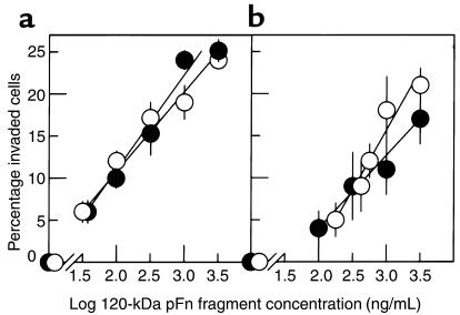
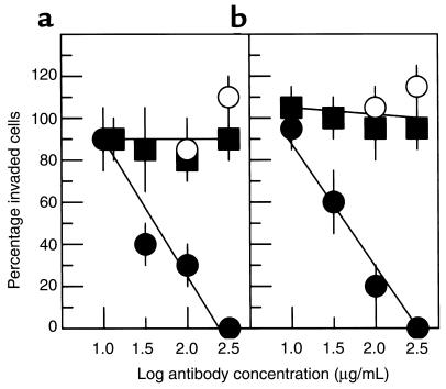
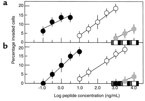
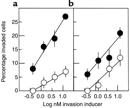
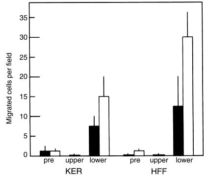
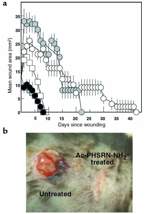
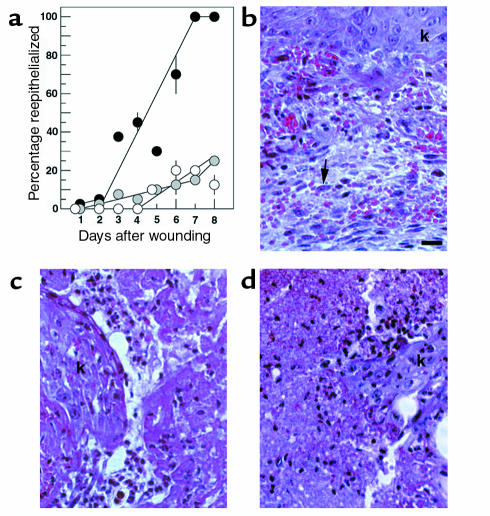
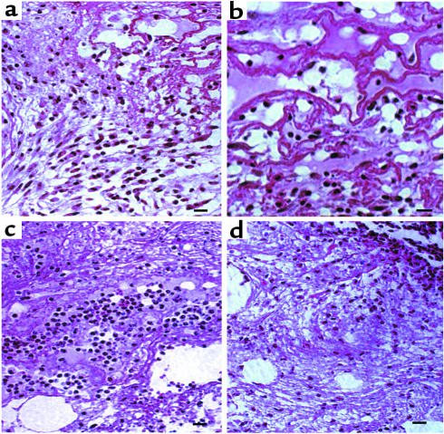
The PHSRN sequence of the plasma fibronectin (pFn) cell-binding domain induces human keratinocytes and fibroblasts to invade the naturally serum-free extracellular matrices of sea urchin embryos. The potency of acetylated, amidated PHSRN (Ac-PHSRN-NH(2)) is significantly increased, making it more active on a molar basis than the 120-kDa cell-binding domain of pFn. Arginine is important to this activity because PHSAN and PHSEN are inactive, as is a randomized sequence peptide, Ac-HSPNR-NH(2). One treatment with Ac-PHSRN-NH(2) stimulates reepithelialization and contraction of dermal wounds in healing-impaired, obese diabetic C57BL6/KsJ db/db mice. Wound closure is equally rapid in treated db/db and db/+ mice and may be more rapid than in untreated nondiabetic db/+ littermates. In contrast, treatment with either Ac-HSPNR-NH(2) or normal saline (NS) has no effect. Analysis of sectioned db/db wounds shows that, in contrast to treatment with Ac-HSPNR-NH(2) or NS, a single Ac-PHSRN-NH(2) treatment stimulates keratinocyte and fibroblast migration into wounds, enhances fibroplasia and vascularization in the provisional matrix, and stimulates the formation of prominent fibers that may be associated with wound contraction.


Figures

Figure 1
Figure 1
Invasive responses of normal keratinocytes and fibroblasts to the 120-kDa pFn fragment. (a) Undifferentiated keratinocytes. (b) Fibroblasts. White circles, serum-free media; black circles, FCS-containing media. Data presented as mean ± SD.
Figure 2
Figure 2
Effects of P1D6 anti-α5β1 or P1B5 anti-α3β1 antibodies on invasion. (a) Keratinocytes. (b) Fibroblasts. Y-axis shows mean percentage of invaded cells (± SD) relative to the mean percentage invaded without antibody. Black circles, P1D6 prebound to cells before exposure to 10 μg/mL 120-kDa pFn fragment and placement on SU-ECM invasion substrates. White circles, P1D6 bound to cells in the presence of 120-kDa fragment. Black squares, P1B5 bound to cells before exposure to the 120-kDa pFn fragment.
Figure 3
Figure 3
Invasive responses of keratinocytes and fibroblasts to derivatives of the PHSRN sequence. (a) Keratinocytes. (b) Fibroblasts. Black circles, Ac-PHSRN-NH2; white circles, PHSRN; gray circles, PHSKN; black squares, PHSAN; white squares, PHSEN; hatched squares, Ac-HSPNR-NH2. Data presented as mean ± SD.
Figure 4
Figure 4
Comparative dose-response curves of Ac-PHSRN-NH2 and the 120-kDa pFn fragment. (a) Keratinocytes. (b) Fibroblasts. Y-axis shows mean percentages of invaded cells (± SD). Black circles, Ac-PHSRN-NH2; white circles, 120-kDa pFn fragment.
Figure 5
Figure 5
Effect of PHSRN on keratinocyte and fibroblast motility in Boyden chambers. X-axis shows cell types and chamber contents. KER, human keratinocytes. Exposure to PHSRN: pre, cells prebound with 1 μg/mL PHSRN (solid bars) or to 0.2% BSA (open bars); upper, cells exposed to 1 μg/mL PHSRN by both prebinding and by placement in upper chambers containing the same PHSRN concentration; lower, cells prebound to 1 μg/mL PHSRN (solid bars) or to 0.2% BSA (open bars) during prebinding, then placed in upper chambers containing 0.2% BSA, and allowed to undergo chemotaxis in response to 1 μg/mL PHSRN in the lower chambers. Data presented as mean ± SD.
Figure 6
Figure 6
Functional activities of Ac-PHSRN-NH2 or Ac-HSPNR-NH2 in db/db and in db/+ dermal wounds. (a) Areas of dermal wounds treated with Ac-PHSRN-NH2, Ac-HSPNR-NH2, or NS during the days required for wound closure. White circles, untreated db/db wounds; black circles, Ac-PHSRN-NH2–treated db/db wounds; gray circles, Ac-HSPNR-NH2–treated db/db wounds; white squares, untreated db/+ wounds; gray squares, Ac-PHSRN-NH2–treated db/+ wounds. (b) Ac-PHSRN-NH2–treated and untreated duplicate wounds in a db/db mouse 7 days after wounding. Data presented as mean ± SD.
Figure 7
Figure 7
Effect of Ac-PHSRN-NH2, Ac-HSPNR-NH2, or NS alone on reepithelialization and fibroplasia in db/db dermal wounds. (a) Effect of Ac-PHSRN-NH2, Ac-HSPNR-NH2, or NS on reepithelialization. Y-axis shows mean percentage (with SD) of wounds in section covered by keratinocytes. Black circles, mean percentage of each Ac-PHSRN-NH2–treated wound reepithelialized; gray circles, mean percentage of each Ac-HSPNR-NH2–treated wound reepithelialized; white circles, mean percentage of each NS-treated wound reepithelialized. (b) A section of an Ac-PHSRN-NH2–treated db/db wound 8 days after wounding, photographed at ×400 using phase-contrast optics. Arrow, active fibroblast; k, keratinocytes. Bar, 20 μm. (c) An Ac-HSPNR-NH2–treated db/db wound 8 days after wounding shown at the same magnification as b; k, keratinocytes. (d) A section of an untreated db/db wound 8 days after wounding shown at the same magnification as b. Identical areas of wound sections are shown in b, c, and d.
Figure 8
Figure 8
Effect of Ac-PHSRN-NH2, Ac-HSPNR-NH2, or NS alone on the presence of fibroblasts in the provisional matrix of db/db dermal wounds 3 days after wounding. (a) A section of an Ac-PHSRN-NH2–treated wound in a db/db mouse 3 days after wounding, Bar, 20 μm. (b) A section of an Ac-PHSRN-NH2–treated wound in a db/db mouse 3 days after wounding. Bar, 20 μm. (c) A section of an NS-treated wound in a db/db mouse 3 days after wounding, Bar, 20 μm. (d) A section of an Ac-HSPNR-NH2–treated wound in a db/db mouse 3 days after wounding. Bar, 20 μm. ad, × 400.

Comment in

References

    1. Clark, R.A.F. 1996. Wound repair: overview and general considerations. In The molecular and cellular biology of wound repair. 2nd edition. R.F. Clark, editor. Plenum Press. New York, New York, USA. 3–50.
    1. Martin P. Wound healing: aiming for perfect skin regeneration. Science. 1997;276:75–81. - PubMed
    1. Mosher DF. Physiology of fibronectin. Annu Rev Med. 1984;35:561–575. - PubMed
    1. Clark RAF, et al. Fibronectin and fibrin provide a provisional matrix for epidermal cell migration during wound reepithelialization. J Invest Dermatol. 1982;70:264–269. - PubMed
    1. Clark RAF, Quinn JH, Winn HJ, Colvin RB. Fibronectin beneath reepithelializing epidermis in vivo: sources and significance. J Invest Dermatol. 1983;80(Suppl.):26S–30S. - PubMed

Publication types