

doi: 10.1073/pnas.97.12.6242.
Glioblastoma multiforme: the terminator
Affiliations
- PMID: 10841526
- PMCID: PMC33993
- DOI: 10.1073/pnas.97.12.6242
Item in Clipboard
Glioblastoma multiforme: the terminator
Proc Natl Acad Sci U S A.
.
No abstract available
Figures
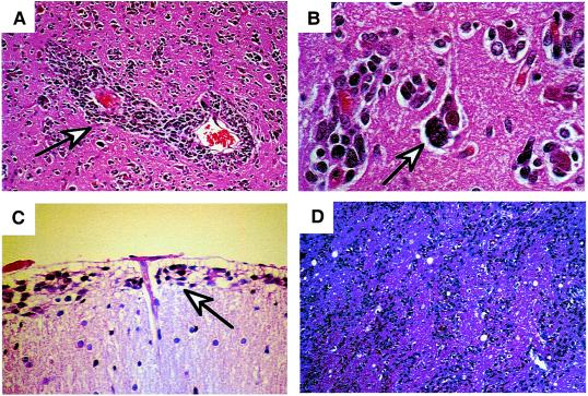
Secondary structures of Scherer demonstrating migration of glioma cells
through normal brain structures. (A) Glioma cells
surrounding blood vessels (perivascular satellitosis) (arrow).
(B) Perineuronal satellitosis (arrow).
(C) Collection of cells below pial surface (subpial
spread) (arrow). (D) Intrafascicular spread of tumor
cells through the corona radiata.
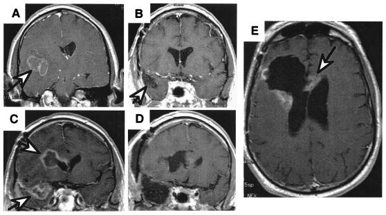
MRI scans of a patient with a right temporal GBM illustrating the
spread of the disease. (A) Presurgical scan, GBM (arrow)
is surrounded with edema. (B) Scan after surgery and
radiation therapy showing “gross total resection” and clear
resection cavity, and (C) six months later, showing
recurrence not only at the resection margin (arrow) but a second focus
of GBM across the Sylvian fissure in the frontal lobe (arrow).
(D) Postresection scans of both recurrent tumors.
(E) Scan 3 months later, showing the tumor recurring at
the resection margin and crossing the corpus callosum to the other
hemisphere (arrow).
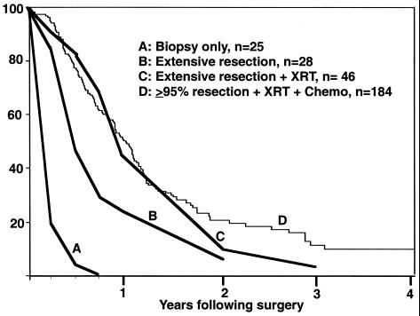
Kaplan–Meier survival plots for patients diagnosed with GBM. Curves
A, B, and C are historical
data from Jelsma and Bucy (8) published in 1967 before the availability
of MRI scans: biopsy only (A), extensive resection
(undefined) (B), and extensive resection followed by
radiation therapy (C). Curve D is current
data from the M. D. Anderson Cancer Center on patients with >95%
resection (by volumetric MRI measurements) followed by both radiation
therapy and chemotherapy. Although there are essentially no long-term
survivors, removal of tumor mass clearly increases longevity.
Comment in
-
Intergeneric poliovirus recombinants for the treatment of malignant glioma.Proc Natl Acad Sci U S A. 2000 Jun 6;97(12):6803-8. doi: 10.1073/pnas.97.12.6803. Proc Natl Acad Sci U S A. 2000. PMID: 10841575 Free PMC article.
References
-
- James C D, Olson J J. Curr Opin Oncol. 1996;8:188–195. - PubMed
-
- Ishii N, Tada M, Hamou M F, Janzer R C, Meagher-Villemure K, Weistler O D, Tribolet N, Van Meier E G. Oncogene. 1999;18:5870–5878. - PubMed
-
- Livingstone L R, White A, Sprouse J, Livanos E, Jacks T, Tlsty T D. Cell. 1992;70:923–935. - PubMed
-
- Scherer H J. Brain. 1940;40:631–635.
MeSH terms
Substances
LinkOut - more resources
Full Text Sources
Other Literature Sources
