. 2000 Jun 6;97(12):6568-73.
doi: 10.1073/pnas.97.12.6568.

Defense by foot adhesion in a beetle (Hemisphaerota cyanea)

Affiliations

Defense by foot adhesion in a beetle (Hemisphaerota cyanea)

T Eisner et al. Proc Natl Acad Sci U S A. .

Abstract

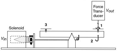
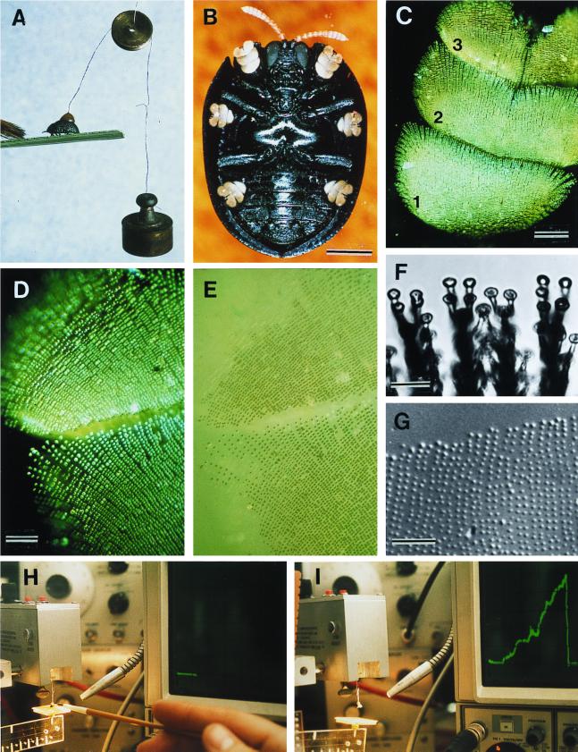
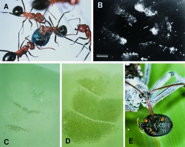
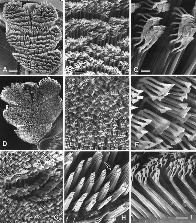
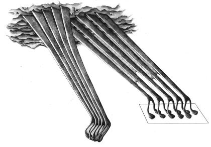
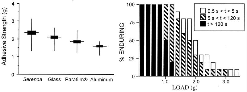
The beetle Hemisphaerota cyanea (Chrysomelidae; Cassidinae) responds to disturbance by activating a tarsal adhesion mechanism by which it secures a hold on the substrate. Its tarsi are oversized and collectively bear some 60,000 adhesive bristles, each with two terminal pads. While walking, the beetle commits but a small fraction of the bristles to contact with the substrate. But when assaulted, it presses its tarsi flatly down, thereby touching ground with all or nearly all of the bristles. Once so adhered, it can withstand pulling forces of up to 0.8 g ( approximately 60 times its body mass) for 2 min, and of higher magnitudes, up to >3 g, for shorter periods. Adhesion is secured by a liquid, most probably an oil. By adhering, the beetle is able to thwart attacking ants, given that it is able to cling more persistently than the ant persists in its assault. One predator, the reduviid Arilus cristatus, is able to feed on the beetle, possibly because by injecting venom it prevents the beetle from maintaining its tarsal hold.


Figures

Figure 1
Figure 1
Apparatus for application of pulling forces to beetle. 1, beetle; 2, hook for suspension of weights; 3, pan for placement of balancing weights.
Figure 2
Figure 2
(A) Beetle withstanding a 2-g pull; brush strokes are causing the beetle to adhere with its tarsi. (B) Ventral view of beetle, showing yellow tarsi. (C) Tarsus (numbers refer to tarsomeres). (D) Tarsus in contact with glass (polarized epi-illumination). (E) Same as preceding, in nonpolarized light; contact points of the bristles are seen to be wet. (F) Bristle pads, in contact with glass. (G) Droplets left on glass as part of a tarsal “footprint.” (H and I) Apparatus diagrammed in Fig. 1. In H, beetle is on platform, before lift is applied (horizontal trace on oscilloscope); in I, the lift has been applied (ascending green trace) to point where beetle has detached (return of trace to baseline). [Bars = 1 mm (B), 100 μm (C), 50 μm (D), 10 μm (F), and 50 μm (G).]
Figure 3
Figure 3
(A) F. exsectoides attacking beetle, on glass surface. (B) Footprints left by a beetle attacked as in A (dark-field illumination). (C) Tarsal contact during ordinary walking: only a few bristles touch the glass. (D) Tarsus pressed down flatly, in defensive response (beetle subjected to electromagnetic pull). (E) A. cristatus feeding on beetle. [Bar = 1 mm (B).]
Figure 4
Figure 4
(A–C) Normal tarsus, and details thereof; the pads are stuck together in clusters (C), which are arranged in rows (A). (D–F) Comparable with preceding, but of a tarsus cleaned of oil by treatment with methanol/chloroform solution. (G) Comparable with E but with some of the bristles clustered where a droplet of oil has been applied. (H) Portion of tarsus where tips of bristles have been cut off, showing how bristle shafts are stuck together in groups; a substance, presumed to be oil, is seen between the bases of the bristles (upper arrow). Lower arrows point to pores from which tarsal oil is presumed to be secreted. (I) Bristles, in profile view, showing the component parts (shaft, bifurcated tip, pads) and oil pores between their bases. [Bars = 100 μm (A), 20 μm (B), 5 μm (C), 10 μm (I).]
Figure 5
Figure 5
Postulated mechanism by which tarsal bristle pads adhere and become prewetted for adherence. Details in Results and Conclusions.
Figure 6
Figure 6
(Left) Pull in g needed to detach beetle from four different substrates (n = 10 beetles per substrate). The four mean values differ significantly from one another (P < 0.01 for all comparisons). (Right) Length of time that a fraction of beetles endured detachment pulls (load in g) of given magnitudes. Ten beetles each were subjected to the full range of loads, beginning with the lightest and proceeding incrementally to the heaviest.

References

    1. Eisner T. Verh Dtsch Zool Ges. 1972;65:123–137.
    1. Baier R E, Shafrin E G, Zisman W A. Science. 1968;162:1360–1368. - PubMed
    1. Nachtigall W. Biological Mechanisms of Attachment. New York: Springer; 1974.
    1. Walker G. Int J Adhes Adhes. 1992;13:3–7.
    1. Walker G, Yule A B, Ratcliffe J. J Zool London. 1985;205:297–307.

Publication types

LinkOut - more resources