. 2000 Jun 6;97(12):6716-21.
doi: 10.1073/pnas.97.12.6716.

Trans-splicing vectors expand the utility of adeno-associated virus for gene therapy

Affiliations

Trans-splicing vectors expand the utility of adeno-associated virus for gene therapy

Z Yan et al. Proc Natl Acad Sci U S A. .

Abstract

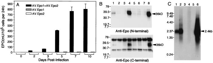
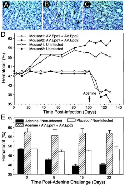
Adeno-associated viral (AAV) vectors have demonstrated considerable promise for gene therapy of inherited diseases. However, with a packaging size of <5 kb, applications have been limited to relatively small disease genes. Based on the finding that AAV genomes undergo intermolecular circular concatamerization after transduction in muscle, we have developed a paradigm to increase the size of delivered transgenes with this vector through trans-splicing between two independent vectors coadministered to the same tissue. When two vectors encoding either the 5' or 3' portions of the erythropoietin genomic locus were used, functional erythropoietin protein was expressed in muscle subsequent to the formation of intermolecular circular concatamers in a head-to-tail orientation through trans-splicing between these two independent vector genomes. These findings will allow for the application of AAV technologies to a wider variety of diseases for which therapeutic transgenes exceed the packaging limitation of present AAV vectors.


Figures

Figure 1
Figure 1
Structure of rAAV vectors and strategy used to generate trans-splicing vectors expressing functional human Epo. Two rAAV vectors, AV.Epo1 and AV.Epo2, encoding either the 5′ or 3′ genomic segment of the human Epo gene, and the diagnostic restriction enzyme sites used to characterize the structure of circular intermediates are shown in A. Additional elements, kanamycin resistance (Kanr), ampicillin resistance (Ampr), and a bacterial replication origin (ori) were included to allow for molecular characterization of circular concatamers after bacterial rescue from muscle DNA extracted by the Hirt procedure. An internal ribosome entry site (IRES) sequence and the EGFP transgene were also incorporated into the 3′ end of the Epo transcript to allow for direct visualization of transgene expression after concatamerization. The locations of 5′ and 3′ Epo DNA probes used for structural analysis in Fig. 4 are marked below each viral construct. (B) Expected structure of a circular concatamer arising from intermolecular recombination that would allow for functional trans-splicing of Epo message. The expected splicing patterns are denoted by dashed lines. (C) Expected heterogeneous nuclear RNA, splicing pattern, and mature mRNA transcripts resulting from circular concatamerization.
Figure 2
Figure 2
Production of Epo protein after coinfection of primary fibroblasts with two independent trans-splicing vectors. Confluent primary fetal fibroblasts (8 × 10⁵ cells) were infected with 7 × 10⁹ particles each of AV.Epo1 and/or AV.Epo2 viral vectors. Epo expression was monitored using ELISA by harvesting culture media 24 h subsequent to media replacement. (A) Results comparing the indicated three vector combinations give the mean (±SEM) Epo levels from three independent experiments normalized to 1 × 10⁵ cells. (B) Samples of fibroblast culture media (1 ml) at 2 days (lanes 1–3) and 10 days (lanes 4–6) postinfection with either AV.Epo1/AV.Epo2 (lanes 1 and 4), AV.Epo1 (lanes 2 and 5), or AV.Epo2 (lanes 3 and 6) were immunoprecipitated with a rabbit polyclonal anti-human Epo Ab (R&D Systems) and resolved by 12% SDS/PAGE. Western blots were then probed with either an N-terminal (Upper) (Santa Cruz Biotechnology) or C-terminal (Lower) (R&D Systems) anti-Epo Ab and detected by ECL. Lanes 7 and 8 represent mock-infected and control cells infected with a full-length cDNA Epo AAV vector, respectively. Arrows indicating the glycosylated Epo protein (36 kDa) and molecular mass markers (kDa) are to the left of each blot. (C) Northern blot analysis of Epo mRNA expression: lane 1, coinfected with AV.Epo1/AV.Epo2 (5 days); lane 2, coinfected with AV.Epo1/AV.Epo2 (10 days); lane 3, infected with AV.Epo1 alone (10 days); lane 4, infected with AV.Epo2 alone (10 days); lane 5, uninfected; lane 6, transfected with pEpoEGFP (as a control for the full-length Epo/EGFP bicistronic message). Molecular lengths (kb) are marked to the left of the gel. Successful transcription and splicing will yield a 2.28-kb mature mRNA + poly(A) tail (marked by arrow at 2.4 kb).
Figure 3
Figure 3
In vivo production of Epo protein from trans-splicing rAAV vectors in muscle. The tibialis muscles of C57BL6 mice were injected with either 6 × 10¹⁰ particles or 4 × 10¹¹ particles by direct coinfection with both AV.Epo1 and AV.Epo2 or bilateral infection of tibialis muscles with each vector individually. Uninfected age-matched controls were also evaluated at the same time. Hematocrits were analyzed by retroorbital bleeding (A), and serum levels of human Epo protein were evaluated by using a human-specific ELISA protocol (B) at various times postinfection. No changes in hematocrit or the level of human Epo protein were detected in animals infected with the individual vectors in separate tibialis muscles (only data for the high dose group are shown). Results depict the mean (±SEM) for n = 6 (virally coinfected low dose group), n = 4 (virally coinfected high dose group), and n = 4 (control groups) independent animals in each group. (C) EGFP expression in AV.Epo1- and/or AV.Epo2-infected muscles at 150 days postinfection. Images show representative fluorescent photomicrographs of whole mount fresh muscle tissues.
Figure 4
Figure 4
Structural and functional analysis of rescued circular AAV intermediates from infected muscle. Hirt DNA was extracted from AV.Epo1- and AV.Epo2-coinfected muscles at 110 days postinfection and used to transform E. coli. Rescued circular intermediate clones were evaluated by restriction enzyme mapping and Southern blotting against three 32P-labeled probes (A). Probe sequences, as diagrammed in Fig. 1, included a 5′Epo 750-bp KpnI/BclI fragment (containing exon 2 and part of intron 3 encoded in the AV.Epo1 vector), a 3′Epo 1.4-kb BclI/KpnI fragment (containing part of intron 3 through the IRES sequence of AV.Epo2), and an EGFP 800-bp SacII fragment. The structures of three representative rescued circular intermediate plasmids (p21, p16, and p19) are evaluated in A, with lanes marked as U (uncut), K (KpnI-digested), and A (AflIII-digested). Molecular length standards (L) are shown in kb to the left of the ethidium bromide gel (EtBr). Probes used for each Southern blot are labeled below each panel. p21 was an Ampr clone with digestion patterns consistent with a circular monomer from AV.Epo1. p19, and p16 were Amp/Kan coresistant clones with digestion patterns consistent with intermolecular dimer concatamers of AV.Epo1 and Av.Epo2 with head-to-head and head-to-tail orientations, respectively. Structures of these clones are diagrammatically depicted in B. These representative clones were transfected into HeLa cells to evaluate Epo expression (C). Epo protein was detected by ELISA assay and normalized by cotransfection with a pCMVluciferase reporter as the internal standard. pEpoEGFP (with the full-length Epo gene) and pEpo(ITR)EGFP (derived from pEpoEGFP with a double-D ITR structure inserted into intron 3 of the Epo gene) were used as comparative positive controls. Results indicate the mean (±SEM) for three independent transfections.
Figure 5
Figure 5
Trans-splicing production of human Epo protects from adenine-induced renal failure anemia. C57BL6 mice were infected with 6 × 10¹⁰ particles of both AV.Epo1 and AV.Epo2 in the same tibialis muscle and challenged with adenine time-release capsules implanted s.c. at 35 or 100 days postinfection. Both adenine-treated/uninfected and placebo-treated/uninfected age-matched animals were also evaluated as controls. Hematoxylin/eosin sections (8 μm) of kidneys from placebo-treated/uninfected (A), adenine-treated/uninfected (B), and adenine-treated AV.Epo1/AV.Epo2 coinfected (C) mice are shown. Comparison of A with B and C demonstrates significant cast formation (arrow) in proximal tubules in both adenine-treated groups. In contrast, as shown in D, animals infected with both AV.Epo1 and AV.Epo2 were provided with significant protection from adenine-induced anemia initiated at 100 days postinfection (arrow). Viral infection was performed at day 0. (E) Mean (±SEM) hematocrit from both combined groups (n = 4 for each condition) for placebo-treated/uninfected, adenine-treated/uninfected, and adenine-treated/AV.Epo1/AV.Epo2 coinfected animals. In the adenine treatment groups of these combined data, 4 animals were challenged at both 35 (n = 2) and 100 (n = 2) days postinfection with AV.Epo1 and AV.Epo2.

Comment in

  • The persistence of alien genomes.
    Tattersall P. Tattersall P. Proc Natl Acad Sci U S A. 2000 Jun 6;97(12):6239-41. doi: 10.1073/pnas.97.12.6239. Proc Natl Acad Sci U S A. 2000. PMID: 10841525 Free PMC article. No abstract available.

References

    1. Muzyczka N. Curr Top Microbiol Immunol. 1992;158:97–129. - PubMed
    1. Carter B J. Curr Opin Biotechnol. 1992;3:533–539. - PubMed
    1. Berns K I, Giraud C. Adeno-Associated Virus (AAV) Vectors in Gene Therapy. Berlin: Springer; 1996.
    1. Summerford C, Samulski R J. J Virol. 1998;72:1438–1445. - PMC - PubMed
    1. Summerford C, Bartlett J S, Samulski R J. Nat Med. 1999;5:78–82. - PubMed

Publication types