Comment
. 2000 Jun 6;97(12):6803-8.
doi: 10.1073/pnas.97.12.6803.

Intergeneric poliovirus recombinants for the treatment of malignant glioma

Affiliations
Comment

Intergeneric poliovirus recombinants for the treatment of malignant glioma

M Gromeier et al. Proc Natl Acad Sci U S A. .

Abstract

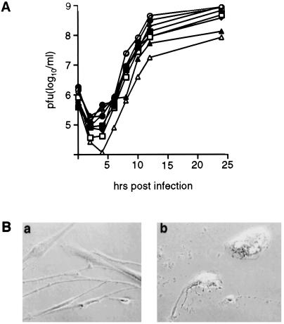
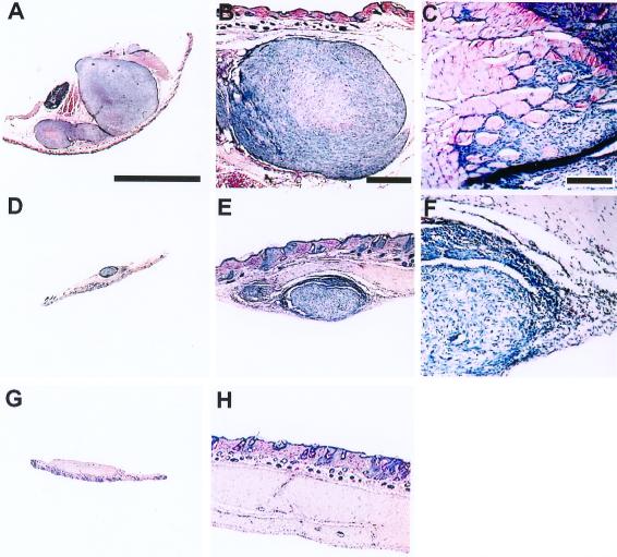
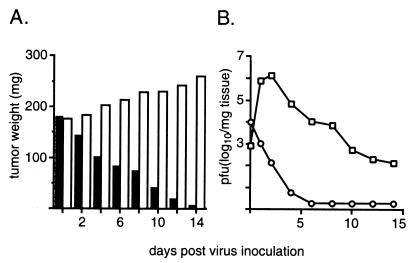
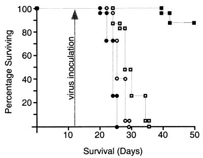
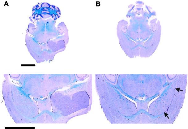
Poliovirus neuropathogenicity depends on sequences within the 5' nontranslated region of the virus. Exchange of the poliovirus internal ribosomal entry site with its counterpart from human rhinovirus type 2 resulted in attenuation of neurovirulence in primates. Despite deficient virus propagation in cells of neuronal origin, nonpathogenic polio recombinants retain excellent growth characteristics in cell lines derived from glial neoplasms. Susceptibility of malignant glioma cells to poliovirus may be mediated by expression of a poliovirus receptor, CD155, in glial neoplasms. Intergeneric polio recombinants with heterologous internal ribosomal entry site elements unfolded strong oncolytic potential against experimentally induced gliomas in athymic mice. Our observations suggest that highly attenuated poliovirus recombinants may have applicability as biotherapeutic antineoplastic agents.


Figures

Figure 1
Figure 1
Propagation and toxicity of PV1(RIPO) in malignant glioma tissue culture cell lines. (A) One-step growth curves of PV1(RIPO) were obtained after synchronized infection of monolayer cultures at a multiplicity of infection of 10 according to previous reports (10). Glioma cell lines tested included SF188 (open square; glioblastoma multiforme), SF295 (closed square; glioblastoma multiforme), SF767 (open triangle; anaplastic astrocytoma), SF763 (closed triangle; anaplastic astrocytoma), HTB-14 (open circle; anaplastic astrocytoma), HTB-15 (closed circle; anaplastic astrocytoma), DU54 (open diamond; glioblastoma multiforme), and DU65 (closed triangle; glioblastoma multiforme). (B) Synchronized infection of primary cultured glioblastoma multiforme cells. Cells were photographed before (a) and 12 h after (b) infection with PV1(RIPO) at a multiplicity of infection of 10.
Figure 2
Figure 2
Effect of PV1(RIPO) on s.c. HTB-15 tumor xenografts in athymic mice. Mice received bilateral implants. When tumors reached an average cross diameter of 8 mm (6–8 weeks after implantation), the mice were given a single i.v. injection of PV1(RIPO) (n = 10) or PBS (n = 10). An s.c. tumor of a PBS-control-treated animal 2 weeks after PBS treatment (A) infiltrated into surrounding tissues (B) and skeletal muscle (C). The tumors regressed 2 weeks after i.v. administration of 2 × 10⁷ pfu PV1(RIPO) (D), revealing remaining tumor mass encircled by necrotic debris and inflammatory infiltrates invading the tumor from the periphery (E and F). At 3 weeks after PV1(RIPO) treatment, the tumors had been replaced by a fibrotic patch (G) with no evidence of residual neoplastic cells (H). Sections are 12 μm thick; hematoxylin/eosin stain. [A, Bar = 8 mm (applies to A, D, and G); B, Bar = 1 mm (applies to B, E, and H); C, Bar = 0.2 mm (applies to C and F)].
Figure 3
Figure 3
Intratumoral propagation of PV1(RIPO) in s.c. HTB-15 xenografts grown in athymic mice. (A) Tumor growth in mock-treated (open bars) animals and xenograft regress in mice treated with PV1(RIPO) (closed bars). Mice (n = 4) with similarly sized tumors (8-mm cross diameter) were treated either with a single i.v. inoculation of 2 × 10⁷ pfu of PV1(RIPO) or with PBS alone and were killed at the indicated intervals. Tumors were dissected, weighed, and processed for determination of the viral load by plaque assay (10). The data shown represent mean values for all four animals comprising an experimental group. At 2 weeks after virus treatment, tumor tissue could no longer be macroscopically discerned (compare Fig. 2). (B) Intratumoral (squares) and i.v. (circles) virus load after i.v. administration of PV1(RIPO) to xenografted athymic mice.
Figure 4
Figure 4
Survival of athymic mice with intracerebral HTB-14 xenografts. Tumor implantation was carried out on day 0, and animals were treated on day 12 (arrow) with a single intramuscular (open circles), i.v. (open squares), or intratumoral (closed squares) inoculation of 2 × 10⁷ pfu PV1(RIPO). Closed circles represent untreated control mice. The number of surviving animals was recorded daily and plotted against time.
Figure 5
Figure 5
Histopathological evaluation of intracerebral tumor implantation sites. (Lower) Details of the respective cross sections of the brain. (A) A transversal midcephalic section reveals a large xenograft originating in the area of the anterolateral fornix of an untreated control mouse 22 days after tumor implantation. (B) A midcephalic section through the brain of an athymic mouse treated with a single injection of PV1(RIPO) 12 days after xenograft implantation. The image shows the brain 50 days after tumor induction and 38 days after virus treatment. A large tissue defect delineating the fornix indicates the region of the implantation site (arrows). Apart from this lesion and the presence of reactive infiltrates within the area, no tissue abnormalities or residual tumor can be distinguished. Sections are 12 μm thick (luxol fast blue/periodic-acid-Schiff/hematoxylin stain). (Bars = 3 mm.)
Figure 6
Figure 6
Growth curve analyses of the original PV1(RIPO) inoculum (squares) and three isolates recovered from treated intracranial glioma xenografts (circles, diamonds, and triangles). One-step growth curves of all three isolates derived from treated xenografts revealed the deficient propagation in SK-N-MC neuroblastoma cells that characterizes the nonneuropathogenic phenotype of PV1(RIPO) (10).

Comment on

  • Glioblastoma multiforme: the terminator.
    Holland EC. Holland EC. Proc Natl Acad Sci U S A. 2000 Jun 6;97(12):6242-4. doi: 10.1073/pnas.97.12.6242. Proc Natl Acad Sci U S A. 2000. PMID: 10841526 Free PMC article. No abstract available.

References

    1. Levin V A, Gutin P H, Leibel S. In: Cancer: Principles & Practice of Oncology. DeVita V, Hellman S, Rosenberg S A, editors. Philadelphia: Lippincott; 1993. pp. 1679–1737.
    1. Martuza R L, Malick A, Markert J M, Ruffner K L, Coen D M. Science. 1991;252:854–856. - PubMed
    1. Coffey M C, Strong J E, Forsyth P A, Lee P W K. Science. 1998;282:1332–1334. - PubMed
    1. Culver K W, Ram Z, Wallbridge S, Ishii H, Oldfield E H, Blaese R M. Science. 1992;256:1550–1552. - PubMed
    1. Eck S L, Alavi J B, Alavi A, Davis A, Hackney D, Judy M, Mollman J, Phillips P C, Wheeldon E B, Wilson J M. Hum Gene Ther. 1996;7:1465–1482. - PubMed

Publication types

LinkOut - more resources