. 2000 Jun 6;97(12):6838-43.
doi: 10.1073/pnas.97.12.6838.

Kainate receptor-mediated synaptic currents in cerebellar Golgi cells are not shaped by diffusion of glutamate

Affiliations

Kainate receptor-mediated synaptic currents in cerebellar Golgi cells are not shaped by diffusion of glutamate

I Bureau et al. Proc Natl Acad Sci U S A. .

Abstract

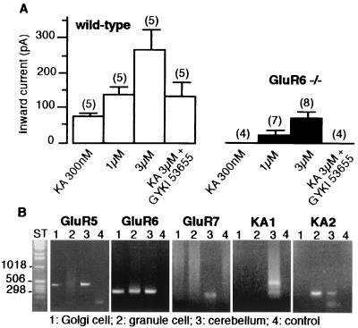
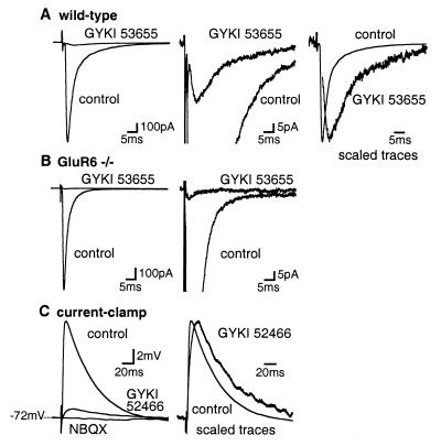
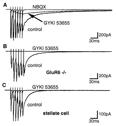
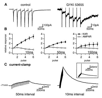
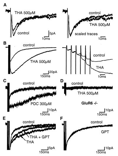
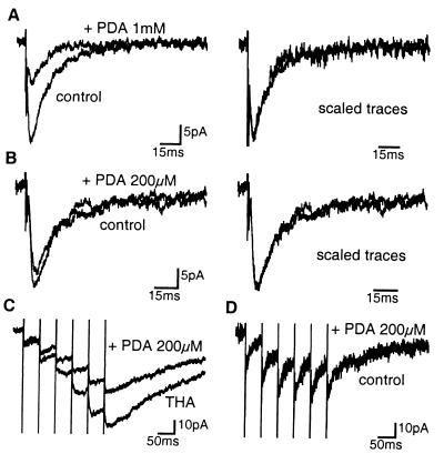
We report the presence of kainate receptors (KARs) in cerebellar Golgi cells of wild-type but not GluR6-deficient mice. Parallel fiber stimulation activates KAR-mediated synaptic currents [KAR-excitatory postsynaptic currents (EPSCs)] of small amplitude. KAR-EPSCs greatly differ from synaptic currents mediated by alpha-amino-3-hydroxy-5-methylisoxazole-4-propionate (AMPA) receptors (AMPAR-EPSCs) at the same synapse. KAR-EPSCs display slow rise and decay time and summate in response to a train of stimulations. By using PDA, a low-affinity competitive antagonist and agents that modify the clearance of glutamate, we show that these properties cannot be explained by diffusion of glutamate outside of the synaptic cleft and activation of extrasynaptic KARs. These data suggest that the slow kinetic of KAR-EPSCs is due to intrinsic properties of KARs being localized at postsynaptic sites. The contrasting properties of KAR- and AMPAR-EPSCs in terms of kinetics and summation offer the possibility for a glutamatergic synapse to integrate excitatory inputs over two different time scales.


Figures

Figure 1
Figure 1
KARs in Golgi cells. (A) Pooled data of the average amplitudes of inward currents activated by kainate in the presence and absence of GYKI 53655 (50 μM) in wild-type (□) and GluR6−/− (■) mice (mean ± SEM values; in parentheses, number of cell tested). Membrane holding potential was −70mV. All solutions were bath applied and contained tetrodotoxin (500 nM), d,l-AP-5 (50 μM), and picrotoxin (100 μM). (B) RT-PCR analysis of mRNA coding for kainate receptor subunits in Golgi cell (lane 1), granule cell (lane 2), total mouse cerebellum mRNA (lane 3), or pipette buffer as a control (lane 4). ST, one kb ladder.
Figure 2
Figure 2
Synaptic activation of KARs in Golgi cells by single stimulations. (A) (Left) EPSCs were largely blocked by GYKI 53655 (50 μM) in Golgi cells of wild-type mice. (Middle) Traces shown at an expanded amplitude scale. (Right) The GYKI-resistant EPSC is scaled to the peak of the control EPSC. Artifact was deleted from the trace of the GYKI-resistant EPSC. (B) (Left) in GluR6−/− mice, EPSCs were completely blocked by GYKI 53655 (50 μM). (Right) Traces shown at an expanded amplitude scale. (C) Current-clamp recordings. (Left) EPSPs were partially inhibited by GYKI 52466 (100 μM) and completely blocked by NBQX (100 μM). (Right) Traces shown at an expanded time scale. In this and the following figures, all solutions contained d,l-AP-5 (100 μM) and picrotoxin (100 μM). Each trace represents the average of 15–20 consecutive sweeps.
Figure 3
Figure 3
Synaptic activation of KARs by repetitive stimulations. (A) Repetitive stimulations (100 Hz) evoked a slowly decaying compound EPSC partially inhibited by GYKI 53655 (50 μM) and completely blocked by NBQX (150 μM) in Golgi cells from wild-type mice. (B) In GluR6−/− mice, GYKI 53655 (50 μM) completely blocked the compound EPSC (C). GYKI 53655 (50 μM) completely blocked EPSCs evoked by train stimulations in stellate cell from wild-type mice.
Figure 4
Figure 4
Summation of KAR-EPSCs during repetitive stimulations. (A) EPSCs evoked in Golgi cell by repetitive stimulations (50-ms interval) of parallel fibers in control conditions (Left) or in the presence of GYKI 53655 (50 μM) (Right). (B) Pooled data of the amplitude of AMPAR-EPSCs (○) measured in control conditions and KAR-EPSCs (●) measured in the presence of GYKI 53655 (50 μM) or GYKI 52466 (100 μM). The amplitude of EPSCs evoked by each pulse is measured in reference to initial baseline and is expressed as a function of the amplitude of the first pulse. Repetitive stimulations were performed with intervals of 10, 50, and 150 ms. (C) Current-clamp recordings. KAR-EPSPs evoked by repetitive stimulations with interval of 50 ms (Left) and 10 ms (Right) in the presence of GYKI 52466 (100 μM). In the inset, depolarization induced by summation of KAR-EPSPs reached the firing threshold.
Figure 5
Figure 5
Impact of the block of glutamate transporters on KAR-EPSCs. (A) (Left ) THA (500 μM) had no effect on rise time and early phase of decay of single KAR-EPSCs. (Right) The EPSC evoked in the presence of THA is scaled to the peak of the control trace. (B) (Left) THA (500 μM) potentiated and prolonged compound KAR-EPSCs evoked by repetitive stimulations (6 pulses, 100 Hz). (Right) Traces shown at an expanded time scale. (C) PDC (300 μM) potentiated and prolonged compound KAR-EPSCs. (D) No additional current was observed during repetitive stimulations in the presence of THA (500 μM) in GluR6−/− mice. (E) GPT (5 units/ml) and pyruvate (2 mM) partially reversed the effect of THA (500 μM) on the decay rate of compound KAR-EPSCs evoked by repetitive stimulations. (F) GPT (5 units/ml) and pyruvate (2 mM) did not affect by itself the compound KAR-EPSCs in control conditions. All solutions contained GYKI 53655 (50 μM) or GYKI 52466 (100 μM).
Figure 6
Figure 6
KAR-EPSCs are not shaped by the build-up of glutamate concentration in the extracellular space. (A) (Left) the low-affinity competitive antagonist PDA (1 mM) partially blocked single KAR-EPSCs. (Right) The PDA-resistant KAR-EPSC is scaled to the peak of the control trace. (B) Same as in A with PDA (200 μM). (C) In the presence of THA (500 μM), PDA (200 μM) induced an increasing block of the successive KAR-EPSCs evoked by repetitive stimulations (6 pulse, 20 Hz). (D) In control conditions, PDA (200 μM) similarly affected successive KAR-EPSCs evoked by repetitive stimulations (6 pulses, 20 Hz). All solutions contained GYKI 53655 (50 μM) or GYKI 52466 (100 μM).

References

    1. Bettler B, Mulle C. Neuropharmacology. 1995;34:123–139. - PubMed
    1. Hollmann M, Heinemann S. Annu Rev Neurosci. 1994;17:31–108. - PubMed
    1. Chittajallu R, Braithwaite S, Clarke V, Henley J. Trends Pharmacol Sci. 1999;20:26–35. - PubMed
    1. Clarke V R, Ballyk B A, Hoo K H, Mandelzys A, Pellizzari A, Bath C P, Thomas J, Sharpe E F, Davies C H, Ornstein P L, et al. Nature (London) 1997;389:599–603. - PubMed
    1. Kamiya H, Ozawa S. J Physiol (London) 1998;509:833–846. - PMC - PubMed

Publication types

Substances

LinkOut - more resources