

Six-month test-retest reliability of MRI-defined PET measures of regional cerebral glucose metabolic rate in selected subcortical structures
- PMID: 10843513
- PMCID: PMC6871851
- DOI: 10.1002/(sici)1097-0193(200005)10:1<1::aid-hbm10>3.0.co;2-o
Six-month test-retest reliability of MRI-defined PET measures of regional cerebral glucose metabolic rate in selected subcortical structures
Abstract
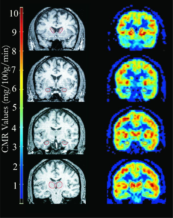
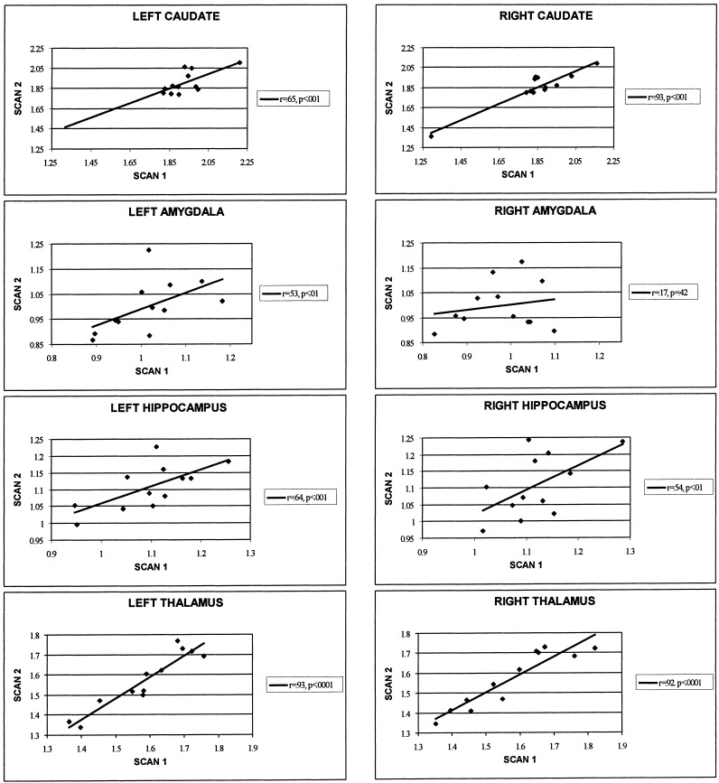
Test-retest reliability of resting regional cerebral metabolic rate of glucose (rCMR) was examined in selected subcortical structures: the amygdala, hippocampus, thalamus, and anterior caudate nucleus. Findings from previous studies examining reliability of rCMR suggest that rCMR in small subcortical structures may be more variable than in larger cortical regions. We chose to study these subcortical regions because of their particular interest to our laboratory in its investigations of the neurocircuitry of emotion and depression. Twelve normal subjects (seven female, mean age = 32.42 years, range 21-48 years) underwent two FDG-PET scans separated by approximately 6 months (mean = 25 weeks, range 17-35 weeks). A region-of-interest approach with PET-MRI coregistration was used for analysis of rCMR reliability. Good test-retest reliability was found in the left amygdala, right and left hippocampus, right and left thalamus, and right and left anterior caudate nucleus. However, rCMR in the right amygdala did not show good test-retest reliability. The implications of these data and their import for studies that include a repeat-test design are considered.
Figures
References
-
- American Psychiatric Association. (1994): Diagnostic and statistical manual of mental disorders (4th Ed.). Washington, D.C.: American Psychiatric Press.
-
- Bartlett EJ, Brodie JD, Wolf AP, Christman DR, Laska E, Meissner M. (1988): Reproducibility of cerebral glucose metabolic measurements in resting human subjects. J Cereb Blood Flow Metab 8: 502–512. - PubMed
-
- Bartlett EJ, Barouche F, Brodie JD, Wolkin A, Angrist B, Rotrosen J, Wolf AP. (1991): Stability of resting deoxyglucose metabolic values in PET studies of schizophrenia. Psychiat Res: Neuroimaging 40: 11–20. - PubMed
-
- Brooks RA, Di Chiro G, Zukerberg BW, Bairamian D, Larson SM. (1987): Test–retest studies of cerebral glucose metabolism using fluorine‐18‐deoxyglucose: Validation of method. J Nucl Med 28: 53–59. - PubMed
-
- Camargo EE, Szabo Z, Links JM, Sostre S, Dannals RF, Wagner HN. (1992): The influence of biological and technical factors on the variability of global and regional brain metabolism of 2‐[18F]Fluoro‐2‐deoxy‐D‐glucose. J Cereb Blood Flow Metab 12: 281–290. - PubMed
Publication types
MeSH terms
Substances
Grants and funding
LinkOut - more resources
Full Text Sources
