. 2000 Jun 15;20(12):4545-54.
doi: 10.1523/JNEUROSCI.20-12-04545.2000.

Postsynaptic scaffolds of excitatory and inhibitory synapses in hippocampal neurons: maintenance of core components independent of actin filaments and microtubules

Affiliations

Postsynaptic scaffolds of excitatory and inhibitory synapses in hippocampal neurons: maintenance of core components independent of actin filaments and microtubules

D W Allison et al. J Neurosci. .

Abstract

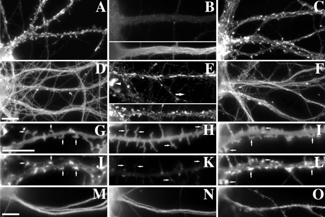
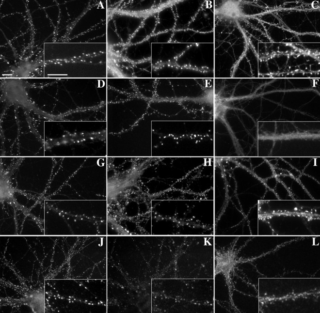
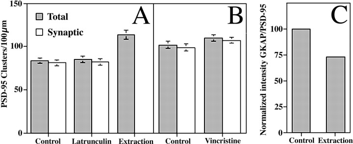
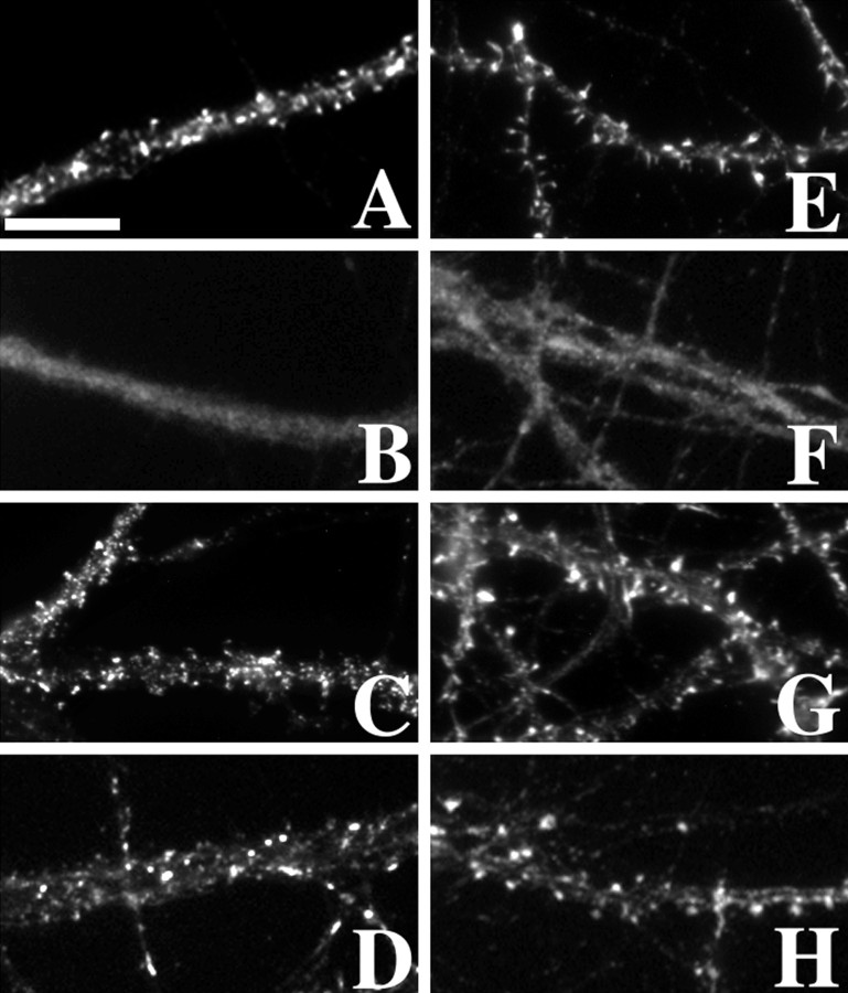
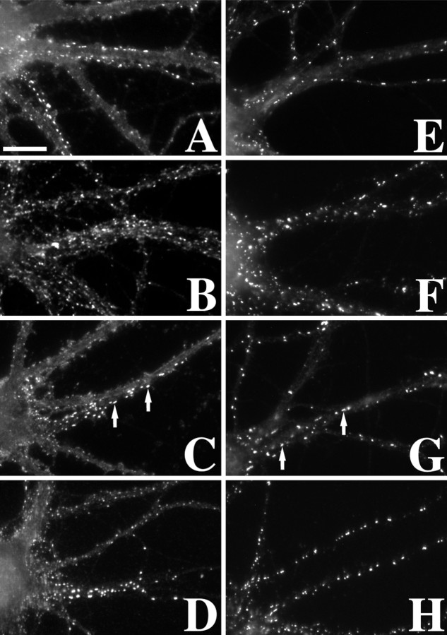
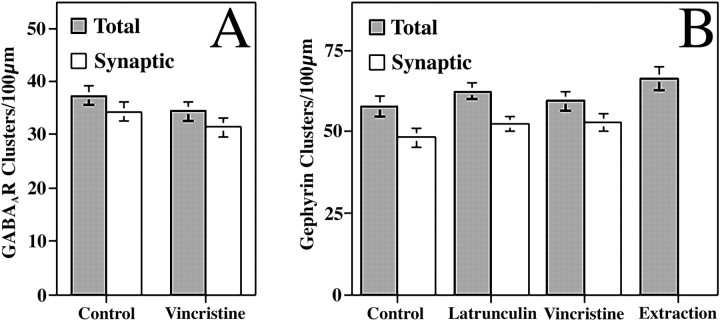
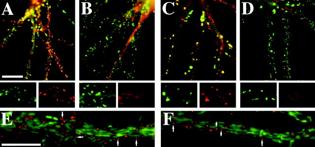
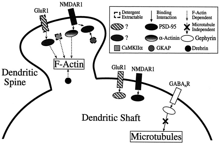
The mechanisms responsible for anchoring molecular components of postsynaptic specializations in the mammalian brain are not well understood but are presumed to involve associations with cytoskeletal elements. Here we build on previous studies of neurotransmitter receptors (Allison et al., 1998) to analyze the modes of attachment of scaffolding and signal transducing proteins of both glutamate and GABA postsynaptic sites to either the microtubule or microfilament cytoskeleton. Hippocampal pyramidal neurons in culture were treated with latrunculin A to depolymerize actin, with vincristine to depolymerize microtubules, or with Triton X-100 to extract soluble proteins. The synaptic clustering of PSD-95, a putative NMDA receptor anchoring protein and a core component of the postsynaptic density (PSD), was unaffected by actin depolymerization, microtubule depolymerization, or detergent extraction. The same was largely true for GKAP, a PSD-95-interacting protein. In contrast, the synaptic clustering of Ca(2+)/calmodulin-dependent protein kinase II (CaMKII)alpha, another core component of the PSD, was completely dependent on an intact actin cytoskeleton and was partially disrupted by detergent. Drebrin and alpha-actinin-2, actin-binding proteins concentrated in spines, were also dependent on F-actin for synaptic localization but were unaffected by detergent extraction. Surprisingly, the subcellular distributions of the inhibitory synaptic proteins GABA(A)R and gephyrin, which has a tubulin-binding motif, were unaffected by depolymerization of microtubules or actin or by detergent extraction. These studies reveal an unsuspected heterogeneity in the modes of attachment of postsynaptic proteins to the cytoskeleton and support the idea that PSD-95 and gephyrin may be core scaffolding components independent of the actin or tubulin cytoskeleton.


Figures

Fig. 1.
Fig. 1.
Treatment with cytoskeletal depolymerizing drugs reversibly disrupted actin microfilaments or microtubules in cultured hippocampal neurons. A, Rhodamine phalloidin staining of 3 week hippocampal neurons shows a high concentration of F-actin within the dendritic spines of control neurons. B, Treatment with latrunculin A eliminates the F-actin within the neurons, while not affecting microtubule staining (below). C, This effect can be reversed by washing out the drug, allowing the actin filaments to repolymerize. D, Tubulin staining after extraction of control neurons reveals tight bundles of microtubules within the dendrites and axons. E, Treatment with the microtubule depolymerizing agent vincristine eliminates the microtubule bundles, leaving only tubulin paracrystals (arrow indicates smaller axonal paracrystals; arrowhead indicates larger somatodendritic paracrystals), which is characteristic of vincristine treatment. This treatment does not affect the F-actin within spines as seen below. F, Within 24 hr after washing out the vincristine, the microtubule bundles repolymerize.GL, DiI membrane labeling (G–I) of random subpopulations of neurons reveals the continued presence of dendritic protrusions after cytoskeletal manipulations, regardless of the local concentrations of F-actin (J–L). Control neurons (G, J) and neurons treated with vincristine (I, L) exhibit spines along the shaft of the dendrites with corresponding concentrations of F-actin. Latrunculin A-treated neurons still show protrusions of membrane coming from the dendritic shafts (H) but lacking F-actin (K). MO, Staining for MAP2 reveals no change after latrunculin A treatment (N) when compared with control (M). Treatment with vincristine does allow MAP2 into the dendritic spines (O) but does not affect polarity. Scale bar, 10 μm.
Fig. 2.
Fig. 2.
Proteins of the postsynaptic density exhibited different modes of cytoskeletal association.AC, PSD-95, GKAP, and CaMKIIα, respectively, were found in clusters within dendritic spines and dendrite shafts. Clusters of each protein were primarily synaptic as found by double-labeling for synaptophysin (data not shown).D, E, After actin depolymerization with latrunculin A, both PSD-95 (D) and GKAP (E) clusters remained largely intact.F, Latrunculin A treatment dispersed the CaMKIIα clusters to a diffuse immunoreactivity throughout the dendrites.GI, Microtubule depolymerization with vincristine had no apparent effect on the distributions of any of these PSD proteins (G, PSD-95; H, GKAP;I, CaMKIIα). J, PSD-95 clusters were resistant to detergent extraction. K, L, GKAP (K) and CaMKIIα (L) clusters, although still present, were reduced in their intensity after detergent extraction, indicating partial extractability. Double staining of PSD-95 and GKAP (J, K) shows the change in relative intensity when compared with controls. In the case of CaMKIIα, it appeared that the staining within the shafts and heads of the spines was extractable, but the staining at the tip within the PSD remained. Scale bars, 10 μm.
Fig. 3.
Fig. 3.
PSD-95 clusters were not disrupted by depolymerization of actin filaments or microtubules or by detergent extraction, but GKAP was partially extractable. A, The graph illustrates the number of PSD-95 clusters/100 μm dendrite length for control, latrunculin A-treated, and detergent-extracted neurons. The number of synaptic clusters was determined as the number of PSD-95 clusters apposed to punctate synaptophysin immunoreactivity. None of the changes represents a significant change in the number of clusters (t test, p > 0.1), except for the number of clusters remaining after extraction (ttest, p < 0.0001). B, A second set of experiments was performed to test the effects of vincristine on PSD-95 distribution. The numbers of total or synaptic clusters of PSD-95 were not significantly different between vincristine-treated and matched control groups (t test, p > 0.1). C, This graph indicates the partial detergent extractability of GKAP. For each cluster of GKAP the average immunofluorescence intensity value was divided by the corresponding intensity of PSD-95 immunofluorescence. With the GKAP to PSD-95 ratio normalized to the control neurons, a 27% decrease was seen after extraction. The difference was significant (t test,p < 0.0001).
Fig. 4.
Fig. 4.
Actin-binding proteins of dendritic spines were dispersed by actin depolymerization but unaffected by depolymerization of microtubules or detergent extraction. A,E, The actin-binding proteins α-actinin-2 (A) and drebrin (E) were found to be abundant within the dendritic spines of hippocampal neurons. B, F, After actin depolymerization with latrunculin A, the clusters dissociated, and both α-actinin-2 (B) and drebrin (F) became diffusely localized within the dendrites. C, D, G,H, Neither microtubule depolymerization with vincristine (C, G) nor detergent extraction (D, H) disrupted the clusters of α-actinin-2 (C, D) or drebrin (G, H). Scale bar, 10 μm.
Fig. 5.
Fig. 5.
Clustering of GABAAR and gephyrin at inhibitory synapses was unaffected by depolymerization of microfilaments or microtubules or by detergent extraction. Immunostaining for the inhibitory neurotransmitter receptor GABAAR β2/3 subunits (A–D) and its putative anchoring protein gephyrin (E–H) was not disrupted by latrunculin A (B, F), vincristine (C, G), or detergent extraction (D, H). Large, elongated, synaptic clusters of both proteins (arrows) still remained, even in the absence of detectable microtubules (as in Fig. 1E), indicating that microtubules are not primarily responsible for anchoring these proteins at inhibitory PSDs. Scale bar, 10 μm.
Fig. 6.
Fig. 6.
Clustering of inhibitory synaptic proteins was not affected by treatment with vincristine to depolymerize microtubules.A, The total number of clusters and the number of synaptic clusters of GABAAR did not change significantly after vincristine treatment (t test,p > 0.1). B, The number of gephyrin clusters was not affected by treatment with latrunculin A, vincristine, or detergent extraction. Similarly, the number of synaptic clusters of gephyrin did not change after latrunculin A or vincristine treatments. There were no significant differences between groups (ttest, p > 0.1).
Fig. 7.
Fig. 7.
Treatment with latrunculin A and detergent extraction affected synaptic clustering of different components of the PSD. A, Double immunostaining with CaMKIIα (red) and NR2A (green) showed that both proteins are concentrated within the PSDs of control neurons.B, After treatment with latrunculin A to depolymerize actin, CaMKIIα clusters dispersed (red), whereas the NR2A clusters remained intact (green).C, Similarly, clusters of GluR1 (red) and PSD-95 (green) were seen within control dendritic spines. D, After detergent extraction, GluR1 clusters were extracted (red), but PSD-95 clusters remained (green). E, F, After treatment with vincristine, neither PSD-95 (E, red) nor gephyrin (F, red) colocalizes with tubulin paracrystals (E, F, green). Scale bar, 10 μm.
Fig. 8.
Fig. 8.
Diagrammatic summary of results. Two different AMPA receptor-binding proteins are postulated to account for the differential detergent extractability and actin dependence of AMPA receptors in spines versus shaft synapses as reported previously (Allison et al., 1998); these may correspond to different forms of GRIP and/or PICK1. The proteins dependent on F-actin for clustering include α-actinin-2, drebrin, CaMKIIα, and AMPAR in spines. GKAP and CaMKIIα are partially detergent extractable, and AMPAR is highly extractable only from spine synapses. All synaptic components were found to be localized independent of microtubules; this is emphasized in the diagram for gephyrin and GABAAR. These results indicate different modes of localization for different components of dendritic spines and suggest that PSD-95 and gephyrin form part of core scaffolds of excitatory and inhibitory synapses maintained independent of association with conventional cytoskeletal systems.

References

    1. Adam G, Matus A. Role of actin in the organisation of brain postsynaptic densities. Brain Res Mol Brain Res. 1996;43:246–250. - PubMed
    1. Allen PB, Ouimet CC, Greengard P. Spinophilin, a novel protein phosphatase 1 binding protein localized to dendritic spines. Proc Natl Acad Sci USA. 1997;94:9956–9961. - PMC - PubMed
    1. Allison DW, Gelfand VI, Spector I, Craig AM. Role of actin in anchoring postsynaptic receptors in cultured hippocampal neurons: differential attachment of NMDA versus AMPA receptors. J Neurosci. 1998;18:2423–2436. - PMC - PubMed
    1. Banker GA, Cowan WM. Rat hippocampal neurons in dispersed cell culture. Brain Res. 1977;126:397–442. - PubMed
    1. Bensch KG, Marantz R, Wisniewski H, Shelanski M. Induction in vitro of microtubular crystals by vinca alkaloids. Science. 1969;165:495–496. - PubMed

Publication types

MeSH terms

LinkOut - more resources