. 2000 Jun 15;20(12):4615-26.
doi: 10.1523/JNEUROSCI.20-12-04615.2000.

Lens injury stimulates axon regeneration in the mature rat optic nerve

Affiliations

Lens injury stimulates axon regeneration in the mature rat optic nerve

S Leon et al. J Neurosci. .

Abstract

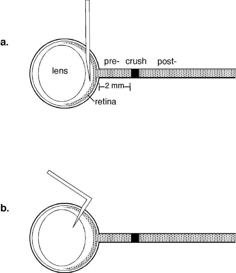
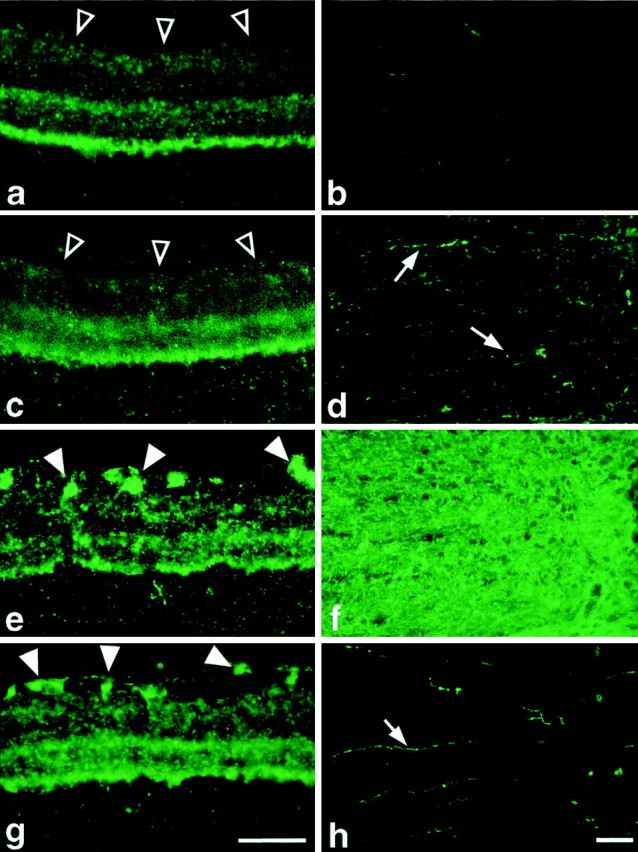
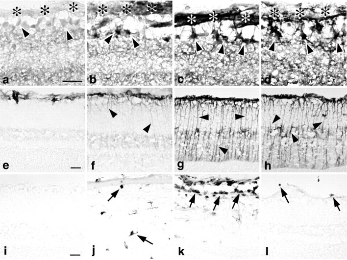
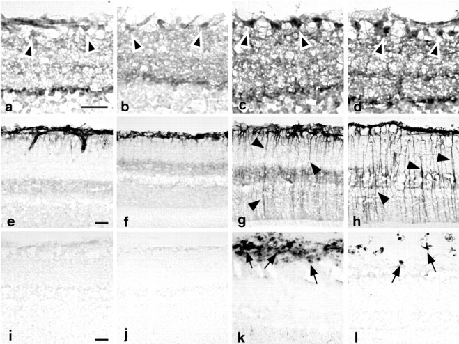
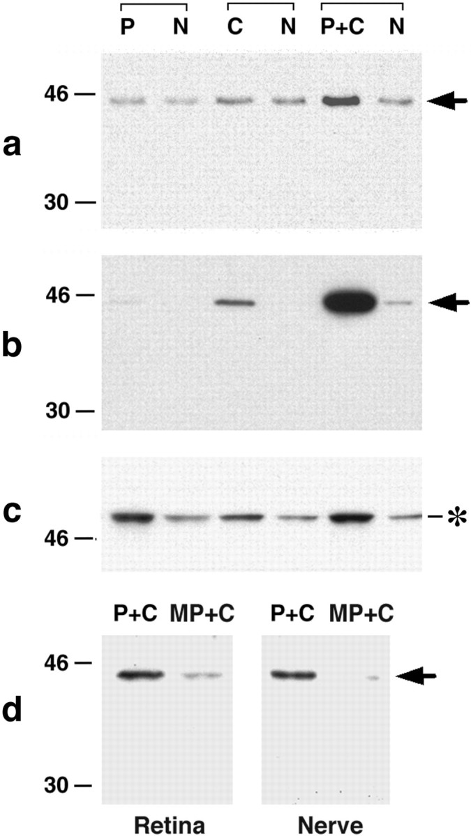
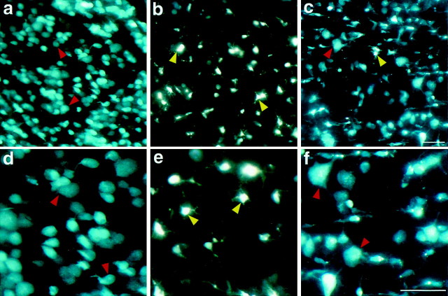
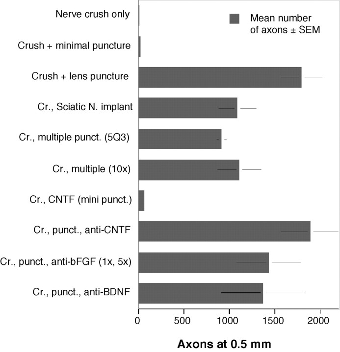
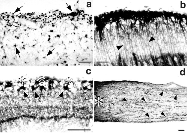
In mature mammals, retinal ganglion cells (RGCs) are unable to regenerate their axons after optic nerve injury, and they soon undergo apoptotic cell death. However, a small puncture wound to the lens enhances RGC survival and enables these cells to regenerate their axons into the normally inhibitory environment of the optic nerve. Even when the optic nerve is intact, lens injury stimulates macrophage infiltration into the eye, Müller cell activation, and increased GAP-43 expression in ganglion cells across the entire retina. In contrast, axotomy, either alone or combined with intraocular injections that do not infringe on the lens, causes only a minimal change in GAP-43 expression in RGCs and a minimal activation of the other cell types. Combining nerve injury with lens puncture leads to an eightfold increase in RGC survival and a 100-fold increase in the number of axons regenerating beyond the crush site. Macrophage activation appears to play a key role, because intraocular injections of Zymosan, a yeast cell wall preparation, stimulated monocytes in the absence of lens injury and induced RGCs to regenerate their axons into the distal optic nerve.


Figures

Fig. 1.
Fig. 1.
Surgical approaches. Optic nerves were crushed 2 mm from the eye. Intraocular injections were made either (a) vertically into the vitreous chamber to avoid puncturing the lens or (b) with a bent needle to puncture the lens to a depth of ∼0.5 mm.
Fig. 2.
Fig. 2.
Lens puncture stimulates axon regeneration in the optic nerve. a, A combination of nerve crush plus lens puncture stimulates RGCs to extend GAP-43-positive fibers through the injury site (*) and several millimeters into the distal portion of the optic nerve. Controls show no GAP-43 in the normal nerve (b) and very little immunostaining in the distal optic nerve after crush injury alone (c) or after nerve crush with a minimally invasive injection that does not infringe on the lens (d). Scale bar, 100 μm;a, c, and d are 21 d after nerve crush.
Fig. 3.
Fig. 3.
Time course of axon growth into the distal optic nerve. After nerve crush combined with lens puncture, the number of axons extending 0.5 or 1.0 mm distal to the injury site rises continuously over the first three weeks, then declines.
Fig. 4.
Fig. 4.
Double-labeling studies of regenerating axons. Double-immunofluorescent labeling shows that, after lens injury, the same axons (arrows) and presumed growth cone (arrowhead) are visualized distal to a crush site with an antibody to GAP-43 (a, arrows) and anti-CTB antibodies (b); c, in the fusion of the two images, yellow reflects a superposition of the two labels. Scale bar, 100 μm.d–f, Double immunostaining for GAP-43 and myelin basic protein. After optic nerve crush plus lens puncture, numerous GAP-43-positive fibers (arrowheads) are seen distal to crush site in myelin-rich regions (e, immunostaining for myelin basic protein). f, Fusion of the two images. Scale bar, 100 μm.
Fig. 5.
Fig. 5.
GAP-43 upregulation in the retina and proximal optic nerve after lens puncture. The normal adult rat retina (a) shows a moderate level of GAP-43 immunofluorescence in the inner plexiform layer (stained bands), but none in the ganglion cell layer (open arrows). Correspondingly, there is no axon staining in the normal optic nerve (b). Twenty-one days after nerve crush alone, there is no change in the pattern of retinal staining (c), although some positively stained axons appear in the optic nerve segment proximal to the crush site (d, arrows). After nerve crush accompanied by lens puncture, there is a marked increase in GAP-43 staining in RGCs (e, arrows) and in axons proximal to the injury site (f). Lens puncture alone leads to a somewhat lesser increase in GAP-43 expression in RGCs (g) and results in a small number of positively stained fibers in the optic nerve (h,arrow). Scale bar, 100 μm.
Fig. 6.
Fig. 6.
Time course of GAP-43, GFAP, and ED1 immunostaining in the retina after nerve crush combined with lens puncture. a–d, GAP-43 immunostaining in RGCs in the normal retina (a) and at 3–21 d after surgery. Three days after nerve crush with lens puncture, RGCs begin to show increased GAP-43 levels in their somata (b,arrows) and in their axons in the overlying optic fiber layer (asterisks); staining intensifies by day 7 (c) and remains high at day 21 (d). e, h, Mueller cells show a parallel activation, as indicated by increased GFAP staining on day 3 (f), which intensifies further on days 7–21 (g, h). i–l, Monocyte activation in the retina is visualized by ED1 staining. Whereas most of the ED-1-positive cells seen at day 3 (j) are within the retinal neuropil, by day 7, most appear on the surface of the retina (k); the number of ED-1-positive cells decreases by day 21 (l). Scale bar, 100 μm.
Fig. 7.
Fig. 7.
Effects of lens puncture or nerve injury alone. Nerve crush alone stimulates only a small change in GAP-43 expression in RGCs on day 7 (a) or day 21 (b) after surgery. In parallel to this, there is no change of GFAP expression in Müller cells (e, f) or in the number of ED-1-positive macrophages (i, j). In contrast, lens puncture alone induces changes in all three cell types by day 7 (c, g,k). The changes in RGCs (d) and Müller cells (h) remain high at day 21, although ED1 staining reveals fewer macrophages at this time point. Scale bar, 100 μm.
Fig. 8.
Fig. 8.
Changes in GAP-43 and GFAP expression: Western blots. Lens puncture combined with nerve crush (P+C) results in increased GAP-43 levels in the retina (a,arrow), and even more strikingly, in the proximal portion of the optic nerve (b, arrow;N represents the normal, contralateral side of the experimental animal; all results are at 14 d after surgery).c, GFAP levels are upregulated equally by lens puncture alone (P) or lens puncture plus nerve crush (P+C). Nerve crush alone (C) causes a lesser change. d, A case in which both optic nerves were crushed but the lens was injured on only one side. There is a much greater increase in GAP-43 in the retina and optic nerve on the side with lens puncture plus nerve crush (P+C) than on the contralateral side having a minimally invasive intraocular puncture that does not infringe on the lens (MP+C).
Fig. 9.
Fig. 9.
Lens puncture enhances RGC survival. RGC survival was evaluated using Fluorogold to retrogradely label RGCs 1 week before surgery. In the normal retina, RGCs appear as oval cells with apale blue–white fluorescence (a, d,red arrows). Optic nerve crush, either alone or with a minimally invasive injection (b, e), results in the death of most RGCs by day 21. Activated microglia pick up label by phagocytosis, and are readily distinguished from RGCs by virtue of their brilliant white fluorescence and multiple, thin processes (green arrows). Nerve crush combined with lens puncture increases RGC survival (c, f, red arrows) and decreases the number of microglia in the retina. Scale bar, 100 μm.
Fig. 10.
Fig. 10.
Quantitation of axon growth after various treatments. The combination of nerve crush plus lens puncture induces nearly 2000 axons to regenerate >500 μm past the injury site. This represents a 100-fold increase relative to animals with nerve crush only. Multiple punctures (Cr., multiple punct.) or an intravitreal sciatic nerve implant (Cr., Sciatic N. implant) stimulate significantly less growth. Intravitreal injections of CNTF without lens injury (Cr., CNTF mini punct.) do not mimic the effects of lens puncture, and anti-CNTF antibodies do not decrease axon growth in lens-puncture cases (Cr., punct., anti-CNTF). Antibodies to BDNF or bFGF also fail to diminish axon growth after lens puncture (Cr., punct., anti-BDNF; Cr., punct., anti-bFGF).
Fig. 11.
Fig. 11.
Macrophage activation mimics the effect of lens puncture. a, A single injection of Zymosan into the vitreous results in a massive infiltration of ED-1-positive monocytes in the retina (arrows). This is paralleled by increased GFAP expression in Müller cells (b,arrowheads), increased GAP-43 levels in RGCs (c, arrowheads) and in their axons in the overlying fiber layer (asterisks), and growth of GAP-43-positive axons past the injury site (d,asterisk) into the distal portion of the optic nerve (arrowheads).

References

    1. Aguayo AJ, Vidal-Sanz M, Villegas-Perez MP, Bray GM. Growth and connectivity of axotomized retinal neurons in adult rats with optic nerves substituted by PNS grafts linking the eye and the midbrain. Ann NY Acad Sci. 1987;495:1–9. - PubMed
    1. Battisti WP, Wang J, Bozek K, Murray M. Macrophages, microglia, and astrocytes are rapidly activated after crush injury of the goldfish optic nerve: a light and electron microscopic analysis. J Comp Neurol. 1995;354:306–320. - PubMed
    1. Batchelor PE, Liberatore GT, Wong JY, Porritt MJ, Frerichs F, Donnan GA, Howells DW. Activated macrophages and microglia induce dopaminergic sprouting in the injured striatum and express brain-derived neurotrophic factor and glial cell line-derived neurotrophic factor. J Neurosci. 1999;19:1708–1716. - PMC - PubMed
    1. Benowitz LI, Lewis ER. Increased transport of 44,000- to 49,000-dalton acidic proteins during regeneration of the goldfish optic nerve: a two-dimensional gel analysis. J Neurosci. 1983;3:2153–2163. - PMC - PubMed
    1. Benowitz LI, Apostolides PJ, Perrone-Bizzozero N, Finklestein SP, Zwiers H. Anatomical distribution of the growth-associated protein GAP-43/B-50 in the adult rat brain. J Neurosci. 1988;8:339–352. - PMC - PubMed

Publication types

LinkOut - more resources