. 2000 Jun 15;20(12):4745-57.
doi: 10.1523/JNEUROSCI.20-12-04745.2000.

Sustained visual attention performance-associated prefrontal neuronal activity: evidence for cholinergic modulation

Affiliations

Sustained visual attention performance-associated prefrontal neuronal activity: evidence for cholinergic modulation

T M Gill et al. J Neurosci. .

Abstract

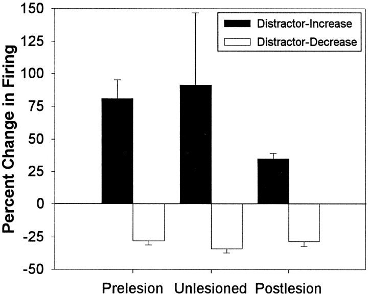
Cortical cholinergic inputs are hypothesized to mediate attentional functions. The present experiment was designed to determine the single unit activity of neurons within the medial prefrontal cortex (mPFC) of rats performing a sustained visual attention task. Demands on attentional performance were varied by the presentation of a visual distractor. The contribution of cholinergic afferents of the mPFC to performance-associated unit activity within this area was determined by recording neuronal activity before and after unilateral cholinergic deafferentation using intracortical infusion of the immunotoxin 192 IgG-saporin. Presentation of the visual distractor resulted in a decrease in the detection of brief, unpredictable visual signals. As predicted, the unilateral loss of cholinergic inputs within the recording area of the mPFC did not affect sustained attentional performance. Cholinergic deafferentation, however, resulted in a decrease in the overall firing rate of medial prefrontal neurons and a substantial reduction in the proportion of neurons whose firing patterns correlated with specific aspects of behavioral performance. Furthermore, cholinergic deafferentation attenuated the frequency and amplitude of increased mPFC neuronal firing rates that were associated with the presentation of the visual distractor. The main findings from this experiment suggest that cholinergic inputs to the mPFC strongly influence spontaneous and behaviorally correlated single unit activity and mediate increases in neuronal activity associated with enhanced demands for attentional processing, all of which may be fundamental aspects in the maintenance of attentional performance.


Figures

Fig. 1.
Fig. 1.
Response rules of the sustained visual attention task. Schematic of the response rules of the sustained visual attention task after the presentation of signal or nonsignal events. Thetop two panels demonstrate the response rules after center panel light illumination (signal event). The bottom two panels demonstrate the response rules after the nonillumination of the center panel light (nonsignal event). A tone was presented 1 sec after signal and nonsignal events initiating a 4 sec response window. Water reinforcement (40 μl) was delivered into a water port affixed to the back wall of the operant chamber.
Fig. 2.
Fig. 2.
Electrode path and 192 IgG-saporin induced AChE-positive fiber loss within the mPFC. A, Schematic of a coronal section through the level of the mPFC (3.20 mm anterior to bregma) illustrating the path and final recording sites of the recording electrodes in the left or right hemispheres.PL, Prelimbic sector of mPFC; IL, infralimbic sector of mPFC. The box outline inA represents the location from which the photomicrograph in B was taken. B, Photomicrograph (5×) illustrating the restricted loss of AChE-positive fibers to mPFC and the final recording site demarcated by the small electrolytic lesion (star) within mPFC layers III–V of the left hemisphere.C, Higher-magnification photomicrograph (13.2×) of the loss of AChE-positive fibers throughout the recording area inB. Note the distinct loss of AChE-positive fiber staining throughout all cortical layers dorsal to the final recording site (star) when compared with the high density of fiber staining throughout the contralateral hemisphere.
Fig. 3.
Fig. 3.
Sustained visual attention performance during baseline testing. Relative number of hits (mean ± SEM) across signal lengths and relative number of correct rejections (mean ± SEM) during prelesion (filled circles andbar) and postlesion (open circles andbar) phases of testing. NS, Nonsignal trials.
Fig. 4.
Fig. 4.
Sustained visual attention performance across trial blocks during standard and distractor testing conditions.A, Relative number of hits (mean ± SEM) across trial blocks under standard and distractor testing conditions during prelesion and postlesion phases of attentional testing.B, Relative number of correct rejections (mean ± SEM) across trial blocks under standard and distractor testing conditions during prelesion and postlesion phases of attentional testing.
Fig. 5.
Fig. 5.
Distractor modulation of mPFC unit activity. Mean firing rates of two mPFC units plotted over the course of a distractor testing session (46 min). A, Neuron that exhibited a distractor-induced increase in firing rate of 98% from 0.89 spikes/sec within trial block 1 to 1.76 spikes/sec during the presence of the visual distractor within trial block 2 that subsequently decreased by 55% to 0.97 spikes/sec after the return to standard testing conditions within trial block 3.B, Neuron that exhibited a distractor-induced decrease in firing rate of 41% from 0.89 spikes/sec within trial block 1 to 0.52 spikes/sec during the presence of the visual distractor that subsequently increased by 38% to 0.72 spikes/sec within trial block 3.
Fig. 6.
Fig. 6.
Amplitude of distractor-induced alterations in mPFC unit activity during sustained visual attention. The graph illustrates that the magnitude (mean ± SEM) of positive alterations (percentage change, trial block 2 relative to trial block 1) in mPFC unit firing exhibited during the presence of the visual distractor was attenuated after the removal of cholinergic input, whereas the magnitude of negative alterations in unit firing during the presence of the visual distractor was unaltered across testing phases.
Fig. 7.
Fig. 7.
Behavioral correlates of mPFC unit activity during sustained visual attention. The PETHs (20 msec per bin) presented are from six different mPFC units at different levels within the prelimbic sector of the same rat. AC, Representative response-related behavioral correlates. Aillustrates a preresponse excitatory correlate with increased unit firing before correct rejection responses (CR, correct rejection); B illustrates a response excitatory correlate with increased unit firing coincident with correct rejection responses; and C illustrates a response inhibitory correlate with decreased unit firing coincident with hit responses.DF, Representative reward-related behavioral correlates. D illustrates an anticipatory excitatory correlate with increased unit firing that began shortly after the emission of hits and remained elevated until the rat reached the water port; E illustrates an anticipatory inhibitory correlate with decreased unit firing after correct rejection responses and during the approach to the water port; and Fillustrates a consumption excitatory correlate with increased unit firing that occurred immediately after entry into the water port and remained elevated throughout the consumption of the reward.WP, Water port entry.
Fig. 8.
Fig. 8.
Trial-type specificity of behaviorally correlated mPFC unit activity during sustained visual attention.AC, Trial type-specific unit activation of the same response-related behavioral correlate presented in Figure7B. A illustrates the PETH of the response excitatory correlate during correct rejections (CR) on nonsignal trials, whereas Billustrates the absence of behaviorally correlated activity from the same mPFC unit to hits on signal trials. Additionally, Cillustrates that responses on the same right lever as correct rejections, but during the intertrial interval (RL-ITI), were not accompanied by increases in unit activity. DF, Trial type-specific unit activation of the same reward-related behavioral correlate after signal trials presented in Figure 7D.D illustrates the PETH of the reward anticipation excitatory correlate after hits on signal trials, whereasE illustrates the absence of reward anticipatory activity after correct rejections on nonsignal trials. Fillustrates that responses on the same left lever as the hits, but during the intertrial interval (LL-ITI), were not followed by increases in unit activity.
Fig. 9.
Fig. 9.
Behaviorally correlated mPFC unit activity during distractor testing sessions. A, PETH of the response excitatory correlate during correct rejections (CR) summed across all three blocks of trials (same unit that was presented in Fig. 7B). BD, PETHs of the same unit separated by trial blocks 1–3. The increase in unit firing during correct rejection responses under standard testing conditions within trial blocks 1 and 3 (B and D, respectively) persisted despite the presence of the visual distractor within trial block 2 (C) and concomitant decrease in the relative number of correct rejections and overall rate of firing (19% decrease in overall firing rate in trial block 2 relative to block 1).

References

    1. Balleine BW, Dickinson A. Goal-directed instrumental action: contingency and incentive learning and their cortical substrates. Neuropharmacology. 1998;37:407–419. - PubMed
    1. Bassant MH, Baleyte JM, Lamour Y. Effects of acetylcholine on single cortical somatosensory neurons in the unanesthetized rat. Neuroscience. 1990;39:189–197. - PubMed
    1. Bucci DJ, Holland PC, Gallagher M. Removal of cholinergic input to rat posterior parietal cortex disrupts incremental processing of conditioned stimuli. J Neurosci. 1998;18:8038–8046. - PMC - PubMed
    1. Chang JY, Zhang L, Janak PH, Woodward DJ. Neuronal responses in prefrontal cortex and nucleus accumbens during heroin self-administration in freely moving rats. Brain Res. 1997;754:12–20. - PubMed
    1. Chiba AA, Bucci DJ, Holland PC, Gallagher M. Basal forebrain cholinergic lesions disrupt increments but not decrements in conditioned stimulus processing. J Neurosci. 1995;15:7315–7322. - PMC - PubMed

Publication types