. 2000 Jun;120(3):503-11.
doi: 10.1046/j.1365-2249.2000.01239.x.

Enhanced apoptosis of T cells in common variable immunodeficiency (CVID): role of defective CD28 co-stimulation

Affiliations

Enhanced apoptosis of T cells in common variable immunodeficiency (CVID): role of defective CD28 co-stimulation

M Di Renzo et al. Clin Exp Immunol. 2000 Jun.

Abstract

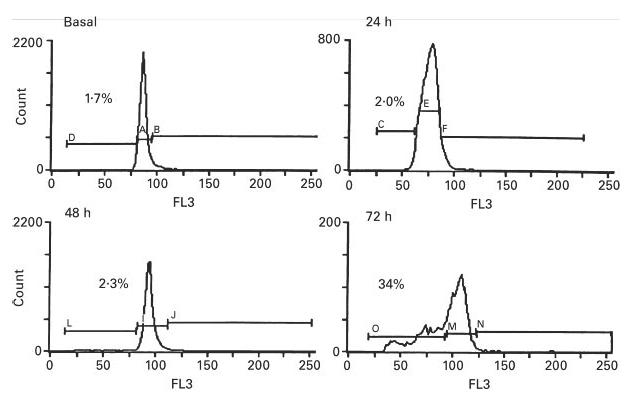
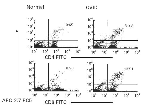
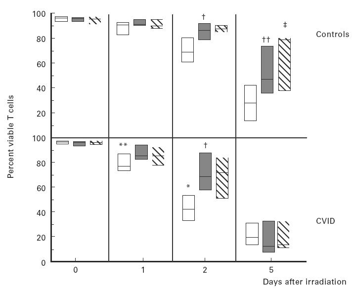
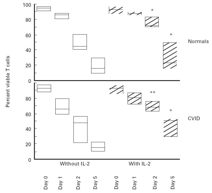
CVID is a primary immune disorder in which hypogammaglobulinaemia may be associated with a number of T cell defects including lymphopenia, anergy, impaired lymphocyte proliferation and deficient cytokine secretion. In this study we show that T cells of CVID subjects, in comparison with control T cells, undergo spontaneous apoptosis in culture and markedly accelerated apoptosis after gamma-irradiation. Although costimulation of the CD28 receptor following engagement of the TCR/CD3 receptor normally provides a second signal necessary for IL-2 secretion, CD28 costimulation in CVID does not significantly increase IL-2 production, nor does this combination of activators enhance the survival of irradiated CVID T cells, as it does for cultured normal T cells. Addition of IL-2 enhances CVID T cell survival, suggesting that the IL-2 signalling pathways are normal. CVID T cells have similar expression of Bcl-2 to control T cells. CD3 stimulation up-regulates T cell expression of bcl-xL mRNA for normal T cells, but anti-CD28 does not augment bcl-xL expression for CVID subjects with accelerated apoptosis. Defects of the CD28 receptor pathway, leading to cytokine deprivation and dysregulation of bcl-xL, could lead to poor T cell viability and some of the cellular defects observed in CVID.
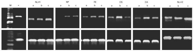

Figures

Fig. 1
Fig. 1
Cell cycle analysis of T cells of one CVID subject after permeabilization and staining with propidium iodide at 0, 24, 48, and 72 h of culture. The percentage of cells in the apoptotic fraction is indicated in each histogram. (Results similar to three other CVID subjects studied; two normal subjects showed 2·3% of apoptotic cells after 72 h of culture).
Fig. 2
Fig. 2
Flow cytometric detection of APO2.7–PECy5 in CD4+ and CD8+ T cells after 24 h of culture. The numbers show the percentages of CD4+ and CD8+ T cells that are positively stained for APO2.7 for one CVID subject (ratio CD4/CD8 = 3·0) in comparison with one representative control (ratio CD4/CD8 = 1·5) (five CVID and four normal controls studied).
Fig. 3
Fig. 3
T cells of 10 normal subjects (upper panel) or 16 CVID subjects (lower panel) were tested for survival before and following γ-irradiation. T cells were cultured in medium alone (□), or stimulated with plate-immobilized anti-CD3 alone (shaded bars) or with anti-CD3 plus anti-CD28 (hatched bars) for 16 h and then exposed to 15 Gy of γ-irradiation. Percent viability of each group was assessed by CD3 staining and propidium iodide exclusion. CVID T cells had significantly poorer T cell viability on days 1 and 2 after irradiation (**P < 0·01; *P < 0·05, respectively). CD3 stimulation enhanced CVID T cell viability on day 2 (†P < 0·05) and normal T cell viability on days 2 and 5 after irradiation (††P < 0·05). CD28 costimulation further increased normal T cell viability on day 5 (‡P < 0·05). The boxes represent the range, the line inside the box represents the median value.
Fig. 4
Fig. 4
Pretreatment with IL-2 enhances the survival of normal (upper panel) and CVID (lower panel) T cells after γ-irradiation. Normal or CVID T cells were cultured overnight and then kept either in the original medium, or in medium with recombinant 100 U/ml IL-2 added prior to irradiation. The data refer to five normal controls and eight CVID subjects. The boxes represent the range, the line inside the box represents the median value. (*P < 0·05; **P < 0·01 difference for IL-2-treated versus no IL-2-treated, significant for both groups).
Fig. 5
Fig. 5
T cells of CVID subjects were examined for up-regulation of bcl-xL gene expression, testing cells incubated with no stimulators, or with anti-CD3, or anti-CD3 plus anti-CD28 in a polymerase chain reaction (PCR) using primers specific for bcl-xL or actin, as a sample control. The positive control was Jurkat cells transfected with bcl-x. The upper panel shows PCR results for two normals and four CVID subjects; a, no stimulation; b, CD3 stimulation; c, CD3 + CD28 stimulation. CVID subjects had enhanced bcl-xL expression after CD3 stimulation which was not augmented by addition of anti-CD28. The actin sample controls are shown (lower panels) for each. M, Molecular weight markers; +, positive control.

Similar articles

Cited by

References

    1. Report of a WHO Scientific Group. Clin Exp Immunol. 1997;109(Suppl.1):1–28. - PubMed
    1. Spickett GP, Farrant J, North ME, Zhang J, Morgan L, Webster Adb. Common variable immunodeficiency: how many diseases? Immunol Today. 1997;18:325–8. - PubMed
    1. Sneller MC. Sneller MC, editor. Clinical spectrum of common variable immunodeficiency. New insights into common variable immunodeficiency. Ann Intern Med. 1993;118:720–30. Moderator. - PubMed
    1. Cunningham-Rundles C, Bodian C. Common variable immunodeficiency: clinical and immunologic features of 248 patients. Clin Immunol. 1999;92:34–48. - PubMed
    1. Hermaszewski RA, Webster Adb. Primary hypogammaglobulinemia: a survey of clinical manifestations and complications. Q J Med. 1993;86:31–42. - PubMed