

Cellular and viral specificities of human immunodeficiency virus type 1 vif protein
- PMID: 10846079
- PMCID: PMC112094
- DOI: 10.1128/jvi.74.13.5982-5987.2000
Cellular and viral specificities of human immunodeficiency virus type 1 vif protein
Abstract
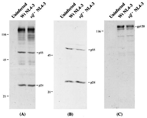
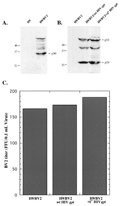
The vif gene of human immunodeficiency virus type 1 (HIV-1) greatly enhances the infectivity of HIV-1 virions that are released from cells classified as nonpermissive (e.g., lymphocytes, macrophages, and H9 leukemic T cells) but is irrelevant in permissive cells (e.g., HeLa or COS cells). Recently, it was reported that vif expression in nonpermissive cells dramatically increases infectivity not only of HIV-1 but also of other enveloped viruses, including murine leukemia viruses (MLVs). This was surprising in part because MLVs and other murine retroviruses lack vif genes yet replicate efficiently in T lymphocytes. To investigate these issues, we first developed improved methods for producing substantial quantities of HIV-1 virions with vif deletions from healthy H9 cells. These virions had approximately the same amounts of major core proteins and envelope glycoproteins as the control wild-type virions but were only approximately 1% as infectious. We then produced H9 cells that contained wild-type or vif deletion HIV-gpt proviruses, which lack a functional env gene. After superinfection with either xenotropic or amphotropic MLVs, these cells released HIV-gpt virions pseudotyped with an MLV envelope plus replication-competent MLV. Interestingly, the pseudotyped HIV-gpt (vif deletion) virions were noninfectious, whereas the MLV virions simultaneously released from the same H9 cells were fully infectious. These results strongly suggest that the Vif protein functions in a manner that is both cell specific and at least substantially specific for HIV-1 and related lentiviruses. In addition, these results confirm that vif deletion HIV-1 virions from nonpermissive cells are blocked at a postpenetration stage of the infection pathway.
Figures

References
-
- Agy M B, Acker R L, Sherbert C H, Katze M G. Interferon treatment inhibits virus replication in HIV-1- and SIV-infected CD4+ T-cell lines by distinct mechanisms: evidence for decreased stability and aberrant processing of HIV-1 proteins. Virology. 1995;214:379–386. - PubMed
-
- Akari H, Sakuragi J, Takebe Y, Tomonaga K, Kawamura M, Fukasawa M, Miura T, Shinjo T, Hayami M. Biological characterization of human immunodeficiency virus type 1 and type 2 mutants in human peripheral blood mononuclear cells. Arch Virol. 1992;123:157–167. - PubMed
-
- Ausubel F M, Brent R, Kingston R E, Moore D D, Seidman J G, Smith J A, Struhl K, editors. Current protocols in molecular biology. New York, N.Y: John Wiley & Sons, Inc.; 1994.
Publication types
MeSH terms
Substances
Grants and funding
LinkOut - more resources
Full Text Sources
Other Literature Sources
