. 2000 Jul;74(13):6050-7.
doi: 10.1128/jvi.74.13.6050-6057.2000.

Retargeting the coxsackievirus and adenovirus receptor to the apical surface of polarized epithelial cells reveals the glycocalyx as a barrier to adenovirus-mediated gene transfer

Affiliations

Retargeting the coxsackievirus and adenovirus receptor to the apical surface of polarized epithelial cells reveals the glycocalyx as a barrier to adenovirus-mediated gene transfer

R J Pickles et al. J Virol. 2000 Jul.

Abstract

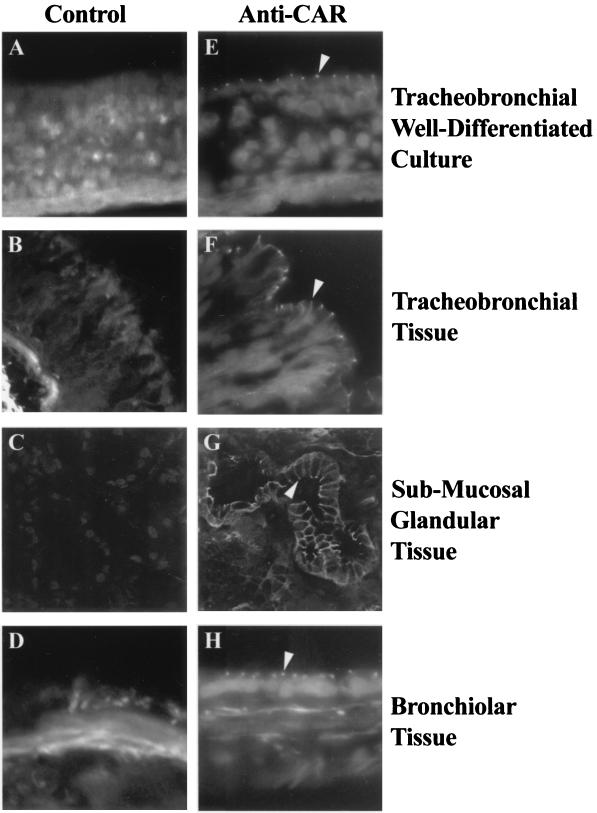
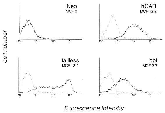
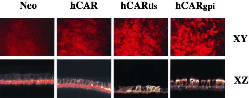
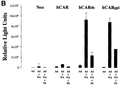
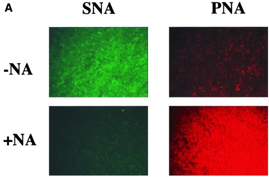
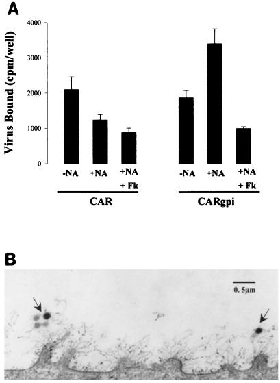
Lumenal delivery of adenovirus vectors (AdV) results in inefficient gene transfer to human airway epithelium. The human coxsackievirus and adenovirus receptor (hCAR) was detected by immunofluorescence selectively at the basolateral surfaces of freshly excised human airway epithelial cells, suggesting that the absence of apical hCAR constitutes a barrier to adenovirus-mediated gene delivery in vivo. In transfected polarized Madin-Darby canine kidney cells, wild-type hCAR was expressed selectively at the basolateral membrane, whereas hCAR lacking the transmembrane and/or cytoplasmic domains was expressed on both the basolateral and apical membranes. Cells expressing apical hCAR still were not efficiently transduced by AdV applied to the apical surface. However, after the cells were treated with agents that remove components of the apical surface glycocalyx, AdV transduction occurred. These results indicate that adenovirus can infect via receptors located at the apical cell membrane but that the glycocalyx impedes interaction of AdV with apical receptors.


Figures

FIG. 1
FIG. 1
Immunolocalization of hCAR expression in human airway epithelial cells. Frozen sections of human non-CF tracheobronchial airway epithelial cells cultured on permeable supports (A and E) (17) or of human non-CF airway tissue derived from the tracheobronchial (B and F), submucosal glandular (C and G), or bronchiolar (D and H) regions were fixed and incubated with either control IgG or the anti-CAR monoclonal IgG antibody RmcB and examined by confocal microscopy as described in Materials and Methods. Specific staining was observed only in those tissues exposed to anti-CAR and was localized to the lateral aspects of all the surface epithelial cells (arrowheads). Magnification, ×40.
FIG. 2
FIG. 2
CAR expression on transfected MDCK cells. Cells transfected with wild-type hCAR, hCARtls, or hCARgpi or with expression vector alone (Neo) were incubated with RmcB (solid line) or with a control IgG (dotted line), followed by fluorescein isothiocyanate-conjugated secondary antibody, and then analyzed for CAR expression by flow cytometry. The mean cell fluorescence (MCF) for each cell line was corrected by subtracting the mean fluorescence of cells stained with control antibody.
FIG. 3
FIG. 3
Immunolocalization of mutant hCAR constructs in polarized MDCK epithelial cells. Polarized MDCK cells transfected with wild-type hCAR, hCARtls, or hCARgpi or with expression vector alone (Neo) were cultured on permeable supports and then fixed, permeabilized, stained with anti-CAR, and examined by confocal microscopy as described in Materials and Methods. No fluorescent staining was seen in controls with nonimmune IgG. Magnification, ×17.6 (XY) and ×35.2 (XZ) (original magnification, ×20 [XY] and ×40 [XZ]).
FIG. 4
FIG. 4
AdV-mediated gene transfer to MDCK cells before and after NA treatment. The apical surfaces of transfected MDCK cells were treated with NA (+NA) or left untreated (−NA) before exposure to AdV. (A) Monolayers exposed to AdV encoding GFP, with transgene expression detected by fluorescent microscopy at 48 h. Magnification, × 4.2 (original magnification, ×5). (B) Monolayers exposed to AdV encoding β-galactosidase, with detection by chemiluminescence assay. Parallel cultures were incubated with excess fiber knob protein (Fk; 25 μg/ml) in addition to AdV. The error bars represent the standard errors of the mean for at least four samples.
FIG. 4
FIG. 4
AdV-mediated gene transfer to MDCK cells before and after NA treatment. The apical surfaces of transfected MDCK cells were treated with NA (+NA) or left untreated (−NA) before exposure to AdV. (A) Monolayers exposed to AdV encoding GFP, with transgene expression detected by fluorescent microscopy at 48 h. Magnification, × 4.2 (original magnification, ×5). (B) Monolayers exposed to AdV encoding β-galactosidase, with detection by chemiluminescence assay. Parallel cultures were incubated with excess fiber knob protein (Fk; 25 μg/ml) in addition to AdV. The error bars represent the standard errors of the mean for at least four samples.
FIG. 5
FIG. 5
Effects of NA treatment on MDCK cell lines. (A) Removal of terminal sialic acid residues. The apical surfaces of MDCK-hCARgpi cells were probed with fluorescein-conjugated SNA lectin (green fluorescence) and rhodamine-conjugated PNA lectin (red fluorescence) in cultures without (−NA) and with (+NA) prior exposure to NA. Similar distribution patterns for SNA and PNA before and after NA treatment were observed for all four transfected MDCK cell lines. Magnification, ×5. (B) Transepithelial resistance measurement before and after NA treatment of the apical surfaces of the respective cell lines. The open bars represent the resistances of the respective cultures before exposure to serum-free medium alone or NA (n = 36); the hatched bars represent the resistances of the respective cultures after exposure to serum-free medium alone (n = 18); and the solid bars represent the resistances of respective cultures after exposure to NA (n = 18).
FIG. 5
FIG. 5
Effects of NA treatment on MDCK cell lines. (A) Removal of terminal sialic acid residues. The apical surfaces of MDCK-hCARgpi cells were probed with fluorescein-conjugated SNA lectin (green fluorescence) and rhodamine-conjugated PNA lectin (red fluorescence) in cultures without (−NA) and with (+NA) prior exposure to NA. Similar distribution patterns for SNA and PNA before and after NA treatment were observed for all four transfected MDCK cell lines. Magnification, ×5. (B) Transepithelial resistance measurement before and after NA treatment of the apical surfaces of the respective cell lines. The open bars represent the resistances of the respective cultures before exposure to serum-free medium alone or NA (n = 36); the hatched bars represent the resistances of the respective cultures after exposure to serum-free medium alone (n = 18); and the solid bars represent the resistances of respective cultures after exposure to NA (n = 18).
FIG. 6
FIG. 6
Nonspecific attachment to and receptor shielding properties of glycocalyx. (A) Radiolabeled AdV attachment to the apical surfaces of MDCK-hCAR and MDCK-hCARgpi cells either without (−NA) or with (+NA) prior treatment with NA. The specificity of the AdV-receptor interaction was determined by coincubation of purified fiber knob protein (Fk; 25 μg/ml) with AdV. Note that virus attachment to CAR cells is reduced after NA pretreatment whereas attachment to hCARgpi is enhanced only after NA pretreatment. (B) Transmission electron micrograph of the surfaces of polarized epithelial cells after exposure to AdV for 2 h at room temperature. The arrows show AdV entangled in the cellular glycocalyx. Note the projection of the glycocalyx from the microvilli extending 0.5 to 1 μm from the cell surface.
FIG. 7
FIG. 7
Specific enzymatic reagents other than NA enhance AdV-mediated gene transfer specifically to MDCK-CARgpi cells. The apical surfaces of the transfected MDCK cells were exposed to serum-free medium alone (control), trypsin, keratanase, or human leukocyte elastase as described in Materials and Methods. After the cells were washed, AdV-GFP was exposed to the apical surfaces. Transgene expression was observed 48 h later by epifluorescence microscopy. The results with MDCK-Neo resembled those with MDCK-hCAR, and MDCK-CARtls showed results similar to those obtained with MDCK-CARgpi (not shown). Magnification, ×5.

References

    1. Arcasoy S M, Latoche J, Gondor M, Watkins S C, Henderson R A, Hughey R, Finn O J, Pilewski J M. MUC1 and other sialoglycoconjugates inhibit adenovirus-mediated gene transfer to epithelial cells. Am J Respir Cell Mol Biol. 1997;17:422–435. - PubMed
    1. Bals R, Xiao W, Sang N, Weiner D J, Meegalla R L, Wilson J M. Transduction of well-differentiated airway epithelium by recombinant adeno-associated virus is limited by vector entry. J Virol. 1999;73:6085–6088. - PMC - PubMed
    1. Bergelson J M, Cunningham J A, Droguett G, Kurt-Jones E A, Krithivas A, Hong J S, Horwitz M S, Crowell R L, Finberg R W. Isolation of a common receptor for Coxsackie B viruses and adenoviruses 2 and 5. Science. 1997;275:1320–1323. - PubMed
    1. Bhaskar K, O'Sullivan D, Opaskar-Hincman H, Reid L, Coles S. Density gradient analysis of secretions produced in vitro by human and canine airway mucosa: identification of lipids and proteoglycans in such secretions. Exp Lung Res. 1986;10:401–422. - PubMed
    1. Boucher R C. Status of gene therapy for cystic fibrosis lung disease. J Clin Investig. 1999;103:441–445. - PMC - PubMed

Publication types

MeSH terms

LinkOut - more resources