. 2000 Jul;74(13):6198-202.
doi: 10.1128/jvi.74.13.6198-6202.2000.

Isolation of human immunodeficiency virus type 1 cores: retention of Vpr in the absence of p6(gag)

Affiliations

Isolation of human immunodeficiency virus type 1 cores: retention of Vpr in the absence of p6(gag)

M A Accola et al. J Virol. 2000 Jul.

Abstract

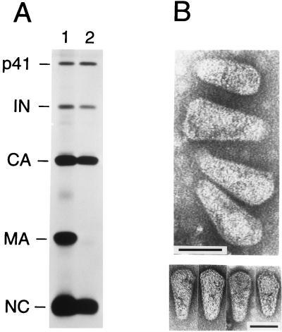
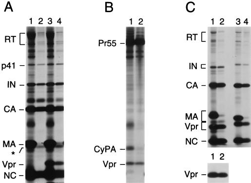
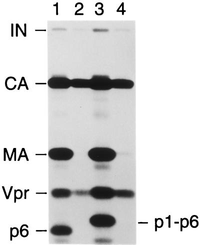
Mature human immunodeficiency virus type 1 (HIV-1) virions contain a typically cone-shaped core that encases the viral genome. In this study, we established conditions which allowed the efficient isolation of morphologically intact HIV-1 cores from virions. The isolated cores consisted mostly of cones which appeared uniformly capped at both ends but were heterogeneous with respect to the shape of the broad cap as well as the dimensions and angle of the cone. Vpr, a nonstructural virion component implicated in the nuclear import of the viral genome, was recovered in core preparations of HIV-1 and simian immunodeficiency viruses from African green monkeys. Unexpectedly, p6(gag), a structural protein required for the incorporation of Vpr, was absent from HIV-1 core preparations. Taken together, our results indicate that the incorporation of Vpr into the virion core is a conserved feature of primate lentiviruses and that the interactions required for the uptake of Vpr into assembling particles differ from those which confine Vpr within the core.


Figures

FIG. 1
FIG. 1
Biochemical and electron microscopic analysis of HIV-1 core preparations. (A) Equal amounts of supernatant containing [35S]cysteine-labeled virus were loaded on top of 20% sucrose layers which either lacked detergent (lane 1) or contained 0.03% Igepal Co-630 (lane 2) and which were supported by 30% sucrose cushions. Following centrifugation, pelleted material was directly analyzed by SDS-PAGE and autoradiography. The product designated p41 is an intermediate Gag cleavage product (2). (B) Electron micrographs of isolated HIV-1 cores prepared by centrifugation through a detergent layer and a 30% sucrose cushion. The lower panel shows examples of cores with angular caps or with a spherical structure that is discernible at the broad end. Bars, 50 nm.
FIG. 2
FIG. 2
Association of Vpr with HIV-1 and SIVagm cores. [35S]cysteine-labeled wild-type HIV-1 virions (A), protease-defective immature HIV-1 virions (B), or wild-type SIVagm virions (C) were spun through step gradients which either lacked detergent (odd-numbered lanes) or contained detergent in the upper layer (even-numbered lanes) as described for Fig. 1. Pelleted material was analyzed directly by SDS-PAGE and autoradiography. Virus-containing supernatants were obtained by transfection of 293T cells with proviral plasmids and metabolic labeling. In panel A, the transfected HIV-1 proviruses were the infectious, vpr-negative molecular clone HXBH10 (lanes 1 and 2) and its vpr-positive variant HXBH10/R+ (lanes 3 and 4). The protease-defective HIV-1 provirus used in panel B was HXBH10/R+/PR (4). The SIVagm proviruses in panel C were SIVagm 155-4 (lanes 1 and 2) and SIVagm gri-1 (lanes 3 and 4). In panel A, the p66 form of RT is partially obscured by a contaminant. The asterisk indicates the position of an unidentified core-associated protein that migrates slightly faster than the bulk of MA.
FIG. 3
FIG. 3
Absence of p6gag from HIV-1 core preparations. [3H]leucine-labeled HIV-1 virions were spun through step gradients which either lacked detergent (odd-numbered lanes) or contained detergent in the upper layer (even-numbered lanes) as described for Fig. 1, and pelleted material was analyzed by SDS-PAGE. Virus was produced in 293T cells, and the transfected HIV-1 proviruses were the infectious, vpr-positive clone HXBH10/R+ (lanes 1 and 2) or a variant with a point mutation at the p1-p6 cleavage site (lanes 3 and 4).

References

    1. Accola M A, Bukovsky A A, Jones M S, Göttlinger H G. A conserved dileucine-containing motif in p6gag governs the particle association of Vpx and Vpr of simian immunodeficiency viruses SIVmac and SIVagm. J Virol. 1999;73:9992–9999. - PMC - PubMed
    1. Accola M A, Höglund S, Göttlinger H G. A putative α-helical structure which overlaps the capsid-p2 boundary in the human immunodeficiency virus type 1 Gag precursor is crucial for viral particle assembly. J Virol. 1998;72:2072–2078. - PMC - PubMed
    1. Bolognesi D P, Montelaro R C, Frank H, Schäfer W. Assembly of type C oncornaviruses: a model. Science. 1978;199:183–186. - PubMed
    1. Bukovsky A, Dorfman T, Weimann A, Göttlinger H G. Nef association with human immunodeficiency virus type 1 virions and cleavage by the viral protease. J Virol. 1997;71:1013–1018. - PMC - PubMed
    1. Bukovsky A A, Weimann A, Accola M A, Göttlinger H G. Transfer of the HIV-1 cyclophilin-binding site to simian immunodeficiency virus from Macaca mulatta can confer both cyclosporin sensitivity and cyclosporin dependence. Proc Natl Acad Sci USA. 1997;94:10943–10948. - PMC - PubMed

Publication types

MeSH terms

Substances