. 2000 Jul;20(13):4532-42.
doi: 10.1128/MCB.20.13.4532-4542.2000.

PKR stimulates NF-kappaB irrespective of its kinase function by interacting with the IkappaB kinase complex

Affiliations

PKR stimulates NF-kappaB irrespective of its kinase function by interacting with the IkappaB kinase complex

M C Bonnet et al. Mol Cell Biol. 2000 Jul.

Abstract

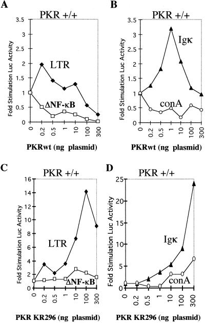
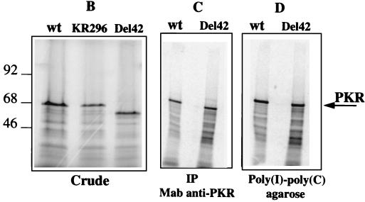
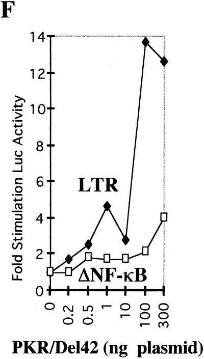
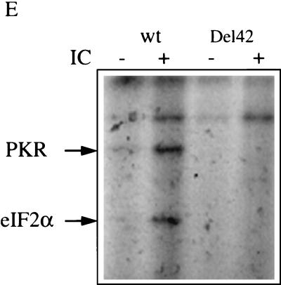
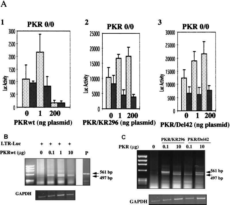
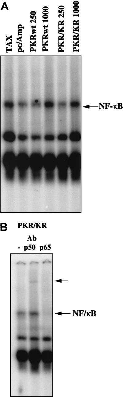
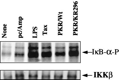
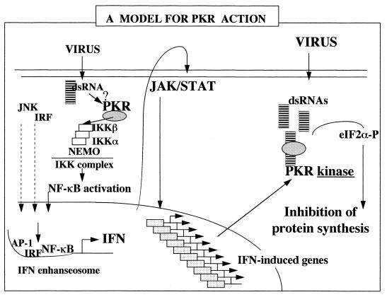
The interferon (IFN)-induced double-stranded RNA-activated protein kinase PKR mediates inhibition of protein synthesis through phosphorylation of the alpha subunit of eukaryotic initiation factor 2 (eIF2alpha) and is also involved in the induction of the IFN gene through the activation of the transcription factor NF-kappaB. NF-kappaB is retained in the cytoplasm through binding to its inhibitor IkappaBalpha. The critical step in NF-kappaB activation is the phosphorylation of IkappaBalpha by the IkappaB kinase (IKK) complex. This activity releases NF-kappaB from IkappaBalpha and allows its translocation to the nucleus. Here, we have studied the ability of PKR to activate NF-kappaB in a reporter assay and have shown for the first time that two catalytically inactive PKR mutants, PKR/KR296 and a deletion mutant (PKR/Del42) which lacks the potential eIF2alpha-binding domain, can also activate NF-kappaB. This result indicated that NF-kappaB activation by PKR does not require its kinase activity and that it is independent of the PKR-eIF2alpha relationship. Transfection of either wild-type PKR or catalytically inactive PKR in PKR(0/0) mouse embryo fibroblasts resulted in the activation of the IKK complex. By using a glutathione S-transferase pull-down assay, we showed that PKR interacts with the IKKbeta subunit of the IKK complex. This interaction apparently does not require the integrity of the IKK complex, as it was found to occur with extracts from cells deficient in the NF-kappaB essential modulator, one of the components of the IKK complex. Therefore, our results reveal a novel pathway by which PKR can modulate the NF-kappaB signaling pathway without using its kinase activity.


Figures

FIG. 1
FIG. 1
NF-κB-dependent stimulation of gene expression by PKRwt and PKR/KR296 in a reporter assay. Different concentrations (0 to 300 ng) of pcDNA1/Amp expressing either PKRwt (A and B) or PKR/KR296 (C and D) were cotransfected in PKR+/+ MEFs with 100 ng of the reporter plasmid expressing luciferase under the control of different promoters: pHIV-1 LTR-luc (LTR; closed diamonds) or pHIV-1 LTRΔNF-κB-luc (ΔNF-κB; open squares) (A and C) and IgκCona-luc (Igκ; closed triangles) or Cona-luc (conA; open circles) (B and D). Each DNA mixture was adjusted to the same final DNA content with the addition of the empty pcDNA1/Amp vector. The luciferase activity per microgram of protein content per well was calculated as the mean of the transfections in four different wells (data not shown). Each value (fold stimulation of luciferase [Luc] activity) corresponds to the ratio of luciferase activity obtained by transfecting PKR with the reporter gene to luciferase activity obtained by transfecting the reporter gene alone.
FIG. 2
FIG. 2
Construction and characterization of the PKR/Del42 mutant. (A) The PKR deletion mutant PKR/Del42 was generated by removing the region between subdomains V and VI (see Materials and Methods). The deletion starts after residue 369 and stops before residue 412. The length of the deletion is 42 amino acids. A schematic presentation of the PKR/Del42 mutant is shown, along with the amino acid sequence of wild-type (wt) PKR residues 357 to 422. The approximate positions of the BclI and AflII restriction sites used for the construction are indicated. DRDB, dsRNA binding domain. (B) RNA preparations were in vitro transcribed from plasmid pcDNA1/Amp expressing PKRwt, PKR/KR296, and PKR/Del42. They were then translated in rabbit reticulocyte lysates in the presence of ProMix and 6 mM 2-aminopurine. After 60 min of incubation at 30°C, a 1/10 volume of the translation mixture was analyzed by SDS-PAGE on 12.5% acrylamide gels. The molecular weights (103) of marker proteins are indicated on the left. (C) Equivalent amounts of in vitro-translated PKRwt and PKR/Del42 (calculated by integrating areas from the different PKR bands shown in panel B) were immunoprecipitated (IP) with 0.2 μl of anti-PKR monoclonal antibody (Mab) 71/10 and 50 μl of protein A/G-agarose. After 18 h of incubation at 4°C and extensive washing with BI buffer, the immunoprecipitated 35S-labeled PKR preparations were analyzed by SDS-PAGE on 12.5% acrylamide gels. (D) Equivalent amounts of in vitro-translated PKRwt and PKR/Del42 (as in panel C) were analyzed for binding to poly(I)-poly(C)–agarose (Pharmacia). After 18 h of incubation at 4°C and extensive washing with BI, the poly(I)-poly(C)-bound 35S-labeled PKR preparations were analyzed by SDS-PAGE on 12.5% acrylamide gels. (E) Preparations corresponding to PKRwt and PKR/Del42 RNAs were in vitro translated in rabbit reticulocyte lysates as described for panel B but in the absence of 35S-labeled amino acids. The proteins were then immunoprecipitated with anti-PKR monoclonal antibody 71/10 as described for panel C. The immunoprecipitated PKR samples were incubated at 30°C in the absence or presence of 1 μg of poly(I)-poly(C) (IC) per ml and in the presence of 0.25 μCi of [γ-32P]ATP per ml. After 15 min of incubation, 2 μl (165 ng) of a purified eIF2 complex preparation was added, and incubation was continued for another 15 min. After the addition of an equal volume of 2× SDS buffer, samples were heated (5 min at 95°C) and analyzed by SDS-PAGE on 12.5% acrylamide gels. Arrows show the positions of PKR (68 kDa) and eIF2α (35 kDa). (F) PKR/Del42 was cotransfected with pHIV-1 LTR-luc (LTR; closed diamonds) or pHIV-1 LTRΔNF-κB-luc (ΔNF-κB; open squares), and the fold stimulation of luciferase (Luc) was measured as described in the legend to Fig. 1.
FIG. 2
FIG. 2
Construction and characterization of the PKR/Del42 mutant. (A) The PKR deletion mutant PKR/Del42 was generated by removing the region between subdomains V and VI (see Materials and Methods). The deletion starts after residue 369 and stops before residue 412. The length of the deletion is 42 amino acids. A schematic presentation of the PKR/Del42 mutant is shown, along with the amino acid sequence of wild-type (wt) PKR residues 357 to 422. The approximate positions of the BclI and AflII restriction sites used for the construction are indicated. DRDB, dsRNA binding domain. (B) RNA preparations were in vitro transcribed from plasmid pcDNA1/Amp expressing PKRwt, PKR/KR296, and PKR/Del42. They were then translated in rabbit reticulocyte lysates in the presence of ProMix and 6 mM 2-aminopurine. After 60 min of incubation at 30°C, a 1/10 volume of the translation mixture was analyzed by SDS-PAGE on 12.5% acrylamide gels. The molecular weights (103) of marker proteins are indicated on the left. (C) Equivalent amounts of in vitro-translated PKRwt and PKR/Del42 (calculated by integrating areas from the different PKR bands shown in panel B) were immunoprecipitated (IP) with 0.2 μl of anti-PKR monoclonal antibody (Mab) 71/10 and 50 μl of protein A/G-agarose. After 18 h of incubation at 4°C and extensive washing with BI buffer, the immunoprecipitated 35S-labeled PKR preparations were analyzed by SDS-PAGE on 12.5% acrylamide gels. (D) Equivalent amounts of in vitro-translated PKRwt and PKR/Del42 (as in panel C) were analyzed for binding to poly(I)-poly(C)–agarose (Pharmacia). After 18 h of incubation at 4°C and extensive washing with BI, the poly(I)-poly(C)-bound 35S-labeled PKR preparations were analyzed by SDS-PAGE on 12.5% acrylamide gels. (E) Preparations corresponding to PKRwt and PKR/Del42 RNAs were in vitro translated in rabbit reticulocyte lysates as described for panel B but in the absence of 35S-labeled amino acids. The proteins were then immunoprecipitated with anti-PKR monoclonal antibody 71/10 as described for panel C. The immunoprecipitated PKR samples were incubated at 30°C in the absence or presence of 1 μg of poly(I)-poly(C) (IC) per ml and in the presence of 0.25 μCi of [γ-32P]ATP per ml. After 15 min of incubation, 2 μl (165 ng) of a purified eIF2 complex preparation was added, and incubation was continued for another 15 min. After the addition of an equal volume of 2× SDS buffer, samples were heated (5 min at 95°C) and analyzed by SDS-PAGE on 12.5% acrylamide gels. Arrows show the positions of PKR (68 kDa) and eIF2α (35 kDa). (F) PKR/Del42 was cotransfected with pHIV-1 LTR-luc (LTR; closed diamonds) or pHIV-1 LTRΔNF-κB-luc (ΔNF-κB; open squares), and the fold stimulation of luciferase (Luc) was measured as described in the legend to Fig. 1.
FIG. 3
FIG. 3
PKR/wt and mutants PKR/KR296 and PKR/Del42 stimulate NF-κB-dependent reporter gene expression in PKR0/0 MEFs. (A) The assay was performed as described in the legend to Fig. 1, except that PKR0/0 MEFs were used instead of PKR+/+ MEFs. The reporter plasmids pHIV-1 LTR-luc (dotted bars) and pHIV-1 LTRΔNF-κB-luc (grey bars) (100 ng each) were transfected either as such (0) or in the presence of 1 or 200 ng of PKRwt, PKR/KR296, or PKR/Del42. Data (luciferase [Luc] activity) are expressed as the mean of four independent transfections ± the standard error. (B) RT-PCR analysis of total RNA from PKR0/0 MEFs (in 10-cm petri dishes) transfected with pGL2 HIV-1 LTR-luc (LTR-Luc) alone or in the presence of increasing concentrations of PKRwt (0.1, 1, and 10 μg of plasmid). The sizes of the PCR products were estimated with a φX174 DNA HaeIII digest as a marker. The upper band (561 bp) corresponds to amplification from the transfected DNA as compared to the size of the PCR product from the pHIV-1 LTR-luc plasmid (P). The lower band (497 bp) is specific for DNA amplified from the reverse-transcribed luciferase mRNA. Control GAPDH RT-PCR is shown below. (C) RT-PCR analysis of total RNA from PKR0/0 MEFs transfected as described for panel B with the vector (LTR-Luc) alone or in the presence of PKR/KR296 or PKR/Del42 (0.1 and 10 μg of plasmid).
FIG. 4
FIG. 4
PKRwt and mutant PKR/KR296 can activate NF-κB in EMSAs. (A) PKR0/0 cells were transfected with 250 ng or 1 μg of plasmid pcDNA1/Amp expressing PKRwt or PKR/KR296 as described in Materials and Methods. The total amounts of plasmids were adjusted to 10 μg with the vector pcDNA1/Amp. All plasmids were prepared with an Endo-free Plasmid kit (Qiagen). Positive controls for NF-κB activation were obtained by transfecting 10 μg of pCMV-Tax (TAX). Background NF-κB activity is shown for cells transfected with pcDNA1/Amp alone (pc/Amp). Five micrograms of nuclear extracts was incubated with 0.2 pmol of 32P-labeled NF-κB probe for 20 min in the presence of 1 μg of poly(dI-dC) · poly(dI-dC) at 20°C and analyzed by an EMSA on 5% native acrylamide gels. Inducible NF-κB complex is indicated by the arrow. (B) Five micrograms of nuclear extracts from PKR/KR296-transfected cells was incubated for 10 min either as such (−) or in the presence of anti-p50 antibody (Ab) (shift indicated by upper arrow) or anti-p65 antibody (disappearance of the complex) prior to the addition of the probe and processed as described above.
FIG. 5
FIG. 5
PKRwt and the PKR mutants activate IKK. PKR0/0 cells were either untreated (None) or transfected with 5 μg of pcDNA1/Amp, alone (pc/Amp) or in the presence of 250 ng of pcDNA1/Amp expressing PKRwt or PKR/KR296. As a positive control, they were also treated for 30 min with 7 μg of LPS (Sigma) per ml or transfected with pCMV-Tax (Tax). Cell extracts (300 μg of protein/sample) were immunoprecipitated with anti-NEMO antibodies and analyzed for IKK activity in an in vitro phosphorylation assay with IκBα as a substrate (see Materials and Methods). The positions of phosphorylated IκBα (IκB-α-P) and IKKβ immunoprecipitated from the different cell extracts and revealed by immunoblotting are indicated.
FIG. 6
FIG. 6
Interaction of PKR with IKKβ. (A) Extracts from HeLa cells treated or not treated with 500 U of IFNα per ml were incubated with protein A/G-agarose previously coated with anti-IKKα or anti-IKKβ antibodies. Although PKR specifically interacts with IKKβ (see text), the antibodies directed against IKKβ (Santa Cruz) that we have used are unable to immunoprecipitate IKKβ. Therefore, they were conveniently used here as a control for nonspecific binding (Blank). After extensive washes, the proteins which were retained on the beads were separated by SDS-PAGE on 12.5% acrylamide gels and analyzed by immunoblotting with anti-PKR antibodies. Each lane represents the analysis of proteins immunoprecipitated (IP) from extracts equivalent to 5 × 10⁶ cells. In parallel, crude extracts were analyzed by immunoblotting for the presence of IKKα or PKR. (B) The proteins IKKβ, IKKα, NEMO, PKR/KR296, and luciferase (Luc) were translated in vitro from 2 μg of their respective plasmids in 25 μl of reticulocyte lysate in the presence of ProMix using a TNT-coupled Reticulocyte Lysate System (Promega). The translation products were analyzed by SDS-PAGE either as such (5 μl; 10% input) or after incubation for 18 h at 4°C with 1.25 μg of GST-PKR or an identical amount of GST-HP1α, a nuclear protein (52). Beads were adjusted to 50 μl in all samples with glutathione-Sepharose 4B (Amersham). (C) Cell extracts from 75 × 106 70Z/3 and 1.3E2 (NEMO-deficient) cells were incubated as described above with GST-PKR or GST-HP1α. Crude extracts corresponding to 7.5 × 10⁶ cells (10% input) and purified extracts were analyzed by immunoblotting with antibodies directed against murine IKKβ after SDS-PAGE.
FIG. 7
FIG. 7
Model for PKR action. The presence of small amounts of dsRNA in cells, such as at the onset of viral infection, may provoke the binding of endogenous PKR to IKK, leading to NF-κB activation and potentiating the induction of IFN genes. For this function, PKR would not need its kinase function. Once IFN is synthesized, it provokes the induction of several genes, through JAK/STAT signaling, including the PKR gene. As the viral infection develops, there is an accumulation in the cytoplasm of dsRNA structures which activate PKR, the levels of which are now increased after IFN induction. As a kinase, PKR phosphorylates eIF2α and provokes the inhibition of protein synthesis, which can limit the propagation of the virus. eIF2α-P, phosphorylated eIF2α; IRF, IFN regulatory factor.

References

    1. Alkalay I, Yaron A, Hatzubai A, Orian A, Ciechanover A, Ben-Neriah Y. Stimulation-dependent IκB phosphorylation marks the NF-κB inhibitor for degradation via the ubiquitin-proteasome pathway. Proc Natl Acad Sci USA. 1995;92:10599–10603. - PMC - PubMed
    1. Baeuerle P, Baichwal V. NF-κB as a frequent target for immunosuppressive and anti-inflammatory molecules. Adv Immunol. 1997;65:111–136. - PubMed
    1. Baldwin A. The NF-κB and IκB proteins: new discoveries and insights. Annu Rev Immunol. 1996;14:649–681. - PubMed
    1. Benkirane M, Neuveut C, Chun R F, Smith S M, Samuel C E, Gatignol A, Jeang K-T. Oncogenic potential of TAR RNA-binding TRBP and its regulatory interaction with protein kinase R. EMBO J. 1997;16:611–624. - PMC - PubMed
    1. Berlanga J J, Santoyo J, De Haro C. Characterization of a mammalian homolog of the GCN2 eukaryotic initiation factor 2α kinase. Eur J Biochem. 1999;265:754–762. - PubMed

Publication types

MeSH terms

LinkOut - more resources