. 2000 Jul;20(13):4859-69.
doi: 10.1128/MCB.20.13.4859-4869.2000.

Arginine N-methyltransferase 1 is required for early postimplantation mouse development, but cells deficient in the enzyme are viable

Affiliations

Arginine N-methyltransferase 1 is required for early postimplantation mouse development, but cells deficient in the enzyme are viable

M R Pawlak et al. Mol Cell Biol. 2000 Jul.

Abstract

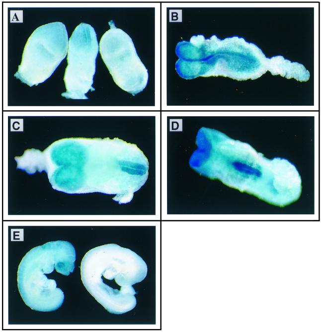
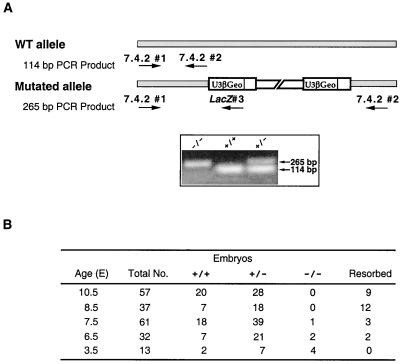
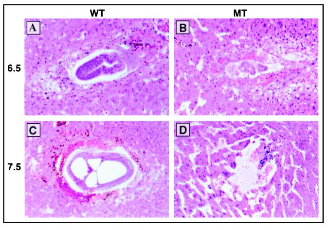
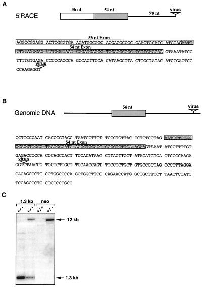
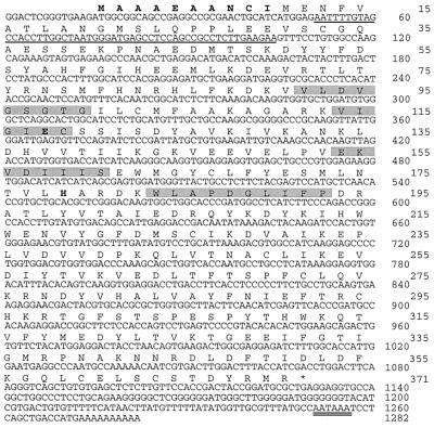
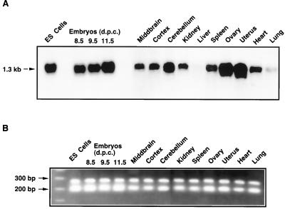
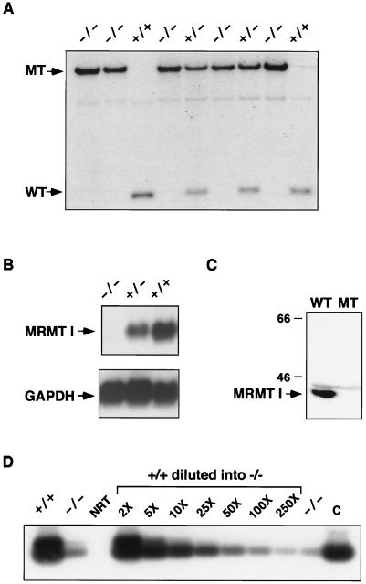
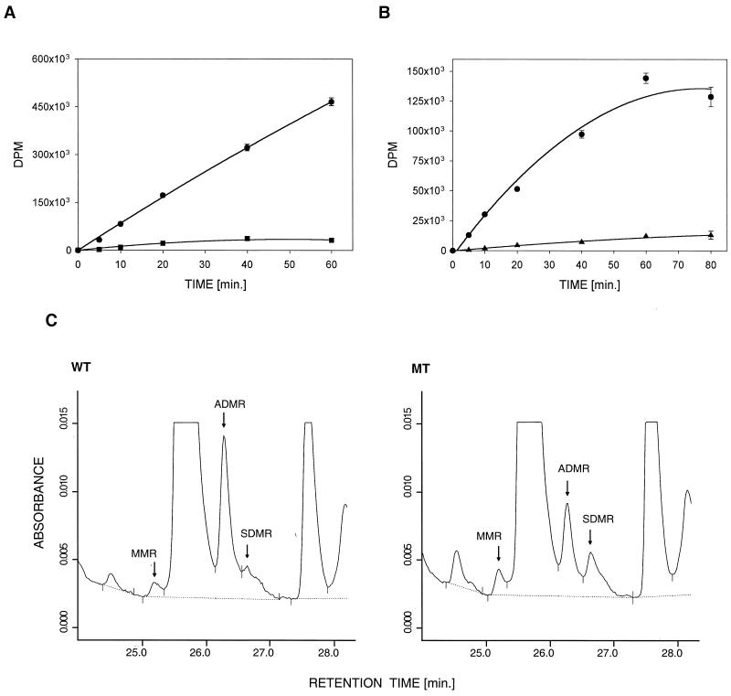
Protein arginine N-methyltransferases have been implicated in a variety of processes, including cell proliferation, signal transduction, and protein trafficking. In this study, we have characterized essentially a null mutation induced by insertion of the U3betaGeo gene trap retrovirus into the second intron of the mouse protein arginine N-methyltransferase 1 gene (Prmt1). cDNAs encoding two forms of Prmt1 were characterized, and the predicted protein sequences were found to be highly conserved among vertebrates. Expression of the Prmt1-betageo fusion gene was greatest along the midline of the neural plate and in the forming head fold from embryonic day 7.5 (E7.5) to E8.5 and in the developing central nervous system from E8.5 to E13.5. Homozygous mutant embryos failed to develop beyond E6.5, a phenotype consistent with a fundamental role in cellular metabolism. However, Prmt1 was not required for cell viability, as the protein was not detected in embryonic stem (ES) cell lines established from mutant blastocysts. Low levels of Prmt1 transcripts (approximately 1% of the wild-type level) were detected as assessed by a quantitative reverse transcription-PCR assay. Total levels of arginine N-methyltransferase activity and asymmetric N(G), N(G)-dimethylarginine were reduced by 85 and 54%, respectively, while levels of hypomethylated substrates were increased 15-fold. Prmt1 appears to be a major type I enzyme in ES cells, and in wild-type cells, most substrates of the enzyme appear to be maintained in a fully methylated state.


Figures

FIG. 1
FIG. 1
Embryonic expression of the 7.4.2-βGeo fusion gene. Spatial and temporal expression of the Prmt1 gene was assessed by staining Prmt1+/− embryos with X-Gal. (A) Left and middle, E7.5 heterozygous embryos showing strong lacZ expression along the midline of the neural plate. X-Gal staining is not detected in the extraembryonic ectoderm. Right, E7.5 wild-type embryo. (B to D) Dorsal (B), rostral (C), and ventral (D) views of E8.5 heterozygous embryos showing lacZ expression in the developing brain and fusing neural folds. Expression extends from the prosencephalon through the mesencephalon and rhombencephalon to the posteriormost aspect of the neural folds. lacZ expression is not detected in floorplate cells. (E) Left, lateral view of an E9.5 heterozygous embryo showing lacZ expression in the developing brain, eye, and closed neural tube; diffuse staining is visible throughout the whole embryo. Right, lateral view of E9.5 wild-type embryo.
FIG. 2
FIG. 2
Timing of embryonic death. Strategy for PCR-based genotyping (A). PCRs used a combination of three primers complementary to cellular (primers 1 and 2) and viral (primer 3) DNA sequences, as shown in schematic representations of the normal and disrupted alleles. Normal and mutant alleles generated PCR products of 114 (primers 1 and 2) and 265 (primers 1 and 3) nt, respectively. The PCR products were visualized following agarose gel electrophoresis and staining with ethidium bromide (box). Primers 1 and 2 did not amplify sequences from the mutant allele, presumably because of the size (10.3 kb) of the intervening U3βGeo provirus. WT, wild type. (B) Genotypes of E3.5 blastocysts and E6.5 to E10.5 embryos. Mice heterozygous for the 7.4.2 provirus were mated, and the resulting embryos were genotyped at various times postcoitus.
FIG. 3
FIG. 3
Phenotype of Prmt1−/− embryos at E6.5 to E7.5. Mice heterozygous for the 7.4.2 provirus were mated, and embryos were examined at various stages of embryonic development. Sections through wild-type (WT) (A and C) and mutant (MT) (B and D) embryos at E6.5 (A and B) and E7.5 (C and D) were stained with hematoxylin and eosin.
FIG. 4
FIG. 4
The 7.4.2 provirus inserts into an intron of the protein arginine N-methyltransferase gene. (A) Fusion transcripts extending into the U3βGeo provirus were isolated by 5′RACE and sequenced. Sequences of 56 and 54 nt (boxed) are from separate exons, while the 79-nt region immediately 5′ of the provirus integration site is intron derived. (B) Genomic sequences containing the site of provirus integration were cloned as a 1.3-kb StuI fragment and partially sequenced. The 54-nt alternatively spliced exon (boxed) is located 79 nt 5′ of the provirus. (C) Southern blot analysis of wild-type (+/+) and heterozygous (+/−) mouse cells. StuI-cleaved cellular DNAs were probed with the StuI fragment that contains the site of provirus integration (lanes 1 and 2) or with a neo-specific probe (lanes 3 and 4). Since StuI does not cleave U3βGeo, the provirus-occupied allele (12 kb) is shifted by the size of the provirus.
FIG. 5
FIG. 5
Nucleotide and deduced amino acid sequences of the murine Prmt1 gene. The 1,282-nt cDNA sequence, including additional sequences obtained by 5′RACE, is shown. The predicted amino acid sequence of the Prmt1 protein is presented above the cDNA sequence. Sequences corresponding to an alternatively spliced 54-nt exon and poly(A) site are underlined. The same exon is located just 5′ of the provirus integration site (Fig. 4B). Conserved arginine N-methyltransferase motifs I, post-I, II, and III are shaded. Differences between the mouse and human proteins are in boldface type.
FIG. 6
FIG. 6
Distribution of Prmt1 transcripts in embryos and tissues. (A) Northern blot hybridization of RNAs from various mouse tissues, embryos, and the D3 ES cell line (as indicated) to a 32P-labeled 1.1-kb Prmt1 cDNA probe. The position of the Prmt1-encoded 1.3-kb transcript is indicated at the left. The autoradiogram was exposed for 24 h. (B) RT-PCR analysis of the alternatively spliced 54-nt exon. Prmt1 transcripts were reverse transcribed from various RNAs (see above) using an oligonucleotide primer complementary to the Prmt1 sequence 3′ of the 54-nt exon and amplified by using primers that flank that exon. RT-PCR products of 266 and 212 bp are expected for transcripts with and without the 54-nt exon, respectively. d.p.c., days postconception.
FIG. 7
FIG. 7
Prmt1 maps to the proximal end of chromosome 7. (A) Maps of Jackson Laboratory BSB and BSS backcrosses showing the proximal end of chromosome 7. The map is depicted with the centromere toward the top and a 3-centimorgan (cM) scale bar. Loci mapping to the same position are listed in alphabetical order. Missing typings were inferred from surrounding data where the assignment was unambiguous. Raw data from The Jackson Laboratory were obtained from the World Wide Web address http://www.jax.org/resources/documents/cmdata. (B) Haplotype of Jackson Laboratory BSB and BSS backcrosses showing the proximal end of chromosome 7 with loci linked to Prmt1. Loci are listed in order, with the most proximal at the top. The black boxes represent the C57BL6/JEi allele, and the white boxes represent the SPRET/Ei allele. The number of animals with each haplotype is at the bottom of each column of boxes. Percent recombination (R) between adjacent loci is given to the right, along with the standard error (SE) of each R value.
FIG. 8
FIG. 8
Prmt1 is not required for cell viability. (A) ES cell lines were derived from E3.5 blastocysts and genotyped by Southern analysis. StuI-digested DNA samples from wild-type (+/+), heterozygous (+/−), and homozygous mutant (−/−) ES cells was analyzed by hybridization to the 1.3-kb StuI fragment containing the site of provirus integration. The genotype of each sample is shown along the top, and the mobility of the wild-type (WT) and mutant (MT) alleles is indicated on the left. (B) Selected cell lines were grown without feeder cells for 2 generations and analyzed by Northern blot hybridization. The murine Prmt1 cDNA detects a single 1.35-kb transcript in the wild-type and heterozygous cell lines. Expression is ablated in mutant cells, indicating that Prmt1 is not essential for cell growth. A glyceraldehyde phosphate dehydrogenase (GAPDH) probe was used to ensure equal loading of RNA for each sample. (C) Western blot analysis of Prmt1 expression. A 200-μg protein sample from Prmt1+/+ (WT) or Prmt1−/− (MT) cells was analyzed by Western blot analysis using an antibody against the rat PRMT1 protein. The values to the left are molecular sizes in kilodaltons. (D) RT-PCR analysis of Prmt1 transcripts in wild-type and mutant ES cells. Cells were grown for 9 passages without feeder layers. Samples analyzed by RT-PCR contained 5 μg of RNA from wild-type (lane +/+) and mutant (left lane −/−) cells, 2.5 μg of RNA from mutant cells (right lane −/−), or 2.5 μg of RNA from mutant cells to which different dilutions of wild-type DNA had been added (lanes with the final dilution factors indicated). For example, lanes 2× and 5× contain 2.5 μg of mutant RNA mixed with 2.5 and 1 μg of wild-type RNA, respectively. Lane NRT shows an analysis of wild-type RNA minus reverse transcriptase. Lane C shows PCR products using 2 ng of Prmt1 cDNA as a positive control. Levels of Prmt1 transcripts in mutant cells appeared to be comparable to 1/100 of the wild-type levels.
FIG. 9
FIG. 9
Reduced Prmt1 activity in Prmt1−/− cells increases the steady-state levels of hypomethylated substrates. (A) Arginine N-methyltransferase activities in wild-type and Prmt1 mutant cells. Extracts (20 μg of protein) from Prmt1+/+ (circles) and Prmt1−/− (squares) cells were assayed for arginine N-methyltransferase activity using a synthetic RGG peptide as a substrate (100 μM). Levels of 3H-methylated substrate were monitored by a filter binding assay. DPM, disintegrations per minute. (B) Hypomethylation of cellular proteins in Prmt1−/− cells. Extracts (20 μg of protein) from wild-type cells were mixed with an equal amount of protein from either Prmt1−/− (circles) or Prmt1+/+ (triangles) cells, and the transfer of methyl-3H groups from S-adenosyl-l-[methyl-3H]methionine into cellular proteins was monitored by a filter binding assay. (C) Analysis of NG-monomethylarginine (MMR), asymmetric NG,NG-dimethylarginine (ADMR), and symmetric NG,N′G-dimethylarginine (SDMR) in wild-type (WT) and Prmt1-deficient mutant (MT) cells. Total cellular protein (20 μg) from Prmt1+/+ and Prmt1−/− cells was subjected to acid hydrolysis and analyzed by high-pressure liquid chromatography.

References

    1. Abramovich C, Yakobson B, Chebath J, Revel M. A protein-ginine methyltransferase binds to the intracytoplasmic domain of the IFNAR1 chain in the type I interferon receptor. EMBO J. 1997;16:260–266. - PMC - PubMed
    1. Altschul S F, Madden T L, Schaffer A A, Zhang J, Zhang Z, Miller W, Lipman D J. Gapped BLAST and PSI-BLAST: a new generation of protein database search programs. Nucleic Acids Res. 1997;25:3389–3402. - PMC - PubMed
    1. Byvoet P, Shepherd G R, Hardin J M, Noland B J. The distribution and turnover of labeled methyl groups in histone fractions of cultured mammalian cells. Arch Biochem Biophys. 1972;148:558–567. - PubMed
    1. Chen D, Ma H, Hong H, Koh S S, Huang S M, Schurter B T, Aswad D W, Stallcup M R. Regulation of transcription by a protein methyltransferase. Science. 1999;284:2174–2177. - PubMed
    1. Chomczynski P, Sacchi N. Single-step method of RNA isolation by acid guanidinium thiocyanate-phenol-chloroform extraction. Anal Biochem. 1987;162:156–159. - PubMed

Publication types

MeSH terms

Substances

Associated data