. 2000 Jul;20(13):4870-8.
doi: 10.1128/MCB.20.13.4870-4878.2000.

Isoform-specific localization of A-RAF in mitochondria

Affiliations

Isoform-specific localization of A-RAF in mitochondria

A Yuryev et al. Mol Cell Biol. 2000 Jul.

Abstract

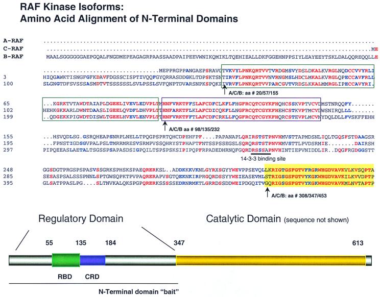
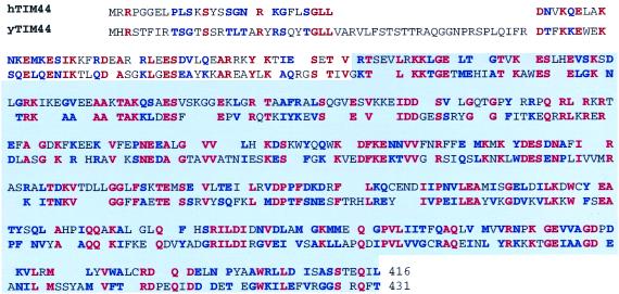
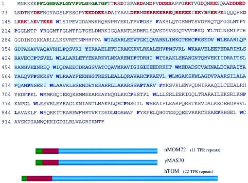
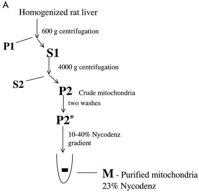
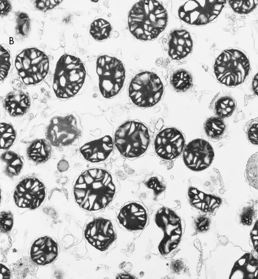
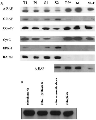
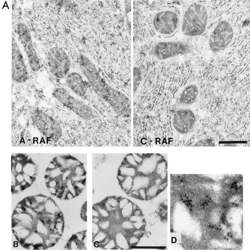
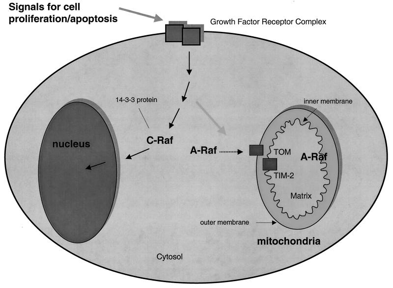
RAF kinase is a family of isoforms including A-RAF, B-RAF, and C-RAF. Despite the important role of RAF in cell growth and proliferation, little evidence exists for isoform-specific function of RAF family members. Using Western analysis and immunogold labeling, A-RAF was selectively localized in highly purified rat liver mitochondria. Two novel human proteins, which interact specifically with A-RAF, were identified, and the full-length sequences are reported. These proteins, referred to as hTOM and hTIM, are similar to components of mitochondrial outer and inner membrane protein-import receptors from lower organisms, implicating their involvement in the mitochondrial transport of A-RAF. hTOM contains multiple tetratricopeptide repeat (TPR) domains, which function in protein-protein interactions. TPR domains are frequently present in proteins involved in cellular transport systems. In contrast, protein 14-3-3, an abundant cytosolic protein that participates in many facets of signal transduction, was found to interact with C-RAF but not with A-RAF N-terminal domain. This information is discussed in view of the important role of mitochondria in cellular functions involving energy balance, proliferation, and apoptosis and the potential role of A-RAF in regulating these systems.


Figures

FIG. 1
FIG. 1
Alignment of the N-terminal regulatory domain of the three RAF isoforms. Amino acid sequences of human A-, C-, and B-isoforms are aligned. Identical residues are colored red, homologous residues are blue, and nonconserved residues are black. The RBD and CRD are indicated (top and bottom). While both domains are relatively conserved across isoforms, important differences exist which may account for isoform-specific protein interactions (see text). The amino acids for each isoform are numbered to the right. The end of the N-terminal domain is indicated by the gold block and corresponds to A/C/B-RAF as number 308/347/453, respectively. Similarly, the RBD and CRD begin at amino acids 14/55/148 and 98/135/232, respectively, for A/C/B-RAF. The N-terminal domain baits used for subsequent experiments end at amino acids (A/C/B) 314/348/460, as detailed in Table 1.
FIG. 2
FIG. 2
Similarity of A-RAF-specific binding protein to TIM mitochondrial transport proteins from lower organisms. The amino acid sequence of hTIM44 as predicted from the gene sequence is presented with its alignment to yeast TIM44. Identical amino acids are shown in red, and conserved substitutions are in blue. The alignment was done using the BESTFIT program with a gap penalty of 0.25, which resulted in a 74% homology score. The 5′-end 77 residues of the hTIM44 gene were cloned by RACE (rapid amplification of cDNA ends) PCR using a human hypocampus Marathon-Ready cDNA (Clontech catalog no. 7419-1). The original two-hybrid clone, which interacted with A-RAF, begins from amino acid 77 and is highlighted.
FIG. 3
FIG. 3
Similarity of A-RAF-specific binding protein to TOM mitochondrial transport proteins from lower organisms. The amino acid sequence of hTOM protein is presented as predicted from the gene sequence. The 5′-end sequence was obtained by RACE PCR as in Fig. 2. TPRs, also found in yeast (yMAS70; also called yTOM70) and N. crassa (nMOM72; also called nTOM72) mitochondrial transport proteins, are separated by breaks. Key repetitive hydrophobic and proline residues (17) are indicated in blue. The predicted transmembrane domain is shown in green. Charged amino acids in the membrane-proximal region are in red. Below the sequence is a schematic comparison of hTOM, yeast MAS70, and N. crassa MOM72 proteins. hTOM resembles nMOM72 and yMAS70 by overall protein domain organization and structure. Among these three proteins, the charged region was 26% and the transmembrane region was 20% homologous. For the TPRs, the best homology was with hTOM amino acids 216 to 904, the 11 C-terminal TPRs. This region was 25% homologous with the TPR regions of yMAS70 and nMOM72. Analysis of overlapping clones from the two-hybrid screen showed that the minimal region required for hTOM interaction with A-RAF contained TPR5 through TPR15; this region is highlighted.
FIG. 4
FIG. 4
Purification of rat liver mitochondria. (A) Outline of the mitochondrial fractionation scheme. Mitochondria from rat liver were isolated as described by Mihara and Omura (19) with subsequent purification by a linear Nycodenz gradient (10) in 0.22 M mannitol–0.075 M sucrose–10 mM HEPES-KOH (pH 7.4)–1 mM EDTA. (B) Electron micrograph of the purified mitochondria, demonstrating a high level of enrichment, estimated at >90% purity.
FIG. 4
FIG. 4
Purification of rat liver mitochondria. (A) Outline of the mitochondrial fractionation scheme. Mitochondria from rat liver were isolated as described by Mihara and Omura (19) with subsequent purification by a linear Nycodenz gradient (10) in 0.22 M mannitol–0.075 M sucrose–10 mM HEPES-KOH (pH 7.4)–1 mM EDTA. (B) Electron micrograph of the purified mitochondria, demonstrating a high level of enrichment, estimated at >90% purity.
FIG. 5
FIG. 5
Western analysis of fractions from rat liver mitochondrial preparation. (A) Western analysis of fractions performed with 50 μg of protein per lane (except the lowest lane, where 100 μg was used) using SDS-PAGE with 10% polyacrylamide gels. Standard procedures were used to blot gels to nitrocellulose membranes and to perform Western analysis with enhanced chemiluminescence detection using appropriate alkaline phosphatase-conjugated secondary antibodies (except with the lowest panel). Purified mitochondria were treated with protease K (10) to judge if A-RAF would be protected. Anti-A-RAF polyclonal antibodies were from Santa Cruz Biotechnologies (catalog no. sc-408). Monoclonal antibodies to various proteins were as follows: C-RAF (Transduction Laboratories catalog no. R19120), ERK-1 (Transduction Laboratories catalog no. M37520), cytochrome c (Cyt C) (PharMingen catalog no. 65981A), cytochrome oxidase subunit IV (COX-IV) (Molecular Probes catalog no. A-6403), and RACK-1 (Transduction Laboratories catalog no. R20620). For the lowest panel, Western analysis was performed as described above except that an iodinated secondary antibody (ICN catalog no. 68086) was used to increase the resolution. This method gave sharper bands than a horseradish peroxidase-conjugated second antibody–chemiluminescence method, where broad bands were detected and the doublet could not be resolved. (B) Experiment to judge the protection of A-RAF immunoreactivity upon preparation of mitoplasts using Western analysis with the technique described for panel A. The mitoplast preparation was performed as described by Glick (9). Note that (low-resolution) chemiluminescence was used, and therefore only a single band of A-RAF was detected.
FIG. 6
FIG. 6
Transmission electron micrographs of thin sections of mitochondrial preparations. (A) Rat liver was treated with either A-RAF or C-RAF antibody and stained with gold as described below for mitochondrial sections. The bar equals 1 μm. (B and C) Purified mitochondrial samples were fixed for immunogold labeling with 4% paraformaldehyde-0.1% glutaraldehyde in 0.1 M sodium cacodylate buffer, dehydrated through a graded series of ethanol, and embedded in LR White resin (London Resin Company). Sections of embedded mitochondria were blocked with TBS containing first 50 mM glycine and then containing 5% BSA and 5% goat serum. After overnight incubation in TBS with 1% BSA containing different primary antibody dilutions of anti-A-RAF polyclonal antibody (Santa Cruz Biotechnologies catalog no. sc-408) (B) or anti-C-RAF polyclonal antibody (Santa Cruz Biotechnologies catalog no. sc-227) (C), sections were washed six times with 1× TBS–1% BSA and incubated for 2 h with a 1:10 dilution of goat anti-rabbit antibody conjugated to 10-nm colloidal gold (Goldmark Biologicals). The bar equals 0.5 μm. (D) Enlargement of a section of panel B showing the gold particles.
FIG. 7
FIG. 7
Model for the localization of RAF isoforms relative to mitochondria. The model summarizes the information from this study showing A-RAF localization inside and outside mitochondria and C-RAF localization uniquely outside mitochondria. The model implies a role of mitochondria A-RAF influencing the proliferation and activity of this organelle (see text). Protein 14-3-3 is an abundant cytosolic protein shown to bind the C- but not A-RAF N-terminal domain.

Similar articles

Cited by

References

    1. Altschul S F, Gish W, Miller W, Myers E W, Lipman D J. Basic local alignment search tool. J Mol Biol. 1990;215:403–410. - PubMed
    1. Ausubel F M, Brent R, Kingston R E, Moore D D, Seidman J G, Smith J A, Struhl K, editors. Current protocols in molecular biology. New York, N.Y: Wiley Interscience; 1992.
    1. Avruch J, Zhang X, Kyriakis J M. Raf meets Ras: completing the framework of signal transduction pathway. Trends Biochem Sci. 1994;19:279–283. - PubMed
    1. Chevray P M, Nathans D. Protein interaction cloning in yeast: identification of mammalian proteins that react with the leucine zipper of Jun. Proc Natl Acad Sci USA. 1992;89:5789–5793. - PMC - PubMed
    1. Das A K, Cohen P W, Barford D. The structure of the tetratricopeptide repeats of protein phosphatase 5: implications for TPR-mediated protein-protein interactions. EMBO J. 1998;17:1192–1199. - PMC - PubMed

MeSH terms

Associated data