. 2000 Jul;20(13):4879-87.
doi: 10.1128/MCB.20.13.4879-4887.2000.

Developmental expression of latent transforming growth factor beta binding protein 2 and its requirement early in mouse development

Affiliations

Developmental expression of latent transforming growth factor beta binding protein 2 and its requirement early in mouse development

J M Shipley et al. Mol Cell Biol. 2000 Jul.

Abstract

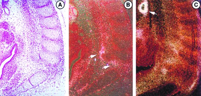
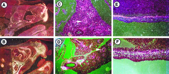
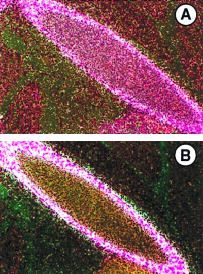
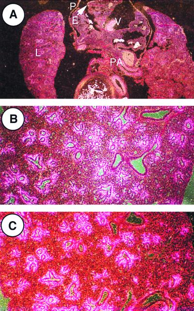
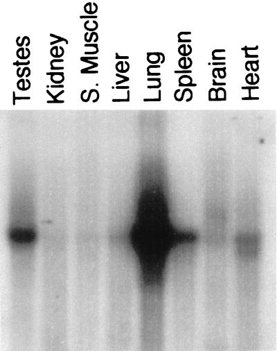
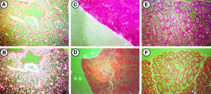
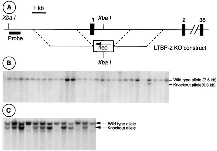
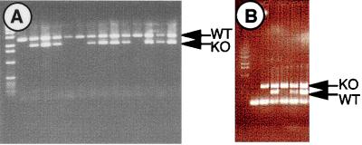
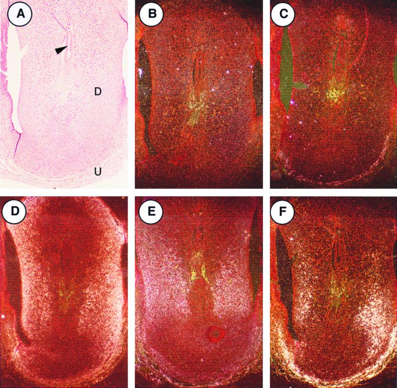
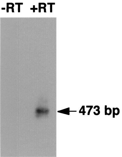
Latent transforming growth factor beta (TGF-beta) binding protein 2 (LTBP-2) is an integral component of elastin-containing microfibrils. We studied the expression of LTBP-2 in the developing mouse and rat by in situ hybridization, using tropoelastin expression as a marker of tissues participating in elastic fiber formation. LTBP-2 colocalized with tropoelastin within the perichondrium, lung, dermis, large arterial vessels, epicardium, pericardium, and heart valves at various stages of rodent embryonic development. Both LTBP-2 and tropoelastin expression were seen throughout the lung parenchyma and within the cortex of the spleen in the young adult mouse. In the testes, LTBP-2 expression was seen within lumenal cells of the epididymis in the absence of tropoelastin. Collectively, these results imply that LTBP-2 plays a structural role within elastic fibers in most cases. To investigate its importance in development, mice with a targeted disruption of the Ltbp2 gene were generated. Ltbp2(-/-) mice die between embryonic day 3.5 (E3.5) and E6.5. LTBP-2 expression was not detected by in situ hybridization in E6.5 embryos but was detected in E3.5 blastocysts by reverse transcription-PCR. These results are not consistent with the phenotypes of TGF-beta knockout mice or mice with knockouts of other elastic fiber proteins, implying that LTBP-2 performs a yet undiscovered function in early development, perhaps in implantation.


Figures

FIG. 1
FIG. 1
Expression of LTBP-2 and tropoelastin mRNAs in E13.5 mouse embryos. In situ hybridization was done to localize expression of each. (A) Bright-field staining of developing ribs. (B) Section hybridized to a LTBP-2 antisense probe showing perichondrial expression (arrows). (C) Section hybridized to a tropoelastin antisense probe, showing perichondrial expression similar to LTBP-2 as well as arterial expression (arrow). Sense probes showed no specific hybridization (not shown). Magnification, ×90.
FIG. 2
FIG. 2
Expression of LTBP-2 and tropoelastin mRNAs in E15.5 mouse embryos. In situ hybridization was done to localize expression of LTBP-2 in the snout (A), lung (C), and dermis (E) and expression of tropoelastin in the snout (B), lung (D), and dermis (F). Perichondrial expression of both mRNAs is seen throughout the snout. Expression in the lung is observable in a limited number of cell layers of the subepithelial mesenchyme of developing airways (arrows), and intense tropoelastin expression in arterial vessels (asterisks) is evident. Both signals are also seen in the dermis. Sense probes showed no specific hybridization (not shown). Magnification for panels A and B, ×36. Magnification for panels C through F, ×180.
FIG. 3
FIG. 3
Expression of LTBP-2 (A) and tropoelastin (B) mRNAs in E17.5 mouse embryos. In situ hybridization was done to localize expression of each. Expression of both mRNAs is seen by medial cells of the descending aorta. Sense probes showed no specific hybridization (not shown). Magnification, ×200.
FIG. 4
FIG. 4
Expression of LTBP-2 and tropoelastin mRNAs in the developing rat thorax. In situ hybridization was done to localize expression of each. (A) Expression of LTBP-2 in the E14 rat thorax. Specific hybridization is seen in the lungs (L), the pericardium (P), the epicardium (E), the heart valves (V), and the pulmonary artery (PA), as well as in other large vessels. The pattern of expression is identical to that seen with tropoelastin probes (data not shown). Magnification, ×36. (B) Expression of LTBP-2 in the E18 rat lung. Expression in the lung is observable in a limited number of cell layers of the subepithelial mesenchyme of developing mid-sized airways, with no or minimal corresponding expression seen in the smallest and largest airways. Magnification, ×90. (C) Expression of tropoelastin in the E18 rat lung. The pattern of expression is identical to that of LTBP-2. Sense probes showed no specific hybridization (data not shown). Magnification, ×90.
FIG. 5
FIG. 5
Northern blot analysis of LTBP-2 expression in adult mouse tissues. A multiple tissue Northern blot including eight adult mouse tissue sources (Clontech) was hybridized to a LTBP-2 cDNA probe. A 7.5-kb mRNA is most abundantly detected in the lung, spleen, and testes.
FIG. 6
FIG. 6
Expression of LTBP-2 and tropoelastin mRNAs in 5-week-old mouse tissues. In situ hybridization was done to localize expression of LTBP-2 in the lung (A) and spleen (C) and expression of tropoelastin in the lung (B) and spleen (D). Coexpression of LTBP-2 and tropoelastin is seen throughout the mesenchyme and vasculature of the lung and the capsule of the spleen. In the testes, LTBP-2 (E) but not tropoelastin (F) is expressed by lumenal cells of the epididymis. Sense probes showed no specific hybridization (data not shown). Magnification for panels A, B, E, and F, ×90. Magnification for panels C and D, ×36.
FIG. 7
FIG. 7
Generation of Ltbp2−/− mice. (A) The LTBP-2 targeting construct. The 5′ and 3′ homologies were derived from the promoter and intron 1, respectively. The intervening region including exon 1, which contains the ATG, is replaced with a PGK-neo cassette. (B) Southern blot of ES cell DNA screened as described for panel A, showing one targeted clone. (C) Live progeny of heterozygous matings were genotyped by Southern blotting. No Ltbp2−/− mice lived to birth.
FIG. 8
FIG. 8
Genotyping of early postimplantation and preimplantation embryos. (A) E6.5 early-postimplantation embryos from heterozygous matings were genotyped by PCR. No Ltbp2−/− embryos were identified at this stage. (B) E3.5 preimplantation blastocysts, in which knockout embryos were identified, were genotyped by PCR in an assay similar to that used for panel A. The lowest band in all lanes corresponds to unincorporated PCR primers. The first lane following the molecular weight standards is a mock PCR in the absence of target DNA. KO, knockout; WT, wild type.
FIG. 9
FIG. 9
Expression of LTBP-2 mRNA at E6.5. LTBP-2 expression by E6.5 embryos was investigated by in situ hybridization. The bright-field image (A) and a section probed with a sense probe (B) are shown. Serial sections were probed for LTBP-2 (C) and other elastic fiber components, including MAGP-2 (D), fibrillin 1 (E), and tropoelastin (F). A signal for all of the elastic fiber components, including LTBP-2, is seen within the maternally derived decidual stroma (D) and the uterine muscle (U) but not within the embryo itself (arrow).
FIG. 10
FIG. 10
Expression of LTBP-2 by preimplantation embryos. LTBP-2 expression by E3.5 preimplantation blastocysts was investigated by RT-PCR. Blastocysts were subjected to reverse transcriptase (+RT) or mock reactions lacking reverse transcriptase (−RT) prior to PCR, as described in Materials and Methods. Southern blotting and hybridization with an LTBP-2 cDNA probe identified a band of the predicted size (473 bp) which was present only in samples treated with reverse transcriptase.

References

    1. Cleary E G. The microfibrillar component of the elastic fibers. Morphology and biochemistry. In: Uitto J, Perejda A J, editors. Connective tissue disease. Molecular pathology of the extracellular matrix. New York, N.Y: Marcel Dekker, Inc.; 1987. pp. 55–81.
    1. Dickson M C, Martin J S, Cousins F M, Kulkarni A B, Karlsson S, Ackhurst R J. Defective hematopoiesis and vasculogenesis in transforming growth factor-beta 1 knockout mice. Development. 1995;121:1845–1854. - PubMed
    1. Dietz H C, Cutting G R, Pyeritz R E, Maslen C L, Sakai L Y, Corson G M, Puffenberger E G, Hamosh A, Nanthakumar E J, Curristan S M, Stetten G, Meyers D A, Francomano C A. Marfan syndrome caused by a recurrent de novo missense mutation in the fibrillin gene. Nature. 1991;352:337–339. - PubMed
    1. Fahrenbach W H, Sandberg L B, Cleary E G. Ultrastructural studies on early elastogenesis. Anat Rec. 1966;155:563–576.
    1. Fang J, Li X, Smiley E, Francke U, Mecham R P, Bonadio J. Mouse latent TGF-β binding protein-2: molecular cloning and developmental expression. Biochim Biophys Acta. 1997;1354:219–230. - PubMed

Publication types

MeSH terms