. 2000 Jul;20(13):4888-99.
doi: 10.1128/MCB.20.13.4888-4899.2000.

Nuclear entry of the circadian regulator mPER1 is controlled by mammalian casein kinase I epsilon

Affiliations

Nuclear entry of the circadian regulator mPER1 is controlled by mammalian casein kinase I epsilon

E Vielhaber et al. Mol Cell Biol. 2000 Jul.

Abstract

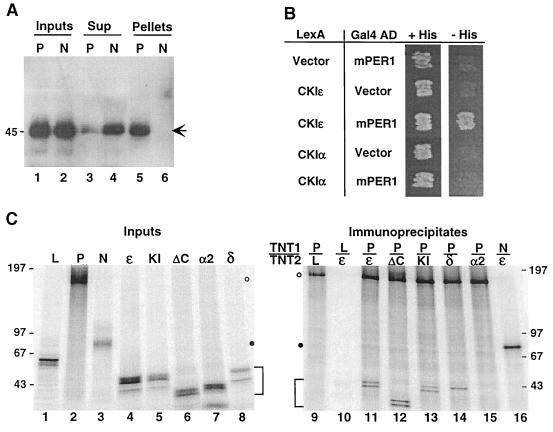
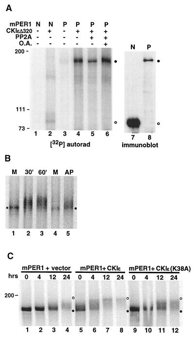
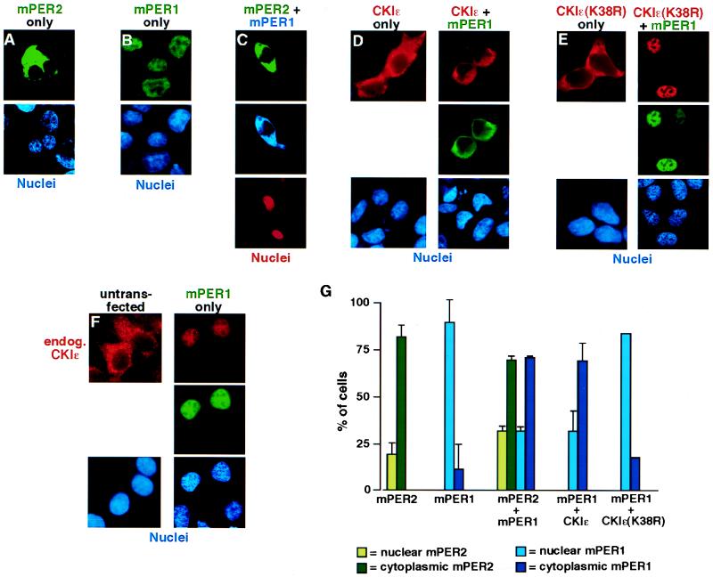
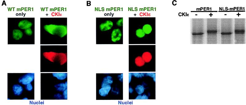
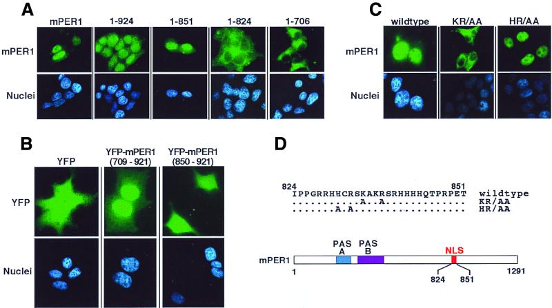
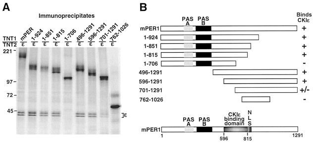
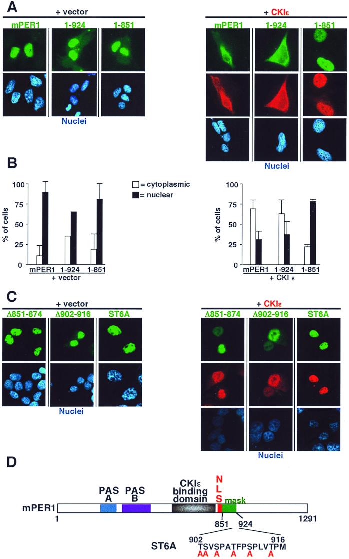
The molecular oscillator that keeps circadian time is generated by a negative feedback loop. Nuclear entry of circadian regulatory proteins that inhibit transcription from E-box-containing promoters appears to be a critical component of this loop in both Drosophila and mammals. The Drosophila double-time gene product, a casein kinase I epsilon (CKIepsilon) homolog, has been reported to interact with dPER and regulate circadian cycle length. We find that mammalian CKIepsilon binds to and phosphorylates the murine circadian regulator mPER1. Unlike both dPER and mPER2, mPER1 expressed alone in HEK 293 cells is predominantly a nuclear protein. Two distinct mechanisms appear to retard mPER1 nuclear entry. First, coexpression of mPER2 leads to mPER1-mPER2 heterodimer formation and cytoplasmic colocalization. Second, coexpression of CKIepsilon leads to masking of the mPER1 nuclear localization signal and phosphorylation-dependent cytoplasmic retention of both proteins. CKIepsilon may regulate mammalian circadian rhythm by controlling the rate at which mPER1 enters the nucleus.


Figures

FIG. 1
FIG. 1
CKIɛ binds to mPER1 in vivo and in vitro. (A) Coimmunoprecipitation of mPER1 and endogenous CKIɛ. HEK 293 cells were transiently transfected with plasmids expressing either full-length Myc-mPER1 (P) or the amino-terminal fragment Myc-mPER1(1-485) (N). The PER proteins were immunoprecipitated from cell lysates with anti-Myc MAb 9E10; 20 μg of cell lysate protein (Inputs; lanes 1 and 2), the equivalent of 20 μg of the cell lysate supernatant following clarification and immunoprecipitation (Sup; lanes 3 and 4), and the immunoprecipitate pellet from 50 μg of cell lysate (Pellets; lanes 5 and 6) were analyzed by SDS-PAGE, followed by immunoblotting with anti-CKIɛ antibody UT31 (14). The arrow indicates the position of endogenous CKIɛ. (B) Specificity of the CKIɛ-mPER1 interaction assessed by two-hybrid assay. Yeast cotransformed with plasmids expressing the indicated proteins fused to either LexA or the Gal4 activation domain (AD) were grown on synthetic medium containing histidine (+His) or on medium containing 5 mM 3-aminotriazole and lacking histidine (−His) as previously described (37). Interaction between the indicated proteins was assessed by growth on −His plates. (C) Specificity of the CKIɛ-mPER1 interaction assessed by coimmunoprecipitation in vitro. In vitro-synthesized [35S]methionine-labeled proteins (Inputs; lanes 1 to 8) luciferase (L), Myc-mPER1 (P), truncated Myc-mPER1(1-485) (N), CKIɛ (ɛ), kinase-inactive CKIɛ(K38R) (KI), truncated CKIɛ(ΔC320) (ΔC), CKIα2 (α2), or CKIδ (δ) were mixed together (TNT1/TNT2) as indicated above lanes 9 to 16. Following a 30-min incubation, the protein mixtures were subjected to immunoprecipitation with anti-Myc MAb 9E10 and analyzed by SDS-PAGE (5 to 15% gel) (lanes 9 to 16). One-tenth of each of the in vitro synthesis reactions used for immunoprecipitation was loaded on the input gel (left). Data were collected and analyzed using a Molecular Dynamics PhosphorImager. Open and closed circles mark the positions of full-length and truncated Myc-mPER1, respectively; brackets mark positions of the various CKI proteins. Here and in subsequent figures, positions of the various protein molecular weight markers are indicated to the side of the gel, with the size of each marker expressed in kilodaltons.
FIG. 2
FIG. 2
Phosphorylation of mPER1 by CKIɛ in vitro and in vivo. (A) CKIɛ phosphorylates immunoprecipitated mPER1. In vitro-synthesized unlabeled mPER1 (P) and an amino-terminal fragment (amino acids 1 to 485; N) were each immunoprecipitated from a single in vitro transcription-translation reaction (200 and 100 μl, respectively) with anti-Myc MAb 9E10, and the immunoprecipitation reactions were divided equally between the various experiments. The immunoprecipitated proteins were then incubated with (lanes 2 and 4 to 6) or without (lanes 1 and 3) 100 ng recombinant CKIɛΔ320 and 25 μM [γ-32P]ATP for 30 min at 37°C in a 20-μl volume. Following the kinase reaction, the samples in lanes 5 and 6 were washed and then incubated with 100 ng of catalytic subunit of PP2A in the absence (lane 5) or presence (lane 6) of 500 nM okadaic acid (O.A.). Phosphorylation was analyzed by SDS-PAGE followed by PhosphorImager analysis. Closed circles indicate the mobility of full-length mPER1; open circles indicate the mobility of the amino-terminal mPER1 fragment. The relative amounts of full-length mPER1 (P; lane 8) and the amino-terminal fragment (N; lane 7) were determined in parallel by immunoblotting with the anti-Myc MAb 9E10. (B) Mobility shift of phosphorylated mPER1 in vitro as assessed by kinase assay. In vitro-synthesized [35S]methionine-labeled mPER1 (from ∼1 mg of reticulocyte lysate) was incubated with 50 ng of CKIɛΔ320 (lanes 2, 3, and 5) for the indicated times, or without added kinase for 60 min (M; lanes 1 and 4). After the kinase reaction, the sample in lane 5 was incubated with 40 U of calf intestinal alkaline phosphatase (New England Biolabs) for an additional 30 min. The samples were then analyzed by SDS-PAGE and phosphorimaging. The closed circles indicate the mobility of unphosphorylated mPER1. (C) Mobility shift of mPER1 in vivo as assessed by pulse-chase experiment. HEK 293 cells expressing Myc-tagged mPER1 only (lanes 1 to 4), mPER1 and full-length active CKIɛ (lanes 5 to 8), or mPER1 and kinase-inactive CKIɛ (lanes 9 to 12) were pulse-labeled with [35S]methionine. At the indicated time points following addition of medium containing unlabeled methionine, cells were lysed and Myc-mPER1 was immunoprecipitated with anti-Myc MAb 9E10. Samples were all run on a single SDS-polyacrylamide gel, and the entire gel was visualized with a PhosphorImager at a constant intensity setting. Closed and open circles indicate the positions of unphosphorylated and phosphorylated mPER1, respectively.
FIG. 3
FIG. 3
mPER2 and CKIɛ regulate mPER1 subcellular localization. (A to F) Representative micrographs illustrating subcellular localization of mPER2, mPER1, and CKIɛ. HEK 293 cells were transiently transfected with constructs encoding FLAG-mPER2 (A). Myc-mPER1 (B), FLAG-mPER2 and Myc-mPER1 (C), HA-CKIɛ without (D, left) or with (D, right) Myc-mPER1, HA-CKIɛ(K38R) without (E, left) or with (E, right) Myc-mPER1, and Myc-mPER1 (F). Forty-eight hours after transfection, the cells were fixed and epitope-tagged proteins were visualized by staining with Alexa 488 (green)-conjugated anti-FLAG (M2) (A and C), Alexa 350 (blue)-conjugated anti-Myc (9E10) (C), Alexa 488 (green)-conjugated MAb 9E10 (B, D, E, and F), Alexa 594 (red)-conjugated anti-HA MAb 12CA5 (D and E), and anti-CKIɛ MAb followed by an Alexa 594-conjugated goat anti-mouse secondary (F). Nuclei were visualized with Hoechst stain (A, B, D, E, and F) or ToPro3 stain (red; C). (G) Quantitation of the experiments illustrated above. Each bar is the result of at least two independent experiments (±standard deviation) in which 40 to 100 cells were counted. All immunofluorescence experiments were done at least twice, but where error bars are omitted experiments were quantitated only once.
FIG. 4
FIG. 4
A heterologous NLS overrides the CKIɛ-dependent cytoplasmic localization of mPER1. (A and B) HEK 293 cells were transiently cotransfected with constructs encoding either 4HA-CKIɛ or empty vector and Myc-mPER1 (A) or NLS-mPER1 (B). Forty-eight hours posttransfection, the epitope-tagged proteins were visualized with Alexa 488 (green)-conjugated anti-Myc MAb 9E10 and Alexa 594 (red)-conjugated anti-HA MAb 12CA5, and the nuclei were visualized with Hoechst staining. WT, wild type. (C) NLS-mPER1 is still a substrate for CKIɛ. In vitro-translated [35S]methionine NLS-mPER1 was incubated without or with added CKI. The addition of the amino-terminal NLS did not interfere with the kinase-dependent electrophoretic mobility shift.
FIG. 5
FIG. 5
Identification of the mPER1 NLS. (A) Progressive truncation of mPER1 reveals a potential NLS. HEK 293 cells were transiently transfected with constructs encoding either full-length Myc-mPER1(mPER1) or one of several carboxyl-terminal truncations of Myc-mPER1 (Fig. 6B). Forty-eight hours posttransfection, the proteins were visualized with Alexa 488 (green)-conjugated anti-Myc MAb 9E10 and the nuclei were visualized with Hoechst staining. Representative micrographs are shown. Quantitation of the nuclear localization of mPER1 and constructs encoding mPER1(1-924) and mPER1(1-851) is shown in Fig. 7. The cytoplasmic localization of mPER1(1-824) and mPER(1-706) was seen in >95% of transfected cells. (B) mPER1 amino acids 709 to 921 are sufficient to direct nuclear localization. HEK 293 cells were transiently transfected with constructs encoding double YFP, either alone or fused to mPER1(709-921) or mPER1(850-921). Double YFP alone and YFP-mPER1(850-921) were diffusely distributed in >95% of transfected cells, while YFP-mPER1(709-921) concentrated in the nuclei of >95% of transfected cells. (C and D) Point mutations identify essential residues in mPER1 NLS. HEK 293 cells were transiently transfected with constructs encoding wild-type or double point mutant K835A/R838A (KR/AA) or H831A/R833A (HR/AA) Myc-mPER1. After transfection, the epitope-tagged mPER1 was visualized as for panel A. A related NLS mutant, K837E/R838D-mPER1, was also cytoplasmic (data not shown). (D) Cartoon demonstrating location of NLS in mPER1 and mutations introduced to create KR/AA and HR/AA Myc-mPER1.
FIG. 6
FIG. 6
(A) Identification of CKIɛ binding site on mPER1. In vitro-synthesized [35S]methionine-labeled Myc-mPER1 (mPER1) and various amino-terminal (1-924, 1-851, 1-815, 1-706) and carboxyl-terminal (496-1291, 596-1291, 701-1291) fragments of mPER1 were mixed with in vitro-synthesized [35S]methionine-labeled full-length CKIɛ. After incubation, mPER1 was immunoprecipitated with the anti-Myc MAb 9E10 and analyzed for the presence of coimmunoprecipitating CKIɛ by SDS-PAGE and PhosphorImager analysis. The data presented are a subset of all the truncations tested; all results were repeated at least three times. (B) Diagrammatic representation of the constructs utilized and the results of the coimmunoprecipitation experiments.
FIG. 7
FIG. 7
CKIɛ-mediated cytoplasmic retention of mPER1 requires a masking domain (amino acids 851-924). (A) Full-length (mPER1) and amino-terminal fragments (1-924 and 1-851) of Myc-mPER1 were expressed in HEK 293 cells without (left) or with (right) HA-CKIɛ. Forty-eight hours posttransfection, the localization of mPER1 and CKIɛ was assessed as described above. The mPER1(1-851) construct contains an NLS and can bind to CKIɛ but failed to relocalize to the cytoplasm. Full-length and mPER1(1-924) were retained in the cytoplasm by coexpression of CKIɛ. Representative micrographs are shown. (B) Quantitation of the experiments shown in panel A. (C) Internal deletions and mutation of potential phosphorylation sites disrupts the function of the masking domain. HEK 293 cells were transfected as above with Myc-mPER1 containing deletion of residues 851 to 874 (Δ851-874) or 902 to 916 (Δ902-916) or with simultaneous mutations of six serine and threonine residues between amino acids 902 to 916 region (ST6A). None of the mutations altered nuclear localization, while all abrogated the ability of CKIɛ to relocalize Myc-mPER1 to the cytoplasm. (D) Cartoon of mPER1 with identification of masking domain and mutant ST6A.

Similar articles

Cited by

References

    1. Albrecht U, Sun Z S, Eichele G, Lee C C. A differential response of two putative mammalian circadian regulators, mper1 and mper2, to light. Cell. 1997;91:1055–1064. - PubMed
    1. Balsalobre A, Damiola F, Schibler U. A serum shock induces circadian gene expression in mammalian tissue culture cells. Cell. 1998;93:929–937. - PubMed
    1. Briggs L J, Stein D, Goltz J, Corrigan V C, Efthymiadis A, Hubner S, Jans D A. The cAMP-dependent protein kinase site (Ser312) enhances dorsal nuclear import through facilitating nuclear localization sequence/importin interaction. J Biol Chem. 1998;273:22745–22752. - PubMed
    1. Brunet A, Bonni A, Zigmond M J, Lin M Z, Juo P, Hu L S, Anderson M J, Arden K C, Blenis J, Greenberg M E. Akt promotes cell survival by phosphorylating and inhibiting a Forkhead transcription factor. Cell. 1999;96:857–868. - PubMed
    1. Cegielska A, Gietzen K F, Rivers A, Virshup D M. Autoinhibition of casein kinase I ɛ (CKIɛ) is relieved by protein phosphatases and limited proteolysis. J Biol Chem. 1998;273:1357. - PubMed

Publication types