. 2000 Jun;11(6):2151-9.
doi: 10.1091/mbc.11.6.2151.

Spatially regulated recruitment of clathrin to the plasma membrane during capping and cell translocation

Affiliations
Free PMC article

Spatially regulated recruitment of clathrin to the plasma membrane during capping and cell translocation

C K Damer et al. Mol Biol Cell. 2000 Jun.
Free PMC article

Abstract

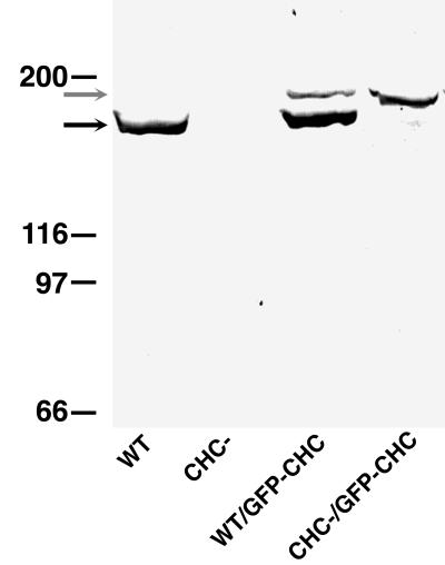
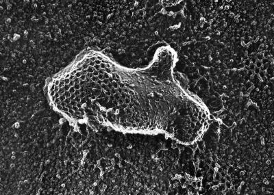
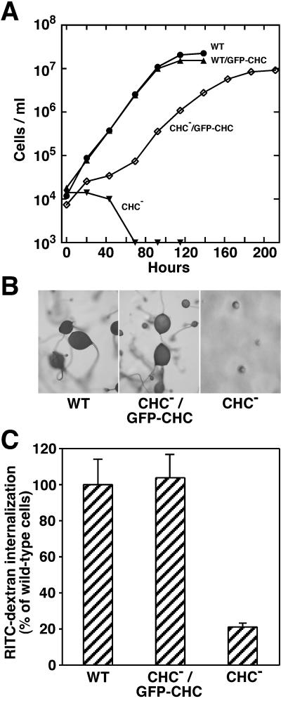
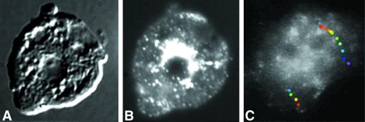
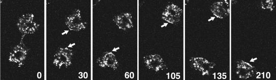
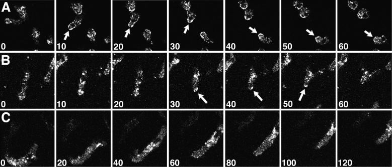
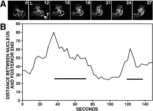
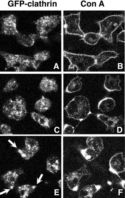
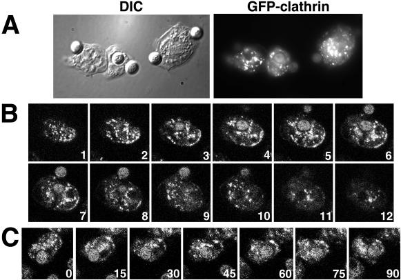
Clathrin-coated vesicles bud from selected cellular membranes to traffic-specific intracellular proteins. To study the dynamic properties of clathrin-coated membranes, we expressed clathrin heavy chain tagged with green fluorescent protein (GFP) in Dictyostelium cells. GFP-clathrin was functional and retained the native properties of clathrin: the chimeric protein formed classic clathrin lattices on cellular membranes and also rescued phenotypic defects of clathrin null cells. GFP-clathrin distributed into punctate loci found throughout the cytoplasm, on the plasma membrane, and concentrated to a perinuclear location. These clathrin-coated structures were remarkably motile and capable of rapid and bidirectional transport across the cell. We identified two local domains of the plasma membrane as sites for clathrin recruitment in motile cells. First, as cells translocated or changed shape and retracted their tails, clathrin was transiently concentrated on the membrane at the back of the cell tail. Second, as cells capped their cell surface receptors, clathrin was recruited locally to the membrane under the tight cap of cross-linked receptors. This suggests that local sites for clathrin polymerization on specific domains of the plasma membrane undergo rapid and dynamic regulation in motile cells.


Figures

Figure 1
Figure 1
Expression of GFP-clathrin in Dictyostelium cells. Wild-type (WT) and clathrin-minus (CHC−) Dictyostelium cells were transformed with the GFP-clathrin (GFP-CHC) expression plasmid. These strains were screened for GFP-clathrin expression by SDS-PAGE followed by Western blot analysis with a polyclonal antibody that recognizes wild-type clathrin (black arrow) and GFP-clathrin.
Figure 2
Figure 2
Coated pits of polymerized GFP-clathrin. Clathrin null cells expressing GFP-clathrin were prepared for electron microscopy using quick freeze and deep etch. An electron micrograph shows that the membranes of these cells display clathrin coats formed from GFP-clathrin that were indistinguishable in size and shape from the clathrin coats found in control wild-type cells. Micrograph kindly provided by Dr. John Heuser (Washington University Medical School, St. Louis, MO).
Figure 3
Figure 3
Rescue of phenotypic defects by GFP-clathrin. (A) Rescue of cytokinesis. Cells from wild-type (WT) and clathrin-minus (CHC−) parental strains and cells expressing GFP-clathrin derived from these strains (WT/GFP-CHC and CHC−/GFP-CHC) were seeded at 1 × 10⁴ cells/ml and grown in suspension culture on a shaking platform. Although CHC− cells failed completely to grow in suspension, CHC−/GFP-CHC cells divided and grew in suspension. (B) Rescue of development. Wild-type (WT), clathrin-minus (CHC−), and clathrin-minus cells expressing GFP-clathrin (CHC−/GFP-CHC) were seeded on a lawn of bacteria. After the bacterial food source was depleted, wild-type cells (WT) and cells expressing GFP-CHC (CHC−/GFP-CHC) developed into fruiting bodies; clathrin-minus (CHC−) cells formed only multicellular aggregates. (C) Rescue of endocytosis. Cells from wild-type (WT) and clathrin-minus (CHC−) parental strains and cells expressing GFP-clathrin (CHC−/GFP-CHC) were incubated with RITC-dextran, a fluid-phase marker, for 1 h. After washing to remove extracellular RITC-dextran, the fluorescence of internalized RITC-dextran was assessed for each sample by fluorescence-activated flow cytometry. The median fluorescence for each sample (8000–9000 cells) is reported as the percentage of median RITC-dextran fluorescence measured in wild-type cells.
Figure 4
Figure 4
Dynamic distribution of GFP-clathrin. Cells expressing GFP-clathrin were fixed and imaged with differential interference contrast (A) and fluorescence microscopy (B). (C) Single frame from images collected at 1-s intervals of a living cell expressing GFP-clathrin. Although most fluorescent spots in this cell moved relatively short distances before disappearing, occasionally fluorescent loci moved relatively long distances. Superimposed on the frame are two paths of fluorescent spots that moved long distances, shown as a series of colored dots. Each dot marks the position of fluorescent spot at 1-s intervals, with the first position marked with a red dot and the last position marked with either a blue dot (the left track) or a purple dot (the right track). These were overlaid on the single image of the cell. See accompanying video. Video is 30 times real time.
Figure 5
Figure 5
Distribution of GFP-clathrin during cytokinesis. Clathrin-minus cells expressing GFP-clathrin were imaged every 15 s with a confocal microscope. Images shown are from 30-s intervals of one cell nearing completion of cytokinesis. GFP-clathrin accumulates at the posterior plasma membranes of the two daughter cells as they move away from each other (arrows). See accompanying video. Video is 120 times real time.
Figure 6
Figure 6
Clathrin localizes to the tail of moving cells. Confocal images were collected from fields of Dictyostelium cells expressing GFP-clathrin during cell locomotion; the numbers indicate the time interval (seconds) between frames. (A and B) During tail retraction, translocating cells displayed enriched GFP-clathrin at their posterior edges (arrows). (C) A cell translocating without tail retraction showed no enrichment of GFP-clathrin in its tail. See accompanying video. Video is 80 times real time.
Figure 7
Figure 7
Clathrin localization to the posterior plasma membrane during tail retraction. (A) Time sequence taken of a translocating cell. The numbers indicate the time interval (seconds) between frames. GFP-clathrin concentrated at the posterior plasma membrane coincident with tail retraction (arrow). (B) The distance between the nucleus and the posterior end of a typical cell undergoing two cycles of lamellipodia extension and tail retraction was measured from confocal images taken every 3 s. GFP-clathrin localization to the posterior plasma membrane (bars) coincided with tail retraction.
Figure 8
Figure 8
GFP-clathrin localization during Con A capping. Clathrin-minus cells expressing GFP-clathrin were treated with Con A labeled with Texas Red and imaged 2 (A and B), 4.5 (C and D), and 6 (E and F) min after Con A treatment for GFP (A, C, and E) and Texas Red (B, D, and F). Initially GFP-clathrin did not colocalize with the C-shaped Texas Red cap formed early in capping (C and D), but once the cap coalesced into a compact spot, GFP-clathrin colocalized with the Texas Red-Con A cap (E, arrows).
Figure 9
Figure 9
GFP-clathrin localization during phagocytosis of yeast cells. Shown are Dictyostelium expressing GFP-clathrin in the process of internalizing yeast cells. (A) Dictyostelium cells fixed 10 min after the addition of yeast cells and imaged with differential interference contrast and fluorescence microscopy. (B) z-series of 1-μm confocal images taken of a fixed Dictyostelium cell. The numbers indicate the focal plane distance (micrometers) from the first image. The cell contains one partially and one fully engulfed yeast cell; both are visible in section 6. (C) Time series of confocal images of a Dictyostelium cell engulfing a yeast cell; the numbers indicate the interval (seconds) between frames.

References

    1. Aggeler J, Werb Z. Initial events during phagocytosis by macrophages viewed from outside and inside the cell: membrane-particle interactions and clathrin. J Cell Biol. 1982;94:613–623. - PMC - PubMed
    1. Aguado-Velasco C, Bretscher MS. Circulation of the plasma membrane in Dictyostelium. Mol Biol Cell. 1999;10:4419–4427. - PMC - PubMed
    1. Anderson RGW, Brown MS, Goldstein JL. Role of the coated endocytic vesicle in the uptake of receptor-bound low density lipoprotein in human fibroblasts. Cell. 1977;10:351–364. - PubMed
    1. Bretscher MS. Endocytosis: relation to capping and cell locomotion. Science. 1984;224:681–686. - PubMed
    1. Bretscher MS. How animal cells move. Sci Am. 1987;257:72–90. - PubMed

Publication types

MeSH terms

LinkOut - more resources