

Organization and cell-cell interaction in starved Saccharomyces cerevisiae colonies
- PMID: 10851012
- PMCID: PMC94568
- DOI: 10.1128/JB.182.13.3877-3880.2000
Organization and cell-cell interaction in starved Saccharomyces cerevisiae colonies
Abstract
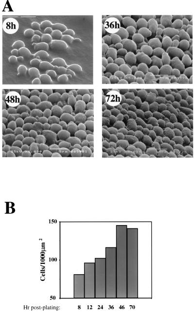
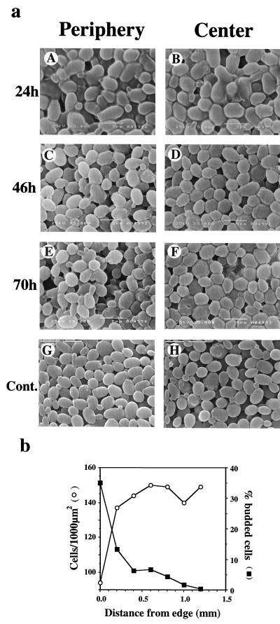
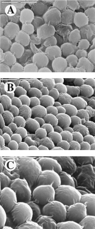
Cell growth in yeast colonies is a complex process, the control of which is largely unknown. Here we present scanning electron micrographs of Saccharomyces cerevisiae colonies, showing changes in the pattern of cell organization and cell-cell interactions during colony development. In young colonies (</=36 h), cell density is relatively low, and the cells seem to divide in a random orientation. However, as the colonies age, cell density increases and the cells seem to be oriented in a more orderly fashion. Unexpectedly, cells in starved colonies form connecting fibrils. A single connecting fibril 180 +/- 50 nm wide is observed between any two neighboring cells, and the fibrils appear to form a global network. The results suggest a novel type of communication between cells within a colony that may contribute to the ability of the community to cope with starvation.
Figures
References
-
- Dworkin M. Fibrils as extracellular appendages of bacteria: their role in contact-mediated cell-cell interactions in Myxococcus xanthus. Bioessays. 1999;21:590–595. - PubMed
-
- Ginocchio C C, Olmsted S B, Wells C L, Galan J E. Contact with epithelial cells induces the formation of surface appendages on Salmonella typhimurium. Cell. 1994;76:717–724. - PubMed
-
- Meunier J-R, Choder M. Saccharomyces cerevisiae colony growth and aging: biphasic growth accompanied by changes in gene expression. Yeast. 1999;15:1159–1169. - PubMed
-
- Palkova Z, Janderova B, Gabriel J, Zikanova B, Pospisek M, Forstova J. Ammonia mediates communication between yeast colonies. Nature. 1997;390:532–536. - PubMed
-
- Parent C A, Devreotes P N. Molecular genetics of signal transduction in Dictyostelium. Annu Rev Biochem. 1996;65:411–440. - PubMed
Publication types
MeSH terms
LinkOut - more resources
Full Text Sources
Molecular Biology Databases
