. 2000 Jun 12;149(6):1215-24.
doi: 10.1083/jcb.149.6.1215.

An essential role for a membrane lipid in cytokinesis. Regulation of contractile ring disassembly by redistribution of phosphatidylethanolamine

Affiliations

An essential role for a membrane lipid in cytokinesis. Regulation of contractile ring disassembly by redistribution of phosphatidylethanolamine

K Emoto et al. J Cell Biol. .

Abstract

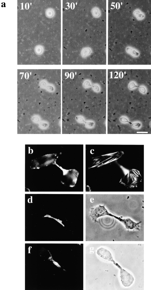
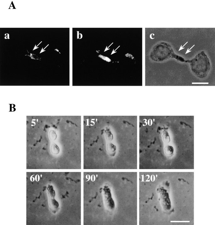
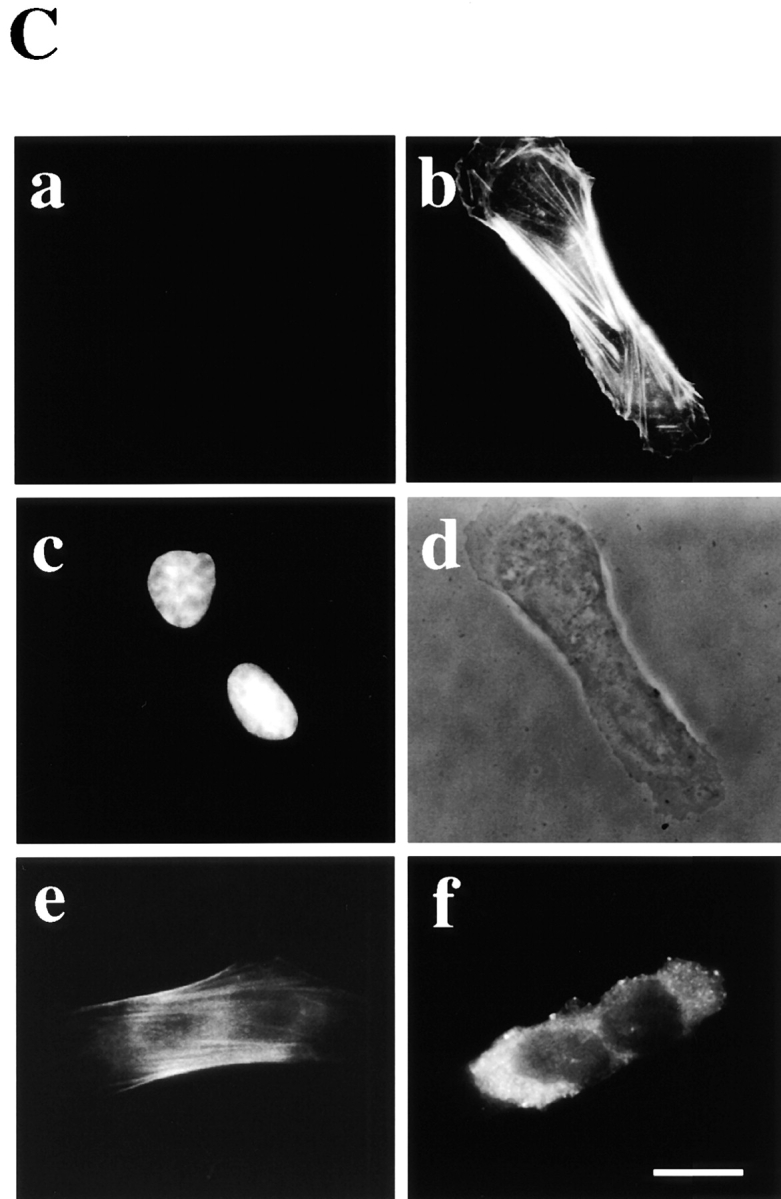
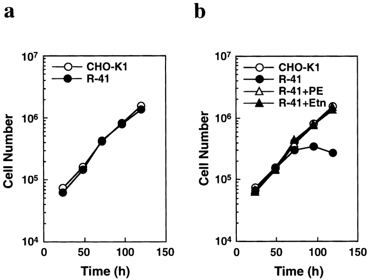
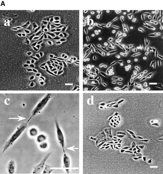
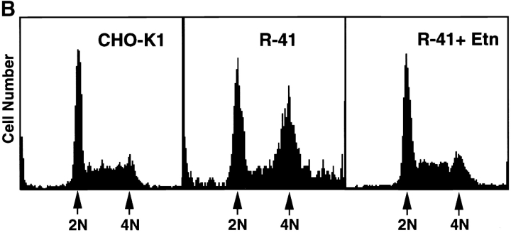
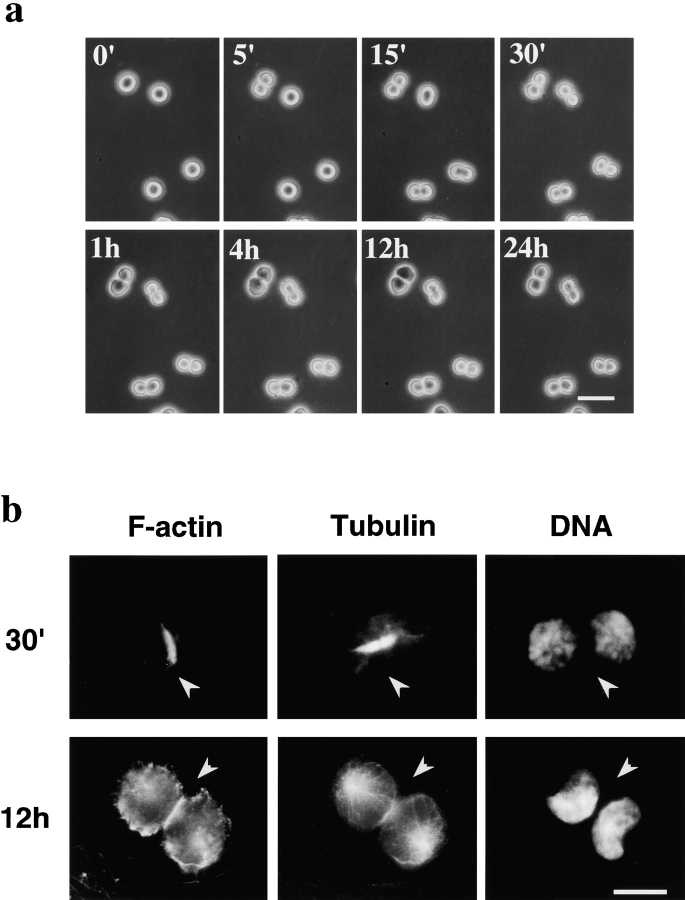
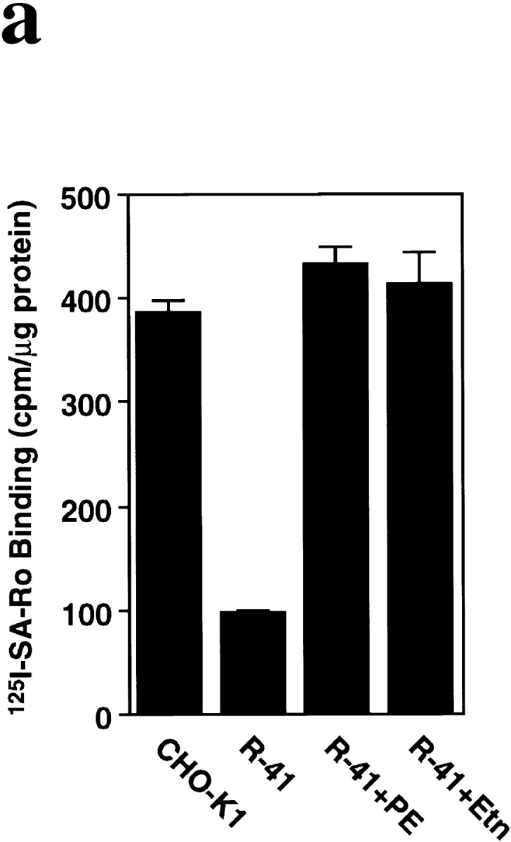
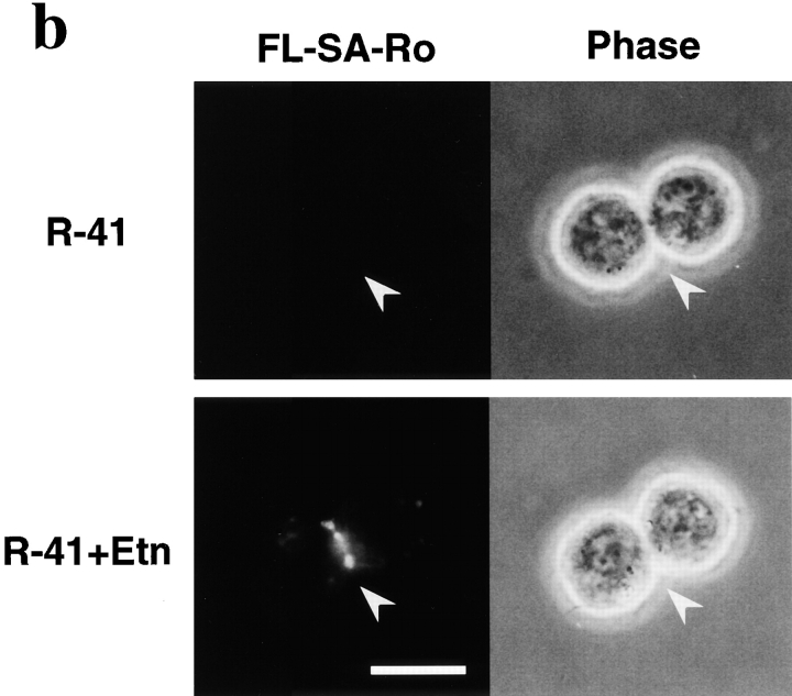
Phosphatidylethanolamine (PE) is a major membrane phospholipid that is mainly localized in the inner leaflet of the plasma membrane. We previously demonstrated that PE was exposed on the cell surface of the cleavage furrow during cytokinesis. Immobilization of cell surface PE by a PE-binding peptide inhibited disassembly of the contractile ring components, including myosin II and radixin, resulting in formation of a long cytoplasmic bridge between the daughter cells. This blockade of contractile ring disassembly was reversed by removal of the surface-bound peptide, suggesting that the PE exposure plays a crucial role in cytokinesis. To further examine the role of PE in cytokinesis, we established a mutant cell line with a specific decrease in the cellular PE level. On the culture condition in which the cell surface PE level was significantly reduced, the mutant ceased cell growth in cytokinesis, and the contractile ring remained in the cleavage furrow. Addition of PE or ethanolamine, a precursor of PE synthesis, restored the cell surface PE on the cleavage furrow and normal cytokinesis. These findings provide the first evidence that PE is required for completion of cytokinesis in mammalian cells, and suggest that redistribution of PE on the cleavage furrow may contribute to regulation of contractile ring disassembly.


Figures

Figure 1
Figure 1
SA-Ro treatment blocks disassembly of the contractile ring. a, Time-lapse observation of cytokinesis of cells treated with SA-Ro. The time shown in the photograph represents time points from the initiation of cell division (min). Bar, 30 μm. b and c, CHO cells synchronized in prometaphase were incubated with 50 μg/ml SA-Ro for 2 h, and then fixed and stained with rhodamine-phalloidin. b, Shows the focal plane of the cytoplasmic bridge where actin bundles remains concentrated. c, Shows the focal plane of the bottom layer of the daughter cells where stress fibers are seen. d–g, Distribution of myosin II (d) and radixin (f) in SA-Ro–treated cells. Phase-contrast of each specimen was shown in e and g, respectively. Bar, 10 μm.
Figure 2
Figure 2
Incubation of the arrested cells with PE liposomes leads to disassembly of the contractile ring. A, Colocalization of SA-Ro bound to the cell surface of PE (a) and actin filaments remaining in the cytoplasmic bridge (b). Cells were incubated with 50 μg/ml SA-Ro for 2 h, and then fixed and double-labeled with either anti-SA antibody (a) or rhodamine-phalloidin (b). The photograph shows the focal plane of the cytoplasmic bridge where actin bundles remain concentrated. Phase-contrast of the specimen is shown in c. The arrows indicate the cytoplasmic bridge. B, Time-lapse observations of the arrested cells incubated in the presence of 10 μM PE liposomes. The time shown in the photograph represents the time points after initiation of the incubation (min). C, The arrested cells were incubated in the presence of 10 μM PE liposomes, and then fixed and triple-stained with anti-SA antibody (a), rhodamine-phalloidin (b), and DAPI (c). Phase-contrast of the same specimen is shown in (d). e and f, The arrested cells were incubated with 10 μM PE liposomes, and then fixed and stained with antimyosin II antibody (e) and antiradixin antibody (f). Bars, 10 μm.
Figure 2
Figure 2
Incubation of the arrested cells with PE liposomes leads to disassembly of the contractile ring. A, Colocalization of SA-Ro bound to the cell surface of PE (a) and actin filaments remaining in the cytoplasmic bridge (b). Cells were incubated with 50 μg/ml SA-Ro for 2 h, and then fixed and double-labeled with either anti-SA antibody (a) or rhodamine-phalloidin (b). The photograph shows the focal plane of the cytoplasmic bridge where actin bundles remain concentrated. Phase-contrast of the specimen is shown in c. The arrows indicate the cytoplasmic bridge. B, Time-lapse observations of the arrested cells incubated in the presence of 10 μM PE liposomes. The time shown in the photograph represents the time points after initiation of the incubation (min). C, The arrested cells were incubated in the presence of 10 μM PE liposomes, and then fixed and triple-stained with anti-SA antibody (a), rhodamine-phalloidin (b), and DAPI (c). Phase-contrast of the same specimen is shown in (d). e and f, The arrested cells were incubated with 10 μM PE liposomes, and then fixed and stained with antimyosin II antibody (e) and antiradixin antibody (f). Bars, 10 μm.
Figure 3
Figure 3
The growth of R-41 mutant cells is dependent on PE or its precursor. a, CHO-K1 and R-41 mutant cells were cultured in normal medium containing 10% newborn calf serum at 39.5°C. b, CHO-K1 and R-41 mutant cells were cultured in ethanolamine-deficient medium at 39.5°C (CHO-K1, R-41), and R-41 mutant cells were cultured in either 30 μM PE (R-41+PE) or 20 μM ethanolamine-supplemented medium (R-41+Etn) at 39.5°C.
Figure 4
Figure 4
R-41 mutant cells have a defect in cytokinesis. A, Morphology of CHO-K1 (a) and R-41 mutant cells (b–d) cultured in normal medium. CHO-K1 and R-41 mutant cells were cultured in normal medium containing 10% newborn calf serum supplemented with (d) or without (a–c) 20 μM ethanolamine at 39.5°C for 3 d. B, Flow cytometric analyses of the DNA contents. CHO-K1 and mutant cells were cultured in ethanolamine-deficient medium supplemented with (R-41+Etn) or without (CHO-K1, R-41) 20 μM ethanolamine at 39.5°C for 4 d. CHO-K1: G1, 41.6%; S, 49.1%; G2/M, 9.3%. R-41: G1, 38.9%; S, 16.3%; G2/M, 44.8%. R-41+Etn: G1, 38.2%; S, 50.5%; G2/M, 11.3%. Bars, 30 μm.
Figure 4
Figure 4
R-41 mutant cells have a defect in cytokinesis. A, Morphology of CHO-K1 (a) and R-41 mutant cells (b–d) cultured in normal medium. CHO-K1 and R-41 mutant cells were cultured in normal medium containing 10% newborn calf serum supplemented with (d) or without (a–c) 20 μM ethanolamine at 39.5°C for 3 d. B, Flow cytometric analyses of the DNA contents. CHO-K1 and mutant cells were cultured in ethanolamine-deficient medium supplemented with (R-41+Etn) or without (CHO-K1, R-41) 20 μM ethanolamine at 39.5°C for 4 d. CHO-K1: G1, 41.6%; S, 49.1%; G2/M, 9.3%. R-41: G1, 38.9%; S, 16.3%; G2/M, 44.8%. R-41+Etn: G1, 38.2%; S, 50.5%; G2/M, 11.3%. Bars, 30 μm.
Figure 5
Figure 5
R-41 mutant cells have a defect in the disassembly of the contractile ring. a, Time-lapse observation of the cell division of R-41 mutant cells cultured in ethanolamine-deficient medium. R-41 mutant cells cultured in ethanolamine-deficient medium for 3 d were synchronized in mitotic phase by nocodazole. The synchronized cells were collected, washed, and then further incubated in the ethanolamine-deficient medium. The time indicates the time point after the initiation of the incubation of synchronized cells. Bar, 30 μm. b, Localization of actin filaments and microtubules in R-41 mutant cells during cytokinesis. The synchronized mutant cells were incubated for 30 min (30') or 12 h in ethanolamine-deficient medium. The cells were then fixed and labeled with rhodamine-phalloidin (for F-actin), antitubulin antibody, or DAPI (for DNA). Bar, 10 μm. The arrowheads indicate the cleavage furrow.
Figure 6
Figure 6
Exposure of PE on the surface of R-41 mutant cells. a, Binding of 125I-SA-Ro to the plasma membrane. CHO-K1 and R-41 mutant cells were incubated in ethanolamine-deficient medium without (CHO-K1, R-41) or with either 30 μM PE (R-41+PE) or 20 μM ethanolamine (R-41+Etn) at 39.5°C for 3 d. Cells were incubated with 125I-SA-Ro (30,000 cpm/ml) on ice for 30 min. The cells were then washed and their radioactivity was measured. The results show the mean ± SEM (n = 3). b, Exposure of PE on the cleavage furrow membrane of R-41 mutant cells. R-41 mutant cells were incubated in ethanolamine-deficient medium with (R-41+Etn) or without (R-41) 20 μM ethanolamine at 39.5°C for 3 d and then synchronized in mitotic phase. The synchronized cells were collected, washed, and then further incubated for 30 min. The cells were washed and stained with FL–SA-Ro for 30 min on ice, then washed and photographed. Bar, 10 μm. The arrowheads indicate the cleavage furrow.
Figure 6
Figure 6
Exposure of PE on the surface of R-41 mutant cells. a, Binding of 125I-SA-Ro to the plasma membrane. CHO-K1 and R-41 mutant cells were incubated in ethanolamine-deficient medium without (CHO-K1, R-41) or with either 30 μM PE (R-41+PE) or 20 μM ethanolamine (R-41+Etn) at 39.5°C for 3 d. Cells were incubated with 125I-SA-Ro (30,000 cpm/ml) on ice for 30 min. The cells were then washed and their radioactivity was measured. The results show the mean ± SEM (n = 3). b, Exposure of PE on the cleavage furrow membrane of R-41 mutant cells. R-41 mutant cells were incubated in ethanolamine-deficient medium with (R-41+Etn) or without (R-41) 20 μM ethanolamine at 39.5°C for 3 d and then synchronized in mitotic phase. The synchronized cells were collected, washed, and then further incubated for 30 min. The cells were washed and stained with FL–SA-Ro for 30 min on ice, then washed and photographed. Bar, 10 μm. The arrowheads indicate the cleavage furrow.

References

    1. Aoki Y., Uenaka T., Aoki J., Umeda M., Inoue K. A novel peptide probe for studying the transbilayer movement of phosphatidylethanolamine. J. Biochem. 1994;116:291–297. - PubMed
    1. Bazzi M.D., Youakin M.A., Nelsestuen G.L. Importance of phosphatidylethanolamine for association of protein kinase C and other cytoplasmic proteins with membranes. Biochemistry. 1992;31:1125–1134. - PubMed
    1. Bi E., Luthenhaus J. FtsZ ring structure associated with division in Escherichia coli . Nature. 1991;354:161–164. - PubMed
    1. Bligh E.G., Dyer W.J. A rapid method of total lipid extraction and purification. Can. J. Biochem. Physiol. 1959;37:911–917. - PubMed
    1. Bluemink J.G., deLaat S.W. New membrane formation during cytokinesis in normal and cytochalasin B-treated eggs of Xenopus laevis. I. Electron microscope observations. J. Cell Biol. 1973;59:89–108. - PMC - PubMed

Publication types