. 2000 Jun 12;149(6):1263-74.
doi: 10.1083/jcb.149.6.1263.

The nonreceptor tyrosine kinase fer mediates cross-talk between N-cadherin and beta1-integrins

Affiliations

The nonreceptor tyrosine kinase fer mediates cross-talk between N-cadherin and beta1-integrins

C Arregui et al. J Cell Biol. .

Abstract

Cadherins and integrins must function in a coordinated manner to effectively mediate the cellular interactions essential for development. We hypothesized that exchange of proteins associated with their cytoplasmic domains may play a role in coordinating function. To test this idea, we used Trojan peptides to introduce into cells and tissues peptide sequences designed to compete for the interaction of specific effectors with the cytoplasmic domain of N-cadherin, and assayed their effect on cadherin- and integrin-mediated adhesion and neurite outgrowth. We show that a peptide mimicking the juxtamembrane (JMP) region of the cytoplasmic domain of N-cadherin results in inhibition of N-cadherin and beta1-integrin function. The effect of JMP on beta1-integrin function depends on the expression of N-cadherin and is independent of transcription or translation. Treatment of cells with JMP results in the release of the nonreceptor tyrosine kinase Fer from the cadherin complex and its accumulation in the integrin complex. A peptide that mimics the first coiled-coil domain of Fer prevents Fer accumulation in the integrin complex and reverses the inhibitory effect of JMP. These findings suggest a new mechanism through which N-cadherin and beta1-integrins are coordinately regulated: loss of an effector from the cytoplasmic domain of N-cadherin and gain of that effector by the beta1-integrin complex.


Figures

Figure 1
Figure 1
Diagram showing the relative positions of the sequences used to generate cell permeable peptide competitors. N-Cadherin is numbered from the beginning of the signal sequence. The model of the nonreceptor protein tyrosine kinase Fer is based on Craig et al. 1999. CC1, CC2, and CC3, coiled-coil domains 1, 2, and 3; SH2, src homology 2 domain; kinase, tyrosine kinase catalytic domain.
Figure 2
Figure 2
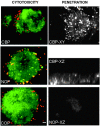
Toxicity and penetration of fusion peptides. Left panels show live (green) and dead (red) cells in the presence of CBP (top), NOP (no peptide, middle) or COP (bottom). Right panels show a retina explant incubated with biotinylated CBP and detected with fluorescein-conjugated avidin. XY and XZ are confocal sections of the explant showing fluorescent cells throughout the explant. Lower right panel shows background fluorescence in a control where peptide incubation is omitted. Bar, ∼20 μm.
Figure 3
Figure 3
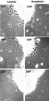
Effect of peptides on neurite outgrowth from explants of E7 neural retina. Phase-contrast of retina explants incubated in the presence of control peptide (COP), catenin-binding peptide (CBP), or the juxtamembrane peptide (JMP) and plated on laminin or N-cadherin. Bar, ∼20 μm.
Figure 4
Figure 4
Quantitative determination of neurite outgrowth by single E7 cells in the presence and absence of peptides. Data are expressed as the percent of neurites in each of three length categories. More than 100 cells bearing neurites were counted for each treatment. NOP, no peptide.
Figure 5
Figure 5
The effect of peptides on cadherin- and integrin-mediated cell adhesion by E7 retina cells. Cells preincubated in the absence or presence of peptides were plated on the indicated substrates, the wells were washed, and adherent cells were counted. Values for each condition represent the percentage of attached cells as compared with controls lacking peptide. Each bar is the average of three measurements, and the error bars represent the SD. The substrate is indicated in the top right of each panel: NCD-2, anti–N-cadherin antibody; LM, laminin; and PL, poly-l-lysine.
Figure 6
Figure 6
Interaction of peptides with presumptive targets. (A) In vitro binding of peptides to the N-cadherin cytoplasmic domain and to β-catenin. 10 μg of recombinant GST-N-cadherin cytoplasmic domain or GST-β-catenin were incubated with 4 μg of biotinylated CBP or JMP. The amount of protein bound to immobilized peptide was determined as described in Materials and Methods. Each point is the average of three measurements and bars represent the SD. (B) The ability of CBP to disrupt the association of cadherin with β-catenin. The ratio of β-catenin to cadherin was measured in neutral detergent extracts in the presence of increasing concentrations of CBP and JMP, and reported as a percentage of the value in the absence of peptide. The results shown are one experiment representative of many. (C) Localization of JMP to regions of cell–cell contact. The left frame labeled DIC (differential interference contrast microscopy) and the middle frame labeled JMP-bio are the same field. The frame on the right shows the distribution of biotin-labeled control peptide (COP-bio). Individual cells are ∼8 μm in diameter.
Figure 7
Figure 7
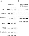
The effect of peptides on the association of proteins with the cytoplasmic domain of N-cadherin (A) and cadherin-free pool of β-catenin (B). (A) Neutral detergent extracts of cells preincubated with peptides were immunoprecipitated with anti–N-cadherin antibody NCD-2, the immunoprecipitates were fractionated by SDS-PAGE, and Western transfers were blotted with the indicated antibodies. (B) The supernatants from the NCD-2 immunoprecipitations were immunoprecipitated with polyclonal anti–β-catenin antibody and processed as in A.
Figure 8
Figure 8
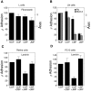
The effect of peptides on adhesion of L-cells (A), L-cells constitutively expressing N-cadherin (LN-cells, B), E7 retina cells (C), and PC-12 cells (D). The substrate is indicated. A585 reflects the number of adherent cells in A and B. In C and D, the indicated peptide pairs were added sequentially with a 1-h incubation following the addition of the first peptide. Each bar represents the average of three measurements and error bars represent the SD.
Figure 9
Figure 9
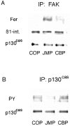
The effect of peptides on the association of Fer with the β1-integrin complex and phosphorylation of p130cas. (A) Neutral detergent extracts of E8 retina cells preincubated with peptides were immunoprecipitated with anti-FAK antibody, the precipitates were fractionated by SDS-PAGE, and Western transfers were blotted with anti-Fer, anti-p130cas, and anti–β1-integrin antibodies. (B) Cells preincubated with peptides were lysed with RIPA buffer, immunoprecipitated with anti-p130cas, and the precipitates were fractionated by SDS-PAGE. Western transfers were blotted with the antiphosphotyrosine mAb PY20 and anti-p130cas antibody.
Figure 10
Figure 10
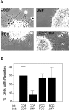
The effect of peptides mimicking specific regions of Fer on inhibition of integrin-mediated neurite outgrowth by JMP. (A) Phase-contrast views of neurite outgrowth from explants of E7 retina. (B) Quantitative determination of neurite outgrowth by single E7 cells after incubation with peptides. Over 100 cells in each treatment group were counted and all cells bearing neurites longer than two cell diameters were considered positive. Bar, ∼20 μm.
Figure 11
Figure 11
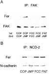
The effect of peptides mimicking specific regions of Fer on its association with the integrin and cadherin complex. (A) Peptide FCC reverses the effect of JMP on targeting of Fer to the integrin complex. The indicated peptide pairs were added to retina cells sequentially with a 1-h incubation after the addition of the first peptide, and a 1-h incubation after the second. The cells were homogenized in neutral detergent and immunoprecipitated with anti-FAK antibody. The immunoprecipitates were fractionated by SDS-PAGE, and Western transfers were blotted with anti-Fer and anti-FAK antibodies. (B) FCC does not affect the association of Fer with the cadherin complex. The indicated peptides were added to retina cells for 1 h, the cells were homogenized in neutral detergent and immunoprecipitated with anti–N-cadherin antibody NCD-2. The immunoprecipitates were fractionated by SDS-PAGE, and Western transfers were blotted with anti-Fer and anticadherin antibodies.

Similar articles

Cited by

References

    1. Aberle H., Schwartz H., Kemler R. Cadherin catenin complexprotein interactions and their implications for cadherin function. J. Cell Biochem. 1996;61:514–523. - PubMed
    1. Aplin A.E., Howe A., Alahari S.K., Juliano R.L. Signal transduction and signal modulation by cell adhesion receptorsthe role of integrins, cadherins, immunoglobulin-cell adhesion molecules, and selectins. Pharm. Rev. 1998;50:199–263. - PubMed
    1. Arregui C., Lilien J., Balsamo J. Impaired integrin-mediated adhesion and signaling in fibroblasts expressing a dominant negative mutant PTP1B. J. Cell Biol. 1998;143:861–873. - PMC - PubMed
    1. Balsamo J., Ernst H., Zanin M.K., Hoffman S., Lilien J. The interaction of the retina cell surface N-acetylgalactosaminylphosphotransferase with an endogenous proteoglycan ligand results in inhibition of cadherin-mediated adhesion. J. Cell Biol. 1995;129:1391–1401. - PMC - PubMed
    1. Balsamo J., Thiboldeaux R., Swaminathan N., Lilien J. Antibodies to the retina N-acetylgalactosaminylphosphotransferase modulate N-cadherin–mediated adhesion and uncouple the N-cadherin transferase complex from the actin-containing cytoskeleton. J. Cell Biol. 1991;113:429–436. - PMC - PubMed

Publication types

MeSH terms