. 2000 Jun 12;149(6):1275-88.
doi: 10.1083/jcb.149.6.1275.

Coordinate regulation of cadherin and integrin function by the chondroitin sulfate proteoglycan neurocan

Affiliations

Coordinate regulation of cadherin and integrin function by the chondroitin sulfate proteoglycan neurocan

H Li et al. J Cell Biol. .

Abstract

N-cadherin and beta1-integrins play decisive roles in morphogenesis and neurite extension and are often present on the same cell. Therefore, the function of these two types of adhesion systems must be coordinated in time and space to achieve the appropriate cell and tissue organization. We now show that interaction of the chondroitin sulfate proteoglycan neurocan with its GalNAcPTase receptor coordinately inhibits both N-cadherin- and beta1-integrin-mediated adhesion and neurite outgrowth. Furthermore, the inhibitory activity is localized to an NH(2)-terminal fragment of neurocan containing an Ig loop and an HA-binding domain. The effect of neurocan on beta1-integrin function is dependent on a signal originating from the cadherin cytoplasmic domain, possibly mediated by the nonreceptor protein tyrosine kinase Fer, indicating that cadherin and integrin engage in direct cross-talk. In the developing chick, neural retina neurocan is present in the inner plexiform layer from day 7 on, and the GalNAcPTase receptor becomes restricted to the inner nuclear layer and the ganglion cell layer (as well as the fiber layer), the two forming a sandwich. These data suggest that the coordinate inhibition of cadherin and integrin function on interaction of neurocan with its receptor may prevent cell and neurite migration across boundaries.


Figures
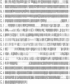
Figure 1
Figure 1
Sequence comparison between the four cloned neurocans: chicken, human, mouse, and rat. Shaded areas represent identical amino acids, and boxed areas represent conservative changes.
Figure 2
Figure 2
(A) Diagram of chick neurocan showing its multiple domains and (B) the regions expressed as fusion peptides. N indicates the position of potential N-linked oligosaccharides, and cs indicates potential chondroitin sulfate attachment sites. RGD indicates the presence of the consensus integrin-binding sequence (Arg-Gly-Asp). SP refers to the signal peptide and CRL to complement regulatory-like domain. Small numbers refer to residue number.
Figure 3
Figure 3
Recombinant chick neurocan is recognized by anti-250kD PG antibody. Lysates of bacteria transformed with plasmids containing the complete chick neurocan cDNA were fractionated by SDS-PAGE and stained with Coomassie brilliant blue (CBB), or transferred to polyvinylidene difluoride membranes and immunoblotted with anti-250kD PG antibody. U, lysate from noninduced bacteria; and I, lysate from bacteria induced with IPTG. Numbers to the left refer to the migration of molecular mass markers (10−3).
Figure 4
Figure 4
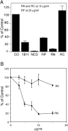
Recombinant neurocan and its NH2-terminal fragment inhibit N-cadherin–mediated adhesion. Single cells were prepared from E9 retinas and added to 96-well plates coated with anti–N-cadherin antibody at 37°C for 30 min in the presence of the indicated additives. The plates were washed and adherent cells determined by staining with crystal violet. Adhesion in the absence of any additives was considered 100%. (A) Co, no additives; 1B11, anti-GalNAcPTase antibody; NCD, anti–N-cadherin antibody; RP, full-length recombinant neurocan; RN, NH2-terminal fragment; and RC, COOH-terminal fragment. (B) Adhesion in the presence of increasing concentrations of RN and RC.
Figure 5
Figure 5
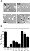
The NH2-terminal fragment of neurocan inhibits N-cadherin–mediated neurite outgrowth. E7 retina cells were plated on anti–N-cadherin–coated coverslips and incubated for 2 h before addition of anti–N-cadherin antibodies (NCD), neurocan NH2-terminal fragment (RN) or COOH-terminal fragment (RC). RN and RC were added to the cultures three more times, after 3-h intervals. The cells were fixed and observed under a microscope. (A) Representative fields of each treatment (RN and RC at 10 μg/ml). (B) The number of cells bearing neurites longer than one cell diameter is expressed as a percentage of the control (number of cells bearing neurites in the absence of additives: 59 ± 7; n = 100). 1B11, anti-GalNacPTase antibody. RN and RC were assayed at 10, 25, and 50 μg/ml.
Figure 6
Figure 6
The NH2-terminal fragment of neurocan inhibits β1-integrin–mediated adhesion. Single cells from E9 retina were plated on laminin-coated 96-well plates and incubated in the presence of the designated additives. Co, no additives; JG22, anti–β1-integrin antibody; NCD, anti–N-cadherin antibody; RN and RC, neurocan NH2- and COOH-terminal fragments respectively at 10 μg/ml. Adhesion was measured as percent of control.
Figure 7
Figure 7
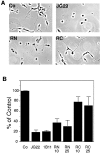
The NH2-terminal fragment of neurocan inhibits β1-integrin–mediated neurite outgrowth. E7 retina cells were plated on laminin-coated glass coverslips and incubated for 2 h before the addition of anti-integrin antibody JG22, RN, or RC, as described in Fig. 5. (A) Representative fields of each treatment (RN and RC at 20 μg/ml). (B) The number of cells bearing neurites longer than one cell in diameter is expressed as a percentage of the control. 1B11, anti-GalNacPTase antibody; JG22, anti–β1-integrin antibody. RN and RC were assayed at 10 and 25 μg/ml.
Figure 9
Figure 9
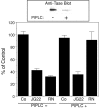
Removal of cell-surface GalNAcPTase with PIPLC eliminates the effect of the neurocan NH2-terminal fragment on integrin-mediated neurite outgrowth. E7 retina cells were incubated with or without 5u/ml PIPLC for 30 min at 30°C. The cells were assayed for neurite growth, as described Fig. 7, and for the presence of the GalNAcPTase by immunoblot after SDS-PAGE, Western transfer, and immunoblot (top).
Figure 8
Figure 8
Neurocan and its NH2-terminal fragment bind to the cell-surface GalNAcPTase. Single cells prepared from E9 retinas were incubated with no additives (CO), RN (20 μg/ml), RC (20 μg/ml), or RP (40 μg/ml) for 30 min at 37°C. The cells were treated with the cross-linking reagent DTSSP. Triton X-100 lysates of cells were immunoprecipitated with anti-GalNAcPTase antibody, fractionated by SDS-PAGE, and Western transfers were prepared. RN/0, cross-linking and lysis at 0 time. (top) Immunoblot with antineurocan antibody. (bottom) Immunoblot with anti-GalNAcPTase antibody.
Figure 10
Figure 10
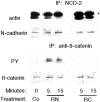
Treatment of cells with neurocan NH2-terminal fragment results in dissociation of N-cadherin from actin and hyperphosphorylation of β-catenin on tyrosine residues. E9 retina cells were incubated with RN or RC at 37°C for 5 and 15 min. The treated cells were lysed in Triton X-100 and immunoprecipitated with anti–N-cadherin antibody NCD-2. The precipitates were fractionated by SDS-PAGE, and Western transfers were prepared and immunoblotted with antiactin antibody and NCD-2 (top). The resulting supernatants were immunoprecipitated with anti–β-catenin antibody (after treatment with 1% SDS to disrupt protein–protein interactions), fractionated by SDS-PAGE, and immunoblotted with anti-PY antibody. The membranes were stripped and reprobed with anti–β-catenin antibody (bottom). The heavy bands in the top panel marked by an asterisk are IgG heavy chains.
Figure 11
Figure 11
Treatment of cells with the neurocan NH2-terminal fragment results in release of the protein tyrosine kinase Fer from the cadherin-associated complex of proteins and its association with the β1-integrin complex. E9 retina cells were treated with 10 μg/ml of recombinant neurocan NH2-terminal fragment (RN) or with anti-GalNAcPTase antibodies 1B11 or 7A2. The cells were lysed in buffer containing nonionic detergent and the lysates were immunoprecipitated with anti-FAK or anti–N-cadherin antibodies. The immunocomplexes were subjected to SDS-PAGE, followed by immunoblotting with the indicated antibodies.
Figure 12
Figure 12
The inhibitory effect of neurocan NH2-terminal fragment on integrin-mediated adhesion is mediated through N-cadherin. E9 retina cells were incubated with 4 μM cell permeable control (COP) or β-catenin–binding (CBP) peptides for 2 h at 4°C. The cells were plated on laminin-coated dishes in the presence or absence of RN (10 μg/ml) and incubated for 2 h at 37°C. Adhesion is expressed as a percentage of the control. COP/N and CBP/N refer to addition of peptide followed by RN.
Figure 13
Figure 13
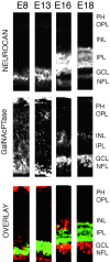
Expression of neurocan and its receptor GalNAcPTase in the developing retina. Frozen sections of retina tissue at several developmental stages were stained with antineurocan and anti-GalNAcPTase antibody, followed by fluorescence-conjugated secondary antibodies. PH, photoreceptor layer; OPL, outer plexiform layer; INL, inner nuclear layer; IPL, inner plexiform layer; GCL, ganglion cell layer; and NFL, nuclear fiber layer.
Figure 14
Figure 14
Amacrine cells express neurocan. E9 retina cells were cultured overnight before staining with antibodies. A shows the morphology of typical cells expressing neurocan. (B) Cells were double stained with antisyntaxin and antineurocan. Bar, 20 μm.
Figure 15
Figure 15
Typical ganglion cells are not reactive with antineurocan antibody. E9 cells cultured overnight were stained with antineurocan and 8D9, a ganglion cell marker, followed by FITC-conjugated secondary antibody. The arrowheads point to cells showing typical ganglion cell morphology, reactive with 8D9, but not antineurocan. The arrows point to cells reactive with both antibodies, but with atypical ganglion cell morphology. Bar, 20 μm.
Figure 16
Figure 16
Pictorial representation of the known protein interactions in the cadherin and integrin complex potentially altered on binding of neurocan to its receptor GalNAcPTase. On the right is a functional cadherin complex showing the known protein interactions. The interaction of β-catenin, p120ctn, and Fer is based on Arregui et al. 2000. On the left, after binding of neurocan, are the alterations induced in the cadherin complex rendering it nonfunctional and potential Fer targets in the integrin complex.

Similar articles

Cited by

References

    1. Arregui C., Balsamo J., Lilien J. Impaired integrin-mediated adhesion and signaling in fibroblasts expressing a dominant negative mutant PTP1B. J. Cell Biol. 1999;143:861–873. - PMC - PubMed
    1. Arregui C., Pathre P., Lilien J., Balsamo J. The nonreceptor tyrosine kinase Fer mediates cross-talk between N-cadherin and β1-integrin. J. Cell Biol. 2000;149:1263–1273. - PMC - PubMed
    1. Balsamo J., Lilien J. N-cadherin is stably associated with, and is an acceptor for, a cell surface N-acetylgalactosaminylphosphotransferase. J. Biol. Chem. 1990;265:2923–2928. - PubMed
    1. Balsamo J., Lilien J. The retina cell-surface N-acetylgalactosaminylphosphotransferase is anchored by a glycophosphatidylinositol. Biochemistry. 1993;32:8246–8250. - PubMed
    1. Balsamo J., Hoffman S., Lilien J. Control of cadherin-mediated cell-cell adhesion through regulated association with the cytoskeleton. J. Braz. Assoc. Adv. Sci. 1996;48:341–346.

Publication types

MeSH terms

Associated data