. 2000 Jun 12;149(6):1297-308.
doi: 10.1083/jcb.149.6.1297.

Signaling by fibroblast growth factors (FGF) and fibroblast growth factor receptor 2 (FGFR2)-activating mutations blocks mineralization and induces apoptosis in osteoblasts

Affiliations

Signaling by fibroblast growth factors (FGF) and fibroblast growth factor receptor 2 (FGFR2)-activating mutations blocks mineralization and induces apoptosis in osteoblasts

A Mansukhani et al. J Cell Biol. .

Abstract

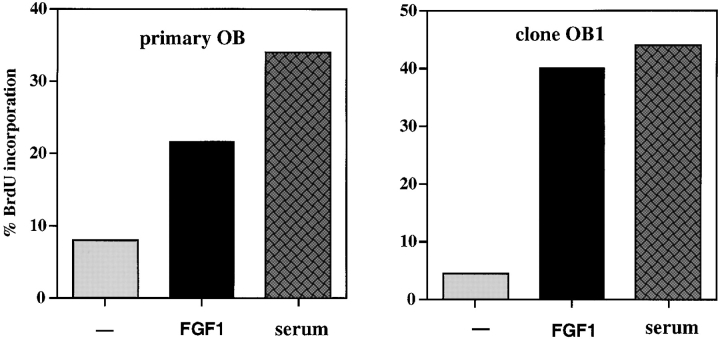
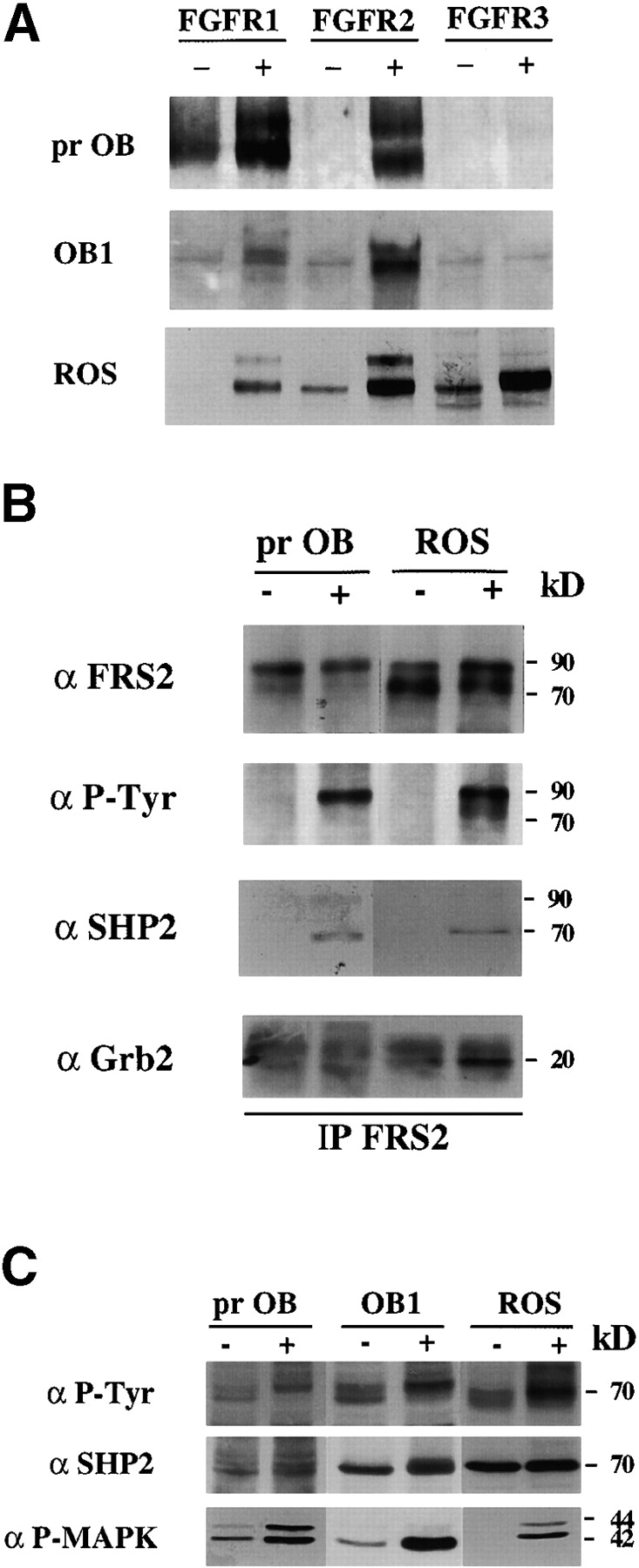
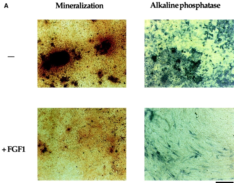
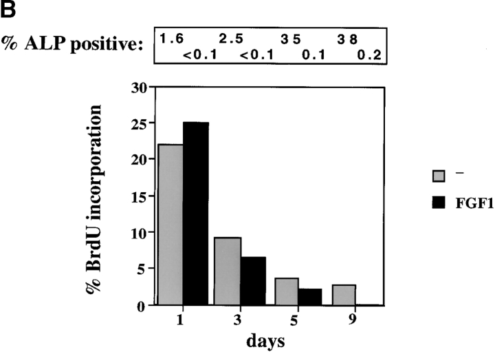
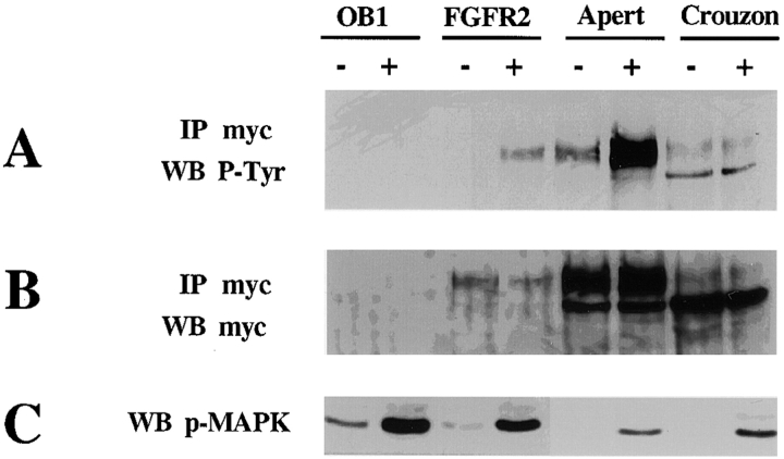
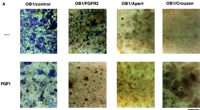
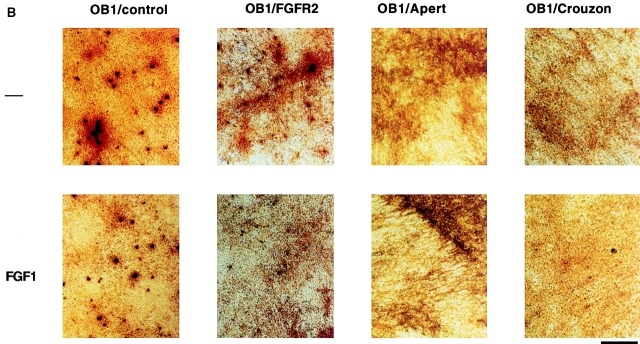
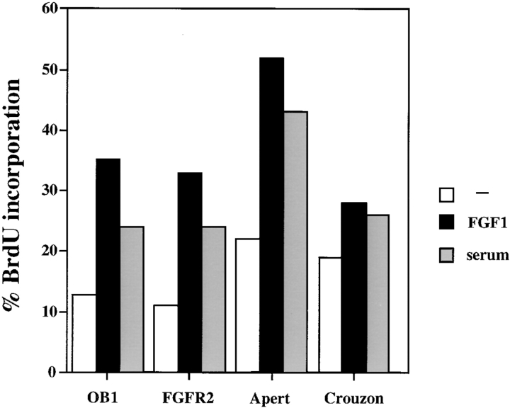
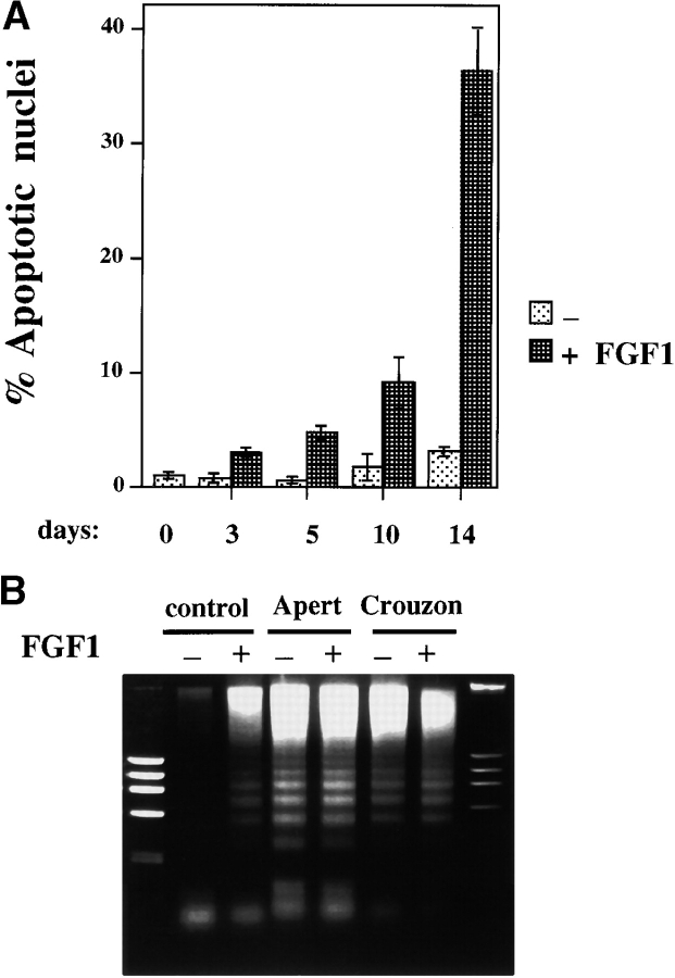
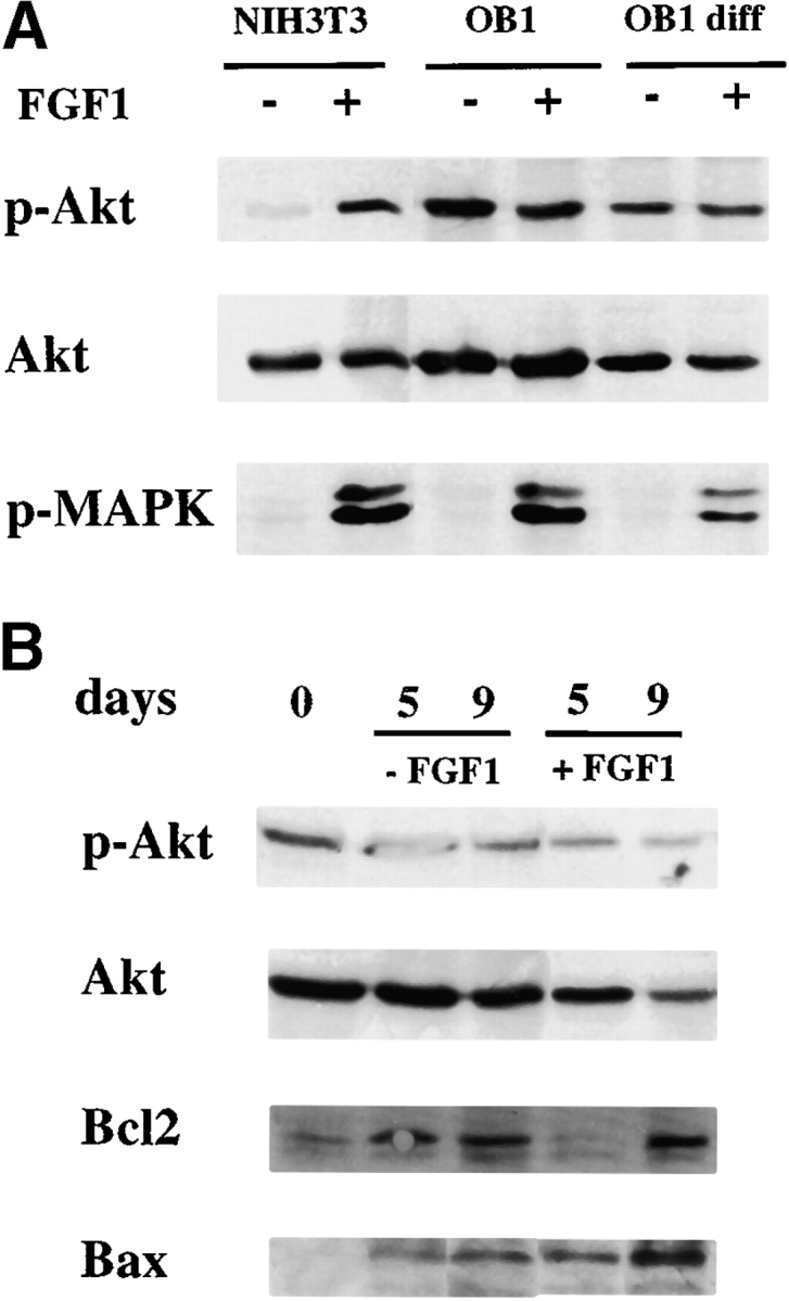
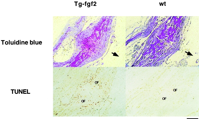
Fibroblast growth factors (FGF) play a critical role in bone growth and development affecting both chondrogenesis and osteogenesis. During the process of intramembranous ossification, which leads to the formation of the flat bones of the skull, unregulated FGF signaling can produce premature suture closure or craniosynostosis and other craniofacial deformities. Indeed, many human craniosynostosis disorders have been linked to activating mutations in FGF receptors (FGFR) 1 and 2, but the precise effects of FGF on the proliferation, maturation and differentiation of the target osteoblastic cells are still unclear. In this report, we studied the effects of FGF treatment on primary murine calvarial osteoblast, and on OB1, a newly established osteoblastic cell line. We show that FGF signaling has a dual effect on osteoblast proliferation and differentiation. FGFs activate the endogenous FGFRs leading to the formation of a Grb2/FRS2/Shp2 complex and activation of MAP kinase. However, immature osteoblasts respond to FGF treatment with increased proliferation, whereas in differentiating cells FGF does not induce DNA synthesis but causes apoptosis. When either primary or OB1 osteoblasts are induced to differentiate, FGF signaling inhibits expression of alkaline phosphatase, and blocks mineralization. To study the effect of craniosynostosis-linked mutations in osteoblasts, we introduced FGFR2 carrying either the C342Y (Crouzon syndrome) or the S252W (Apert syndrome) mutation in OB1 cells. Both mutations inhibited differentiation, while dramatically inducing apoptosis. Furthermore, we could also show that overexpression of FGF2 in transgenic mice leads to increased apoptosis in their calvaria. These data provide the first biochemical analysis of FGF signaling in osteoblasts, and show that FGF can act as a cell death inducer with distinct effects in proliferating and differentiating osteoblasts.


Figures

Figure 1
Figure 1
Effect of FGF1 on BrdU incorporation: primary osteoblasts and OB1 cells. The cells were starved for 48 h in 0.4% FCS then either untreated (−) or treated with FGF1 (10 ng/ml) and heparin (10 ng/ ml) for 24 h, and BrdU (4 μg/ ml) was added from 18–24 h. 10% FCS was included as a positive control. The data show the percentage of cells incorporating BrdU. Data shown are mean ± SD from a representative experiment.
Figure 2
Figure 2
FGF Signaling in osteoblasts. (A) Activation of FGFRs in primary osteoblast, OB1 (prOB), and ROS cells. Cells were serum starved overnight in 0.4% FCS and were untreated (−), or FGF1-treated (+), at 100 ng/ml for 10 min and lysed. Total cell extract was subjected to immunoprecipitation with the anti-FGFR antibody indicated and blotted with anti-phosphotyrosine antibody 4G10. (B) Complex of FRS2, Shp2 and Grb2 in osteoblasts. Total cell extract from untreated (−) or FGF1-treated (+) primary osteoblasts and ROS cells was subjected to immunoprecipitation (IP) with anti-FRS2 antibodies. Immunoprecipitates were Western blotted (WB) with anti-phosphotyrosine (αP-Tyr), or with the indicated antibodies. 90-kD FRS2, 70-kD Shp2, and 20-kD Grb-2 bands are marked. (C) Phosphorylation of Shp2 and MAP kinase. 40 μg of total lysates was blotted with anti–P-Tyr, anti-Shp2, or anti-phospho MAP kinase (P-MAPK) antibodies.
Figure 3
Figure 3
Effect of FGF on osteoblast differentiation. (A) Inhibition of alkaline phosphatase (ALP) and mineralization. Primary calvarial osteoblasts were maintained in differentiation medium for three weeks. FGF1 (10 ng/ml) and heparin were added from the start of the experiments and the differentiation medium was replaced every 3 d. Duplicate plates were fixed and stained for alkaline phosphatase enzymatic activity using Fast blue RR (Sigma) and for mineralization by Von Kossa staining. Positive cells stain purple for ALP expression and black areas indicate areas of mineralization. (B) Effect of FGF on osteoblast differentiation. 10,000 cells/well of primary osteoblasts were plated on coverslips in 10% FCS and differentiation medium was added to the wells at time 0. FGF1 was added at 10 ng/ml. BrdU (4 μg/ml) was added during the last 6 h of each time point. Cells were stained for alkaline phosphatase expression and for BrdU incorporation. Percentage of cells incorporating BrdU are shown. The percentage of cells that were positive for alkaline phosphatase were counted for each time point and are indicated on top of the graph. Data represent the mean of duplicate experiments. Bar, 800 μm.
Figure 3
Figure 3
Effect of FGF on osteoblast differentiation. (A) Inhibition of alkaline phosphatase (ALP) and mineralization. Primary calvarial osteoblasts were maintained in differentiation medium for three weeks. FGF1 (10 ng/ml) and heparin were added from the start of the experiments and the differentiation medium was replaced every 3 d. Duplicate plates were fixed and stained for alkaline phosphatase enzymatic activity using Fast blue RR (Sigma) and for mineralization by Von Kossa staining. Positive cells stain purple for ALP expression and black areas indicate areas of mineralization. (B) Effect of FGF on osteoblast differentiation. 10,000 cells/well of primary osteoblasts were plated on coverslips in 10% FCS and differentiation medium was added to the wells at time 0. FGF1 was added at 10 ng/ml. BrdU (4 μg/ml) was added during the last 6 h of each time point. Cells were stained for alkaline phosphatase expression and for BrdU incorporation. Percentage of cells incorporating BrdU are shown. The percentage of cells that were positive for alkaline phosphatase were counted for each time point and are indicated on top of the graph. Data represent the mean of duplicate experiments. Bar, 800 μm.
Figure 4
Figure 4
Expression and activation of FGFR2 and FGFR2 mutant receptors in clone OB1. Parental OB1 cells as well as cells stably expressing the FGFR2 wild-type, Apert, or Crouzon mutants were serum starved for 24 h and left untreated (−) or treated (+) with 100 ng/ml FGF1 for 10 min. Expression and activation of the myc-tagged receptors was tested by immunoprecipitation with anti-myc antibodies followed by Western blotting as indicated. (A) Western blot with anti-phosphotyrosine antibodies (P-Tyr); (B) Western blot of with anti-myc antibodies; (C) Whole cell extracts blotted with anti-phospho–MAP kinase antibodies.
Figure 6
Figure 6
Alkaline phosphatase and Von Kossa staining in OB1 cells expressing mutated FGFR2. Confluent cells were maintained in differentiation medium for three weeks (100 μg/ml ascorbic acid and 4 mM β-glycerophosphate). FGF1 (10 ng/ml) and heparin (10 μg/ml) were added where indicated after the first week. Medium was replaced every 3 d. (A) Plates were fixed and stained for alkaline phosphatase enzymatic activity using Fast blue RR (Sigma). Positive cells stain purple. (B) Plates were fixed and stained with silver nitrate for mineralization by the Von Kossa method. Bone nodules stain black and the matrix stains red when counterstained with Safranin O. Bars, 800 μm.
Figure 6
Figure 6
Alkaline phosphatase and Von Kossa staining in OB1 cells expressing mutated FGFR2. Confluent cells were maintained in differentiation medium for three weeks (100 μg/ml ascorbic acid and 4 mM β-glycerophosphate). FGF1 (10 ng/ml) and heparin (10 μg/ml) were added where indicated after the first week. Medium was replaced every 3 d. (A) Plates were fixed and stained for alkaline phosphatase enzymatic activity using Fast blue RR (Sigma). Positive cells stain purple. (B) Plates were fixed and stained with silver nitrate for mineralization by the Von Kossa method. Bone nodules stain black and the matrix stains red when counterstained with Safranin O. Bars, 800 μm.
Figure 5
Figure 5
BrdU incorporation of OB1 cells, or cells expressing FGFR2, Apert, or Crouzon mutations. Cells were serum starved for 24 h in 0.4% FCS, then treated for 24 h with FGF (10 ng/ml) + heparin, and with 10% FCS as a positive control. BrdU was added from 13–24 h. The data show the percentage of cells incorporating BrdU.
Figure 7
Figure 7
Apoptosis in differentiating osteoblasts treated with FGF. (A) Cells were plated on coverslips in 24-well plates and cultured in differentiation medium for 14 d in the absence (−) or presence (+) of FGF1 (10 ng/ml + heparin). Cells were fixed on the indicated days. Hoechst dye–stained nuclei from at least seven different microscope fields were counted and apoptotic nuclei calculated as a percentage of total cells. Data shown are means ± SD from a representative experiment. (B) Cells were cultured for 10 d in differentiation medium. FGF1 was added at 10 ng/ml in the (+) lanes and the medium was changed every 3 d. DNA was extracted, electrophoresed on 0.8% agarose gel and visualized with ethidium bromide. Control, OB1 cells with vector alone.
Figure 8
Figure 8
Expression activation of AKT, bax and bcl2 in osteoblasts. (A) Effect of FGF on AKT activation in osteoblasts. Total cell extracts were prepared from cells that were serum starved overnight in 0.4% FCS and were either left untreated (−) or treated with FGF1 (+) at 100 ng/ml for 15 min. OB1 diff, cells were differentiated for 7 d. Antibodies used are indicated. Anti-phospho AKT (anti–P-AKT) or anti-phospho MAPK (P-MAPK). (B) Effect of FGF on expression of AKT, bax, and bcl2 in differentiating osteoblasts. Whole cell extracts of cells that had been maintained in differentiation medium with 10% FCS for the indicated days in the absence (−FGF1) or presence (+FGF1) at 10 ng/ml. Antibodies used are indicated.
Figure 9
Figure 9
Apoptosis in mouse calvarial sections. Sections of 10-d-old post-frontal suture of FGF2 transgenic (Tg) and wild-type (wt) littermates. Tissues were starved with Toluidine blue (upper panel) and for apoptosis using the TUNEL assay (lower panel). Cells in apoptosis are stained brown. Upper panel stained with Toluidine blue. OF, osteogenic front. The arrows point to the endocranial surface of the sections. Bar, 200 μm.

Similar articles

Cited by

References

    1. Anderson J., Burns H.D., Enriquez-Harris P., Wilkie A.O.M., Heath J.K. Apert syndrome mutations in fibroblast growth factor receptor 2 exhibit increased affinity for FGF ligand. Human Mol. Genet. 1998;7:1475–1483. - PubMed
    1. Arman E., Haffner-Krausz R., Chen Y., Heath J.K., Lonai P. Targeted disruption of fibroblast growth factor (FGF) receptor 2 suggests a role for FGF signaling in pregastrulation mammalian development. Proc. Natl. Acad. Sci. USA. 1998;95:5082–5087. - PMC - PubMed
    1. Bellosta P., Zhang Q., Goff S.P., Basilico C. Signaling through the ARK tyrosine kinase receptor protects from apoptosis in the absence of growth stimulation. Oncogene. 1997;15:2387–2397. - PubMed
    1. Burke D., Wilkes D., Blundell T.L., Malcolm S. Fibroblast growth factor receptorslessons from the genes. Trends Biochem. Sci. 1998;23:259–262. - PubMed
    1. Coffin J.D., Florkiewicz R.Z., Neumann J., Mort-Hopkins T., Dorn G.W., II, Lightfoot P., German R., Howles P.N., Kier A., O'Toole B.A. Abnormal bone growth and selective translational regulation in basic fibroblast growth factor (FGF-2) transgenic mice. Mol. Biol. Cell. 1995;6:1861–1873. - PMC - PubMed

Publication types

MeSH terms

Substances