. 2000 Jun 12;149(6):1309-23.
doi: 10.1083/jcb.149.6.1309.

Regulation of cell invasion and morphogenesis in a three-dimensional type I collagen matrix by membrane-type matrix metalloproteinases 1, 2, and 3

Affiliations

Regulation of cell invasion and morphogenesis in a three-dimensional type I collagen matrix by membrane-type matrix metalloproteinases 1, 2, and 3

K Hotary et al. J Cell Biol. .

Abstract

During tissue-invasive events, migrating cells penetrate type I collagen-rich interstitial tissues by mobilizing undefined proteolytic enzymes. To screen for members of the matrix metalloproteinase (MMP) family that mediate collagen-invasive activity, an in vitro model system was developed wherein MDCK cells were stably transfected to overexpress each of ten different MMPs that have been linked to matrix remodeling states. MDCK cells were then stimulated with scatter factor/hepatocyte growth factor (SF/HGF) to initiate invasion and tubulogenesis atop either type I collagen or interstitial stroma to determine the ability of MMPs to accelerate, modify, or disrupt morphogenic responses. Neither secreted collagenases (MMP-1 and MMP-13), gelatinases (gelatinase A or B), stromelysins (MMP-3 and MMP-11), or matrilysin (MMP-7) affected SF/HGF-induced responses. By contrast, the membrane-anchored metalloproteinases, membrane-type 1 MMP, membrane-type 2 MMP, and membrane-type 3 MMP (MT1-, MT2-, and MT3-MMP) each modified the morphogenic program. Of the three MT-MMPs tested, only MT1-MMP and MT2-MMP were able to directly confer invasion-incompetent cells with the ability to penetrate type I collagen matrices. MT-MMP-dependent invasion proceeded independently of proMMP-2 activation, but required the enzymes to be membrane-anchored to the cell surface. These findings demonstrate that MT-MMP-expressing cells can penetrate and remodel type I collagen-rich tissues by using membrane-anchored metalloproteinases as pericellular collagenases.


Figures
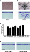
Figure 1
Figure 1
Invasive and tubulogenic properties of resting and SF/HGF-stimulated MDCK cells cultured atop a type I collagen matrix. A, Unstimulated MDCK cells on a collagen gel form a monolayer with cells confined to the gel surface (shown en face) in the top panel and as an H and E stained cross-section in the bottom panel. Bar, 100 μm. The double-headed arrow in the cross-section marks the boundaries of the collagen gel. B, After stimulation for 12 d with SF/HGF (50 ng/ml), MDCK cells invaded the collagen gel and formed a complex meshwork of branching tubular structures. C, MDCK invasion by SF/HGF-stimulated cells was unaffected by inhibitors of serine proteinases (200 μg/ml aprotinin, 100 μg/ml SBTI), cysteine proteinases (100 μM E-64), aspartate proteinases (50 μM pepstatin), aminopeptidases (10 μM bestatin), or serum plasminogen depletion (−Plg) in the course of a 12-d culture period. In contrast, BB-94 (5 μM) or recombinant TIMP-2 (200 ng/ml) completely blocked invasion. Results are shown as the mean number of invasive foci ± 1 SD in ten randomly selected fields in a single representative experiment of four performed. D, Representative cross-sections of MDCK cells stimulated with SF/HGF in the presence of either BB-94 or TIMP-2 for 12 d. Bar, 100 μm.
Figure 2
Figure 2
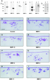
MDCK cells overexpressing secreted MMPs display normal invasive and morphogenic activities. A, Western blots showing the relative expression levels of the identified MMPs in control (C) and transfected (T) MDCK cells. Brackets indicate the positions of the glycosylated and nonglycosylated forms of proMMP-1, proMMP-3, and proMMP-11, respectively, whereas asterisks indicate active glycosylated and nonglycosylated forms of MMP-3, or active forms of MMP-2 and MMP-11. B–I, After stimulation with SF/HGF for 12 d, the invasion patterns of control transfectants (B) and the clones overexpressing each of the indicated MMPs (C–I) were indistinguishable. Neither control nor MMP transfectants displayed invasive activity in the absence of SF/HGF (data not shown). Bar, 100 μm.
Figure 3
Figure 3
Characterization of MT1-MMP–overexpressing MDCK cells. A, Proliferative response of wild-type (▪), vector control-transfected (•), full-length MT1-MMP (□), cytosolic tail-deleted MT1-MMP (MT1-MMPct; ▵), and soluble MT1-MMP (ΔMT1-MMP; ○) during a 7-d culture period. Cell number at each time point was measured in triplicate and the error bars represent the SD of a single representative experiment of three performed. The inset shows Western blots of MT1-MMP expression in either control or MT-MMP transfectants as indicated. Asterisks (*) mark the position of active MT1-MMP species whereas the arrowheads mark the position of a membrane-anchored, catalytically inactive fragment of MT1-MMP (Mr ∼43 kD; Lehti et al. 1998). MT1-MMP stable transfectants did not express detectable levels of MT2-MMP or MT3-MMP, as assessed by Western or Northern blot analysis, respectively. B, Epithelial morphology, E-cadherin staining, and SF/HGF-induced scattering responses of control or MT1-MMP–transfected cells cultured as described in Materials and Methods in either the absence or presence of SF/HGF. C, In the presence of SF/HGF, control vector-transfected MDCK cells cultured atop type I collagen gels for 3 d display widespread, but shallow foci of invasion (arrows) as viewed en face (top; bar, 100 μm), in cross-section (middle; bar, 100 μm), or by TEM (bottom; bar, 5 μm). In contrast, MDCK cells overexpressing MT1-MMP generated numerous large pits in the collagen matrix (arrows) extending well below the surface monolayer as viewed en face (top), in cross-section by light microscopy (middle) or by TEM (bottom). D, After 12 d, MT1-MMP overexpressing cells cultured in the absence of SF/HGF formed pits in the underlying collagen gel whereas SF/HGF-stimulated cells generated interconnecting cyst-like structures extending to the surface of the underlying polycarbonate membrane (compare to Fig. 1, A–D). Invasion by MT1-MMP transfectants was inhibited completely by TIMP-2, but not TIMP-1. Bar, 100 μm.
Figure 4
Figure 4
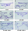
Regulation of MT1-MMP–dependent invasion and tubulogenesis. MDCK cell clones transfected with a control vector (A), MT1-MMP (B), MT1-MMP lacking the cytosolic domain (MT1-MMPct; C) or soluble MT1-MMP (ΔMT1-MMP; D) were cultured atop type I collagen gels for 12 d in complete medium with SF/HGF. Invasive activity of MT1-MMP overexpressing cells was not altered when cocultured in a 1:1 ratio with MDCK clones overexpressing MMP-2 (E) or MMP-13 (F). Bar, 100 μm.
Figure 5
Figure 5
Characterization of MT2-MMP and MT3-MMP overexpressing MDCK cells. A, Western blot analysis of MT2-MMP protein (arrow) in Triton X-114 cell extracts of transfected cells. The expression of the MT3-MMP transcript in transfected cells was shown by Northern blotting. A 36B4 control was run to confirm RNA integrity and equal loading. MT2-MMP stable transfectants did not express detectable levels of MT1-MMP or MT3-MMP (Western and Northern blot analysis, respectively), whereas MT3-MMP stable transfectants did not express detectable levels of MT1-MMP or MT2-MMP, as assessed by Western blot. B, Cell morphology and scattering response to SF/HGF was unaffected by either MT2-MMP or MT3-MMP overexpression (compare to Fig. 3 B). C, Unstimulated MT2-MMP–overexpressing cells formed a confluent monolayer on collagen as assessed in cross-sections (top left-hand corner) or by TEM (bottom left-hand corner). Bar, 1 μm. When stimulated with SF/HGF for 12 d, MT2-MMP transfectants formed a pseudostratified-like epithelial layer with basilar extensions intruding into the underlying collagen (arrows in light section and TEM in the middle panel of the bottom row). The extension of these processes into the collagen was blocked by BB-94. D, Unstimulated MT3-MMP transfectants formed a phenotypically normal confluent monolayer on type I collagen as depicted in a 12-d-old culture. When stimulated with SF/HGF, MT3-MMP overexpressors invaded shallowly and formed cyst-like structures (shown en face, and in cross-section) distinctly different from control cell invasion (see Fig. 1B and Fig. D). The invasive activity of MT3-MMP transfectants was blocked completely by BB-94. Bar, 100 μm. E, Invasion and tubulogenesis by control vector-, MT1-MMP–, MT2-MMP–, or MT3-MMP–transfected cells cultured atop Matrigel with SF/HGF for 12 d in complete media. Bar, 100 μm.
Figure 6
Figure 6
Invasion of peritoneal tissue by MDCK transfectants in vivo. A, Vector control-transfected MDCK cells cultured on the peritoneal matrix (arrow) did not express invasive activity after stimulation with SF/HGF for 12 d in complete media. Bar, 100 μm. The inset shows the peritoneal matrix by TEM after the mesothelial cell layers were removed. The tissue contains numerous collagen and elastin fibrils sandwiched between two discontinuous basement membranes. Bar, 1 μm. B, MT1-MMP–overexpressing cells degraded large blocks of tissue and invaded into the underlying collagen after stimulation with SF/HGF for 12 d. A small segment of remaining peritoneal tissue is indicated by the arrow. C, In the presence of BB-94, MT1-MMP transfectants failed to invade the peritoneum after 12 d in culture (arrow). D, TEM analysis of peritoneal tissue invasion by control or MT1-MMP transfectants cultured for 12 d with SF/HGF in the absence or presence of BB-94 as indicated. Only a portion of the peritoneum can be identified after culture with MT1-MMP–overexpressing cells (arrow). Bar, 1 μm. E, MT2-MMP–overexpressing cells completely degraded entire tracts of the peritoneum after stimulation with SF/HGF for 12 d as shown in an en face view of a live culture. The addition of BB-94 to these cultures blocked tissue dissolution and maintained an intact peritoneal matrix. Bar, 100 μm.
Figure 7
Figure 7
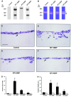
Collagen-invasive activity of MT-MMPs in transiently transfected COS-1 cells. A, Western blot analysis of COS-1 cells transiently transfected with FLAG epitope-tagged MT1-, MT2-, and MT3-MMP. Triton X-114 extracts were immunoblotted with anti-FLAG M2 mAb. B, MT-MMP activity in transiently transfected COS-1 cells was assessed by monitoring the activation of proMMP-2. The pro, intermediate, and active forms of MMP-2 are indicated by the arrow, clear arrowhead, and black arrowhead, respectively. C, Transverse light sections of control vector-transfected COS-1 cultured atop type I collagen gels for 5 d in the presence of SF/HGF were unable to express invasive activity, save for the extension of short processes (arrow) into the underlying substratum. COS-1 cells overexpressing MT1-MMP (D) or MT2-MMP (E) displayed invasive activity whereas MT3-MMP transfectants (F) were indistinguishable from control-transfected COS-1 cells. The number of invasive foci (G) and invasion depth (H) were assessed in transverse light sections of COS-1 cultures after a 5-d incubation period with SF/HGF in the absence or presence of BB-94 (shaded and open bars, respectively). Results are expressed as the mean ± 1 SD in ten randomly selected cross-sections in a single representative experiment of three performed. Bar, 50 μm.

Similar articles

Cited by

References

    1. Aimes R.T., Quigley J.P. Matrix metalloproteinase-2 is an interstitial collagenase. J. Biol. Chem. 1995;270:5872–5876. - PubMed
    1. Belien A.T.J., Paganetti P.A., Schwab M.E. Membrane-type 1 matrix metalloproteinase (MT1-MMP) enables invasive migration of glioma cells in central nervous system white matter. J. Cell Biol. 1999;144:373–384. - PMC - PubMed
    1. Benbow U., Schoenermark M.P., Mitchell T.I., Rutter J.L., Shimokawa K., Nagase H., Brinckerhoff C.E. A novel host/tumor cell interaction activates matrix metalloproteinase 1 and mediates invasion through type I collagen. J. Biol. Chem. 1999;274:25371–25378. - PubMed
    1. Birchmeier C., Birchmeier W. Molecular aspects of mesenchymal–epithelial interactions. Annu. Rev. Cell Biol. 1993;9:511–540. - PubMed
    1. Birkedal-Hansen H. Catabolism and turnover of collagenscollagenases. Meth. Enzymol. 1987;144:140–167. - PubMed

Publication types

MeSH terms