. 2000 Jun;12(6):917-32.
doi: 10.1105/tpc.12.6.917.

Reduced levels of chloroplast FtsH protein in tobacco mosaic virus-infected tobacco leaves accelerate the hypersensitive reaction

Affiliations

Reduced levels of chloroplast FtsH protein in tobacco mosaic virus-infected tobacco leaves accelerate the hypersensitive reaction

S Seo et al. Plant Cell. 2000 Jun.

Abstract

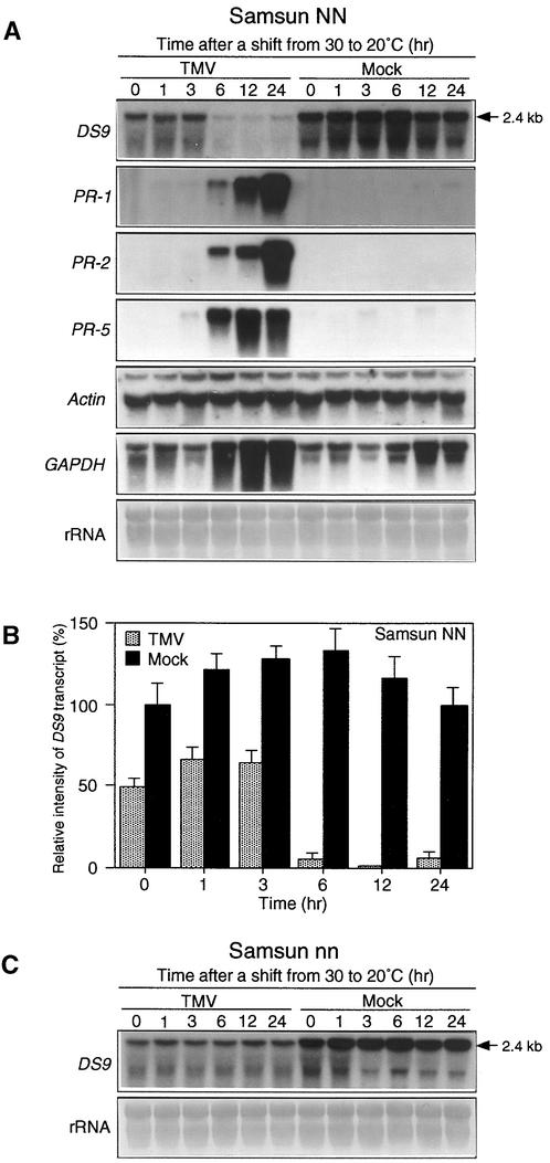
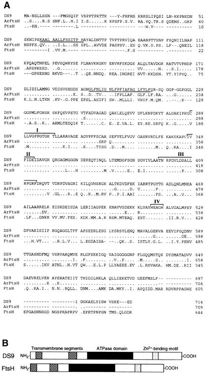
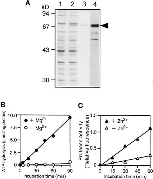
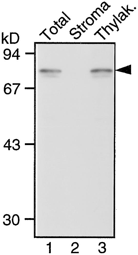
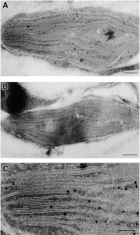
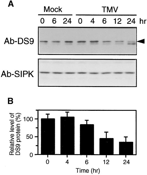
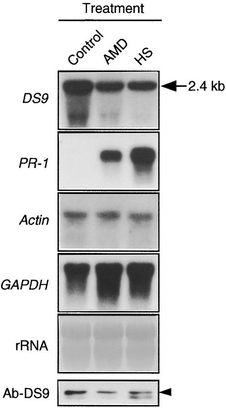
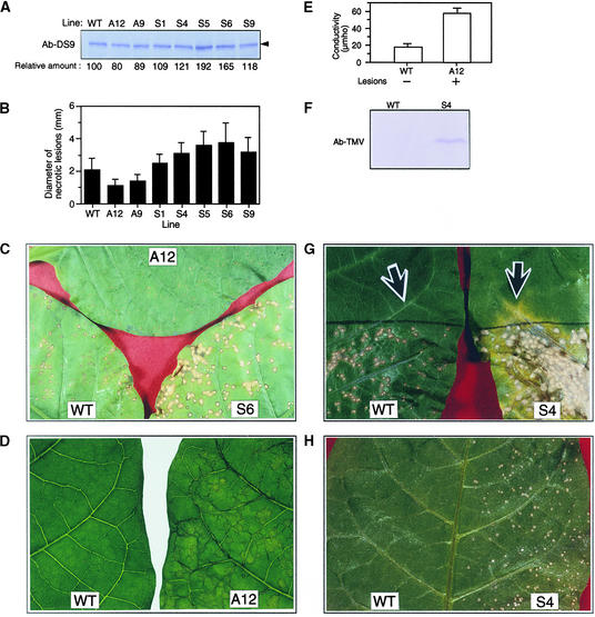
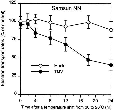
In tobacco cultivars resistant to tobacco mosaic virus (TMV), infection results in the death of the infected cells accompanying the formation of necrotic lesions. To identify the genes involved in this hypersensitive reaction, we isolated the cDNA of tobacco DS9, the transcript of which decreases before the appearance of necrotic lesions. The DS9 gene encodes a chloroplastic homolog of bacterial FtsH protein, which serves to maintain quality control of some cytoplasmic and membrane proteins. A large quantity of DS9 protein was found in healthy leaves, whereas the quantity of DS9 protein in infected leaves decreased before the lesions appeared. In transgenic tobacco plants containing less and more DS9 protein than wild-type plants, the necrotic lesions induced by TMV were smaller and larger, respectively, than those on wild-type plants. These results suggest that a decrease in the level of DS9 protein in TMV-infected cells, resulting in a subsequent loss of function of the chloroplasts, accelerates the hypersensitive reaction.


Figures

Figure 1.
Figure 1.
RNA Gel Blot Analysis of the DS9, PR-1, PR-2, PR-5, Actin, and GAPDH Genes in TMV- and Mock-Inoculated Tobacco Leaves after Temperature Shift. Detached healthy leaves of 50-day-old Samsun NN and 70-day-old Samsun nn tobacco plants were inoculated with TMV (10 μg/mL) or buffer only (Mock), incubated at 30°C for 40 hr, and then shifted to 20°C at time 0. (A) Leaves from Samsun NN tobacco plants were harvested at the indicated times after the temperature shift and used for total RNA extraction. Aliquots of 20 μg of total RNA per lane were fractionated by gel electrophoresis, transferred to nylon membranes, and successively subjected to hybridization with the indicated radioactively labeled probes after stripping the former probe. To standardize RNA loading, the blot was stained with methylene blue (rRNA). The 2.4-kb RNA represents the length of the DS9 gene. (B) Relative intensity of DS9 transcripts as shown in (A). The amounts of DS9 transcript are expressed as a ratio with that present in the control sample (0 hr after the temperature shift of mock-inoculated leaves), which was set equal to 100%. Values are the mean ±sd from four independent experiments. (C) Leaves from Samsun nn tobacco plants were harvested at the indicated times after the temperature shift and used for total RNA extraction. RNA gel blot hybridization was performed as described in (A), using the DS9 cDNA probe. The experiment was repeated twice with similar results. The 2.4-kb RNA represents the length of the DS9 gene.
Figure 2.
Figure 2.
Comparison of the Deduced Amino Acid Sequences of DS9, Arabidopsis FtsH Homolog, and E. coli FtsH. (A) Lines at top, center, and below represent the amino acid sequence of DS9 (GenBank accession number AB017480), an Arabidopsis FtsH homolog (Lindahl et al., 1996), and E. coli FtsH (Tomoyasu et al., 1993a), respectively. The underlined amino acids represent putative membrane-spanning regions. Dots represent identical amino acid residues, and dashes indicate gaps introduced to maximize alignment. The roman numerals indicate two regions of ATP binding motif (I and II), a second region of homology(III), and a Zn2+ binding motif (IV). (B) Diagrammatic alignment of the primary structures of DS9 and E. coli FtsH. Hatched, black, and stippled boxes represent two putative transmembrane segments, the ATPase domain, and the putative Zn2+ binding motif, respectively. The ATPase domain contains an ATP binding motif and a second region of homology (Confalonieri and Duguet, 1995). The putative Zn2+ binding motif is HEXXH, where X indicates nonconserved amino acid residues.
Figure 3.
Figure 3.
Expression and Enzymatic Activity of the Recombinant DS9 Protein. (A) Expression and purification of the recombinant HisDS9 protein from E. coli cells. Total cellular extracts prepared from cells not treated with IPTG (lane 1) and IPTG-treated cells (lane 2) from E. coli were separated by electrophoresis on a 10% SDS–polyacrylamide gel; the gel was stained with Coomassie Brilliant Blue R 250. The recombinant HisDS9 protein (arrowhead) was purified by affinity chromatography (lane 3) and subjected to protein gel blot analysis (lane 4) with antibodies raised against DS9ΔN. Size markers are indicated at left in kilodaltons. (B) Mg2+-dependent ATP-hydrolyzing activity in the recombinant HisDS9 protein. Reactions were performed in the presence (closed circles) or absence (open circles) of 5 mM MgCl2. (C) Zn2+-stimulated casein-hydrolyzing activity in the recombinant HisDS9 protein. Fluorescein isothiocyanate–labeled casein was used as the substrate. Reactions were performed in the presence (closed triangles) or absence (open triangles) of 12.5 μM zinc acetate.
Figure 4.
Figure 4.
Protein Gel Blot Analysis of the DS9 Protein in Tobacco Leaf Tissue. Total protein extracts (lane 1), a stroma fraction (lane 2), and a thylakoid (Thylak.) fraction (lane 3) prepared from healthy leaves of Samsun NN tobacco plants were subjected to protein gel blot analysis with antibodies raised against DS9ΔN. Lanes 1, 2, and 3 contain total protein extracts containing 20 μg of total protein, the stroma fraction equivalent to the volume of thylakoid membrane fraction used, and the thylakoid membrane fraction containing 5 μg of chlorophyll, respectively. The arrowhead indicates the relative molecular masses of ∼78 kD. Size markers are indicated at left in kilodaltons.
Figure 5.
Figure 5.
Immunoelectron Microscopy in Frozen Sections of Leaf Tissue. (A) Immunogold localization of the DS9 protein in a tobacco mesophyll cell treated with antibodies raised against DS9ΔN and 10-nm gold-conjugated anti–rabbit IgG. formula image. (B) A tobacco mesophyll cell treated with preimmune serum and 10-nm gold-conjugated anti–rabbit IgG. formula image. (C) Greater magnification of (A). formula image.
Figure 6.
Figure 6.
Protein Gel Blot Analysis of the DS9 Protein in TMV- and Mock-Inoculated NN Tobacco Leaves after the Temperature Shift. (A) Detached healthy leaves of Samsun NN tobacco plants were inoculated with TMV (10 μg/mL) or buffer only (Mock) and shifted from 30 to 20°C at time 0. Leaves were harvested at the indicated times after the temperature shift and used for protein extraction. Twenty micrograms of total protein was immunoblotted with antibodies raised against DS9ΔN (Ab-DS9). The arrowhead indicates the relative molecular masses of ∼78 kD. The blots were reprobed with antibodies (Ab-SIPK) against a synthetic peptide corresponding to the N-terminal 24–amino acid sequence of SIPK as a loading control. (B) Relative amount of the DS9 protein in TMV-inoculated leaves as shown in (A), expressed as a ratio with that present in the control sample (0 hr after the temperature shift), which was set equal to 100%. Values are the mean ±sd from three independent experiments.
Figure 7.
Figure 7.
RNA Gel Blot Analysis of the DS9, PR-1, Actin, and GAPDH Genes and Protein Gel Blot Analysis of the DS9 Protein in TMV-Inoculated Leaves Treated with Actinomycin D or Heat Shock. Detached leaves of Samsun NN tobacco plants were inoculated with TMV (10 μg/mL), incubated at 30°C for 40 hr, treated with actinomycin D (AMD; 200 μg per gram [fresh weight] of leaf tissues) or subjected to heat shock (HS) at 50°C for 2 min, reincubated at 30°C for 18 hr, and then used for total RNA extraction and protein extraction. Aliquots of RNA (20 μg per lane) were subjected to RNA gel blot analysis with the indicated cDNA probes. The 2.4-kb RNA (arrow) represents the length of the DS9 gene. Twenty micrograms of total protein was subjected to protein gel blot analysis with antibodies raised against DS9ΔN (Ab-DS9). TMV-inoculated leaves that were not treated with actinomycin D or subjected to heat shock were used as the control. The arrowhead indicates the relative molecular masses of ∼78 kD.
Figure 8.
Figure 8.
Analysis of Transgenic Tobacco Plants Containing Various Amounts of the DS9 Protein. The upper, fully expanded healthy leaves of 3-month-old wild-type Samsun NN (WT) and transgenic tobacco plants (S4, S6, and A12) were used for each experiment. (A) Total protein was extracted from the wild-type and transgenic tobacco plants carrying antisense (lines A12 and A9) and sense (lines S1, S4, S5, S6, and S9) constructs. Twenty-five micrograms of total protein from each line was subjected to protein gel blot analysis with antibodies raised against DS9ΔN (Ab-DS9). The numbers below the gel indicate the amount of DS9 protein expressed as a ratio of that present in the wild type, which was set equal to 100%. The arrowhead indicates the relative molecular masses of ∼78 kD. (B) Leaves were inoculated with TMV (2 μg/mL) and incubated at 20°C under 120 μmol of photons m−2 sec−1 fluorescence illumination. In each line, the diameter of 60 local lesions 5 days after inoculation was measured with a stereoscopic microscope. Each bar represents the mean ±sd. (C) Necrotic lesions that had formed on the leaves of a wild-type plant and the S6 and A12 lines, as described in (B). (D) Leaves of line A12 that had been inoculated with TMV (10 μg/mL) and incubated for 40 hr at 30°C were shifted to 20°C, where they were incubated for 3.5 hr and returned to 30°C. As a control, leaves of wild-type plants were used. A photograph was taken 24 hr after returning the leaves to 30°C. The experiment was repeated twice with similar results. (E) After photographing the leaves as shown in (D), five leaf discs were punched from each leaf and measured for electrolyte leakage. Values are the mean ±sd from three independent experiments. +, lesions appeared; −, no lesions appeared. (F) Protein gel blot analysis of TMV coat protein. The experiment was repeated twice with similar results. Ab-TMV, anti-TMV antibody. (G) The bottom half of leaves of wild-type and S4 plants were inoculated with TMV (2 μg/mL) and incubated at 20°C. Seven days after inoculation, the leaves were photographed. Two discs (0.7 cm in diameter each) from each leaf were punched out from the sites indicated by the arrows, which were 1.5 cm away from the black borderline that indicates the edge of the inoculated region. One of the two discs from each leaf was used for protein gel blot analysis with the anti-TMV antibody (F), and the other was used for the TMV bioassay (H). (H) TMV bioassay. For the TMV bioassay, crude leaf extracts from wild-type plants and the S4 line were inoculated onto the left and right half, respectively, of Samsun NN tobacco leaves. Five days after inoculation, the leaf was photographed. The experiment was repeated twice with similar results.
Figure 9.
Figure 9.
Changes in the Rate of Photosynthetic Electron Transport after the Temperature Shift. Detached healthy leaves of Samsun NN tobacco plants were inoculated with TMV (10 μg/mL; closed circles) or buffer only (Mock; open circles) and shifted from 30 to 20°C at time 0. Thylakoid membranes were isolated from leaves harvested at the indicated times after the temperature shift and were used to measure the whole-chain electron transport. Activity was expressed as the percentage of the control sample (0 hr after the temperature shift of mock-inoculated leaves). Values are the mean ±sd of three independent measurements.

Similar articles

Cited by

References

    1. Adam, Z. (1996). Protein stability and degradation in chloroplasts. Plant Mol. Biol. 32, 773–783. - PubMed
    1. Akiyama, Y., Shirai, Y., and Ito, K. (1994). Involvement of FtsH in protein assembly into and through the membrane. II. Dominant mutations affecting FtsH functions. J. Biol. Chem. 269, 5225–5229. - PubMed
    1. Akiyama, Y., Kihara, A., and Ito, K. (1996. a). Subunit a of proton ATPase F0 sector is a substrate of the FtsH protease in Escherichia coli. FEBS Lett. 399, 26–28. - PubMed
    1. Akiyama, Y., Kihara, A., Tokuda, H., and Ito, K. (1996. b). FtsH (HflB) is an ATP-dependent protease selectively acting on SecY and some other membrane proteins. J. Biol. Chem. 271, 31196–31201. - PubMed
    1. Allen, L.J., MacGregor, K.B., Koop, R.S., Bruce, D.H., Karner, J., and Bown, A.W. (1999). The relationship between photosynthesis and a mastoparan-induced hypersensitive response in isolated mesophyll cells. Plant Physiol. 119, 1233–1241. - PMC - PubMed

Publication types

MeSH terms

Associated data

LinkOut - more resources