. 2000 Jun;12(6):933-47.
doi: 10.1105/tpc.12.6.933.

Syncytial-type cell plates: a novel kind of cell plate involved in endosperm cellularization of Arabidopsis

Affiliations

Syncytial-type cell plates: a novel kind of cell plate involved in endosperm cellularization of Arabidopsis

M Otegui et al. Plant Cell. 2000 Jun.

Abstract

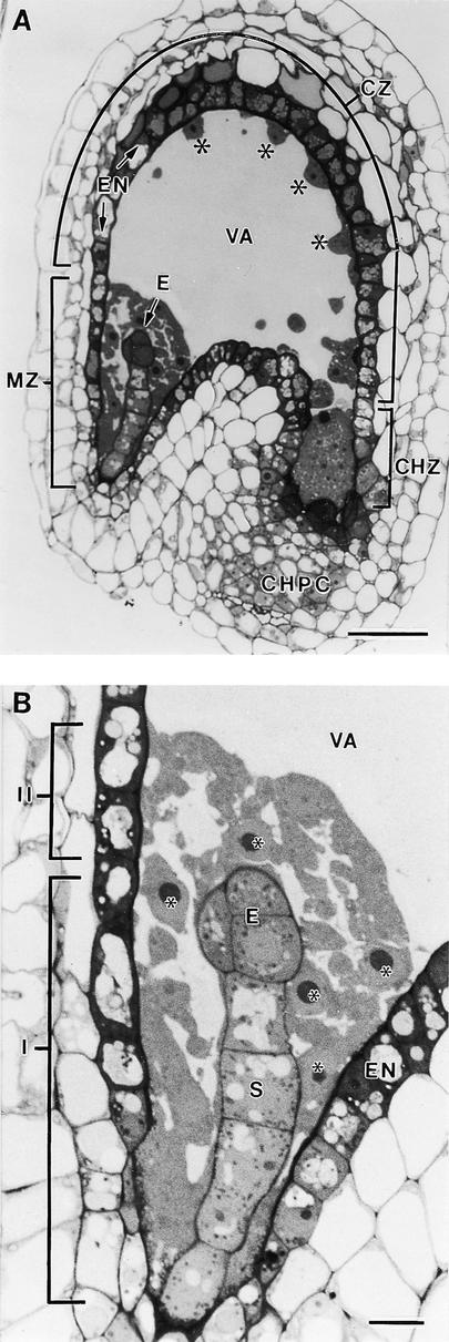
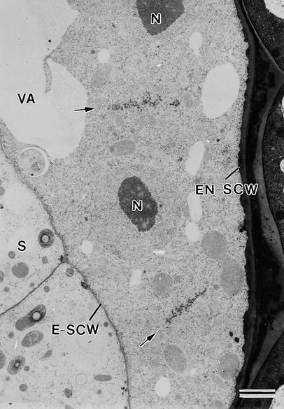
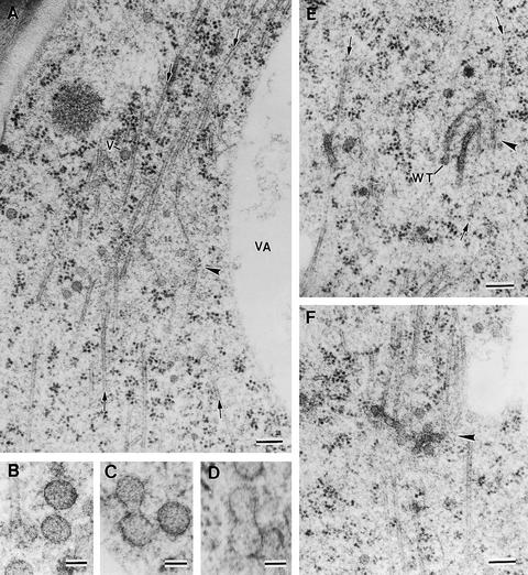
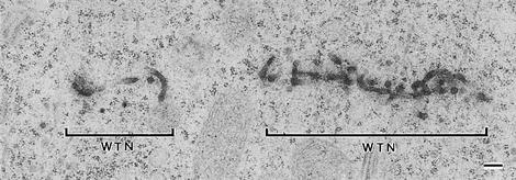
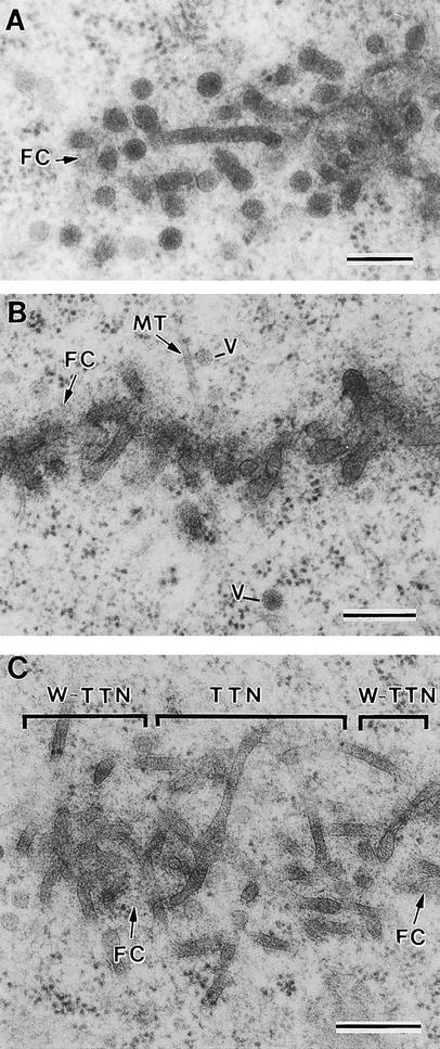
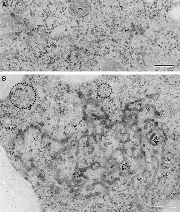
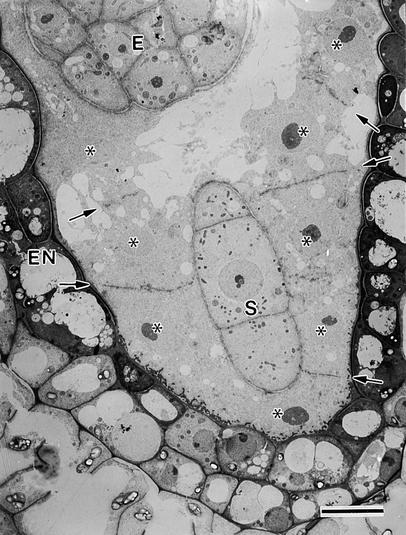
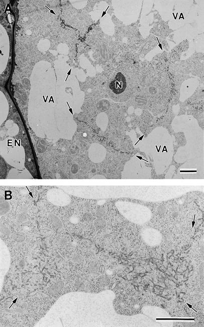
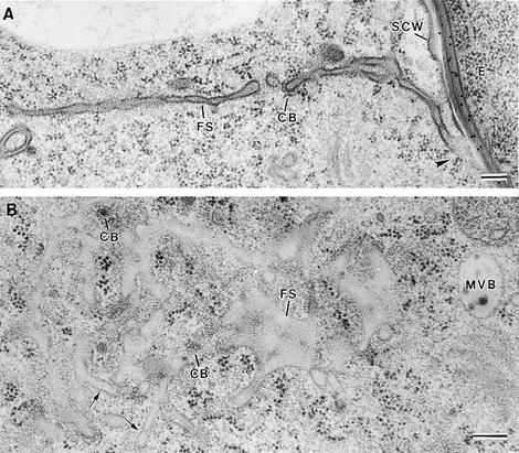
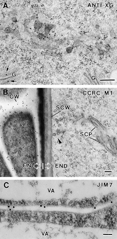
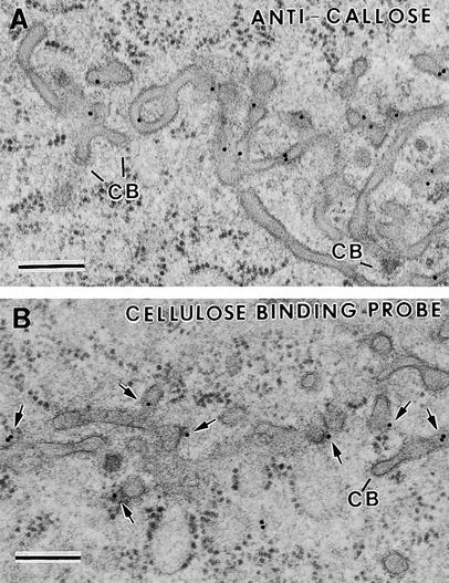
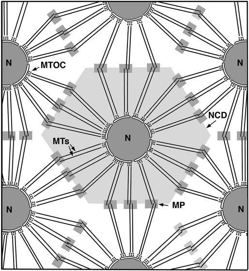
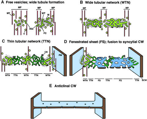
Cell wall formation in the syncytial endosperm of Arabidopsis was studied by using high-pressure-frozen/freeze-substituted developing seeds and immunocytochemical techniques. The endosperm cellularization process begins at the late globular embryo stage with the synchronous organization of small clusters of oppositely oriented microtubules ( approximately 10 microtubules in each set) into phragmoplast-like structures termed mini-phragmoplasts between both sister and nonsister nuclei. These mini-phragmoplasts produce a novel kind of cell plate, the syncytial-type cell plate, from Golgi-derived vesicles approximately 63 nm in diameter, which fuse by way of hourglass-shaped intermediates into wide ( approximately 45 nm in diameter) tubules. These wide tubules quickly become coated and surrounded by a ribosome-excluding matrix; as they grow, they branch and fuse with each other to form wide tubular networks. The mini-phragmoplasts formed between a given pair of nuclei produce aligned tubular networks that grow centrifugally until they merge into a coherent wide tubular network with the mini-phragmoplasts positioned along the network margins. The individual wide tubular networks expand laterally until they meet and eventually fuse with each other at the sites of the future cell corners. Transformation of the wide tubular networks into noncoated, thin ( approximately 27 nm in diameter) tubular networks begins at multiple sites and coincides with the appearance of clathrin-coated budding structures. After fusion with the syncytial cell wall, the thin tubular networks are converted into fenestrated sheets and cell walls. Immunolabeling experiments show that the cell plates and cell walls of the endosperm differ from those of the embryo and maternal tissue in two features: their xyloglucans lack terminal fucose residues on the side chain, and callose persists in the cell walls after the cell plates fuse with the parental plasma membrane. The lack of terminal fucose residues on xyloglucans suggests that these cell wall matrix molecules serve both structural and storage functions.


Figures

Figure 1.
Figure 1.
Light Microscopy of a Longitudinal Section of a Developing Arabidopsis Seed. (A) Overview. Three distinct regions can be recognized in the developing endosperm: the micropylar zone (MZ) in which the embryo (E) is located, the central zone (CZ), and the chalazal zone (CHZ). The central zone consists of a thin peripheral layer of cytoplasm with regularly spaced nuclei (asterisks) and a large central vacuole (VA). The chalazal zone is partially enveloped by the chalazal proliferating cells (CHPC). The endosperm is surrounded by the endothelium (EN). formula image. (B) Detail of the micropylar zone. Two different domains can be distinguished according to their relation with the central vacuole. One cytoplasmic domain (I) surrounds part of the embryo proper (E) and the suspensor (S) but does not abut the central vacuole (VA). The other domain (II) is bordered by the remaining part of the embryo proper and the central vacuole. Endosperm nuclei are indicated by asterisks. EN, endothelium. formula image.
Figure 2.
Figure 2.
Syncytial-Type Cell Plates Form between Nonsister Nuclei at the Onset of Cellularization in the Micropylar Zone. Cell plates are aligned fairly perpendicular to the syncytial cell walls facing the embryo (E-SCW) and the endothelium (EN-SCW) (anticlinal orientation). Arrows indicate syncytial-type cell plates. N, nuclei; S, suspensor; VA, vacuole. formula image.
Figure 3.
Figure 3.
Examples of Assembly of Mini-Phragmoplasts at the Onset of Endosperm Cellularization and Vesicle Fusion Steps. (A) Mini-phragmoplast (arrowhead) consisting of two sets of opposed microtubules (arrows). Golgi-derived vesicles of ∼63 nm (V) are associated with the mini-phragmoplast microtubules. VA, vacuole. (B) to (D) Vesicle fusion steps showing two Golgi-derived vesicles together (B), an hourglass-shaped intermediate resulting from the fusion of two vesicles (C), and a transient structure between an hourglass-shaped intermediate and a wide tubule (D). (E) Wide tubules (WT) are aligned in a parallel orientation to the microtubules (arrows) in a mini-phragmoplast (arrowhead). (F) Fused and branched wide tubules in a mini-phragmoplast (arrowhead). formula image; formula image.
Figure 4.
Figure 4.
Two Wide Tubular Networks Formed in Two Adjacent Mini-Phragmoplasts. WTN, wide tubular network. formula image.
Figure 5.
Figure 5.
Developmental Stages in Syncytial-Type Cell Plate Formation. (A) Early stages in vesicle aggregation and wide tubule consolidation. Wide tubules are already covered by a fuzzy coat (FC). (B) Wide tubular network covered by a dense fuzzy coat. Some microtubules (MT) and vesicles (V) are associated with the wide tubular network. (C) Different membranous domains in a syncytial-type cell plate showing the transition from the wide tubular network to the thin tubular network (W-TTN) and some noncoated thin tubules corresponding to the thin tubular network (TTN). Note that the fuzzy coat is disassembled during the conversion of wide tubules into thin tubules. formula image.
Figure 6.
Figure 6.
Syncytial-Type Phragmoplast and Cell Plate. (A) Transverse section of a syncytial-type cell plate. Wide tubules (WT) are seen in the tubular network. Golgi-derived vesicles (V) are associated with mini-phragmoplast microtubules (arrows) at the growing cell plate margin. Note that vesicles do not seem to fuse directly with the cell plate network but instead fuse first with each other (arrowhead). A mitochondrion (M) and a Golgi stack (G) seem close to the cell plate. (B) Tangential section of a syncytial-type cell plate. Mini-phragmoplasts consisting of 4 to 12 microtubules (dotted circles) are located discontinuously along the margin of the growing cell plate. Some single microtubules (arrows) are interspersed among tubular elements. The nonuniform growth and maturation of these cell plates are evidenced by the presence of thin tubules (TT) between wide tubular domains (WT). Wide tubule to thin tubule transition sites are indicated by open arrows. Clathrin-coated membrane buds (CB) are associated with thin tubules. FC, fuzzy coat. formula image.
Figure 7.
Figure 7.
Syncytial-Type Cell Plates Ready to Fuse with a Syncytial Cell Wall in the Cytoplasmic Domain That Does Not Abut the Central Vacuole. See Figure 1 for an overview. Endosperm nuclei are indicated by asterisks. Syncytial-type cell plates are marked by arrows. E, embryo proper; EN, endothelium; S, suspensor. formula image.
Figure 8.
Figure 8.
Fusion of Syncytial-Type Cell Plates. (A) Syncytial-type cell plates (arrows) forming simultaneously around an endosperm nucleus (N). All of the cell plates exhibit a tubular network architecture, and where they have joined, they give rise to Y- or T-shaped junctional domains. EN, endothelium; VA, vacuole. (B) Detail of the network (arrows) resulting from the fusion of several syncytial-type cell plates. formula image.
Figure 9.
Figure 9.
Fusion of Syncytial-Type Cell Plates with the Syncytial Cell Wall. (A) Transverse section of a flattened fenestrated sheet (FS) that has already fused (arrowhead) with the syncytial cell wall (SCW) facing the embryo (E). (B) Tangential section of a syncytial-type cell plate at the fenestrated sheet stage. Some tubules (arrows) are still observed. Clathrin-coated structures associated with the cell plate and multivesicular bodies (MVB) are commonly observed at this stage. CB, clathrin-coated bud; FS, fenestrated sheet. formula image.
Figure 10.
Figure 10.
Labeling Cell Plates and Cell Walls with Anti-Xyloglucan and Anti-Pectic Polysaccharide Antibodies. (A) Transverse section of a wide tubular network and Golgi stacks (G) labeled with anti-xyloglucan antibody (ANTI-XG). Arrows indicate labeled Golgi stack. (B) Section labeled with CCRC-M1 (anti-xyloglucan fucose residues) antibodies. Note the lack of label on the syncytial-type cell plate (SCP) and the syncytial cell wall (SCW) and the strong labeling of the adjacent endothelium cell walls (CW). The arrowhead indicates the site of fusion between the syncytial-type cell plate and the syncytial cell wall. EN, endothelium; END, endosperm. (C) Transverse section of a recently formed anticlinal cell wall labeled with JIM7. VA, vacuole. formula image.
Figure 11.
Figure 11.
Labeling Cell Plates and Cell Walls with Anti-Callose Antibodies and CBHI-Gold Probe. (A) Tangential section of a thin tubular network and some wide tubules labeled with anti-callose antibodies. Two clathrin-coated membrane structures (CB) are seen budding off some tubules in the cell plate. (B) Transverse section of a wide tubular network labeled with CBHI-gold (arrows). formula image.
Figure 12.
Figure 12.
Diagram Showing Microtubule Organization in the Syncytial Endosperm at the Onset of Cellularization and the Putative Origin of the Mini-Phragmoplasts from Opposing Overlapping Clusters of Microtubules. Microtubules radiating from microtubule-organizing centers (MTOCs) in the nuclear envelope define the nuclear cytoplasmic domains (NCDs) and the sites of syncytial-type cell plate formation. MP, mini-phragmoplasts; N, nucleus.
Figure 13.
Figure 13.
Stages of Syncytial-Type Cell Plate and Cell Wall Formation. The model depicts the main stages of syncytial-type cell plate and cell wall formation in Arabidopsis endosperm, with a newly formed anticlinal cell wall connecting the syncytial cell walls facing the endothelium and the embryo (E). CB, clathrin bud; CW, cell wall; FC, fuzzy coat; HGI, hourglass intermediate; M, ribosome-excluding matrix; MP, mini-phragmoplast; MT, microtubules; PM, plasma membrane; SCW, syncytial cell wall; V, vesicles; WT, wide tubules.

Comment in

  • Cytokinesis: the art of partitioning.
    Jürgens G. Jürgens G. Plant Cell. 2000 Jun;12(6):827-8. doi: 10.1105/tpc.12.6.827. Plant Cell. 2000. PMID: 10970140 Free PMC article. No abstract available.

References

    1. Baskin, T.I., and Cande, W.Z. (1990). The structure and function of the mitotic spindle in flowering plants. Annu. Rev. Plant Physiol. 41, 277–315.
    1. Berger, F. (1999). Endosperm development. Curr. Opin. Plant Biol. 2, 28–32. - PubMed
    1. Brown, R.C., and Lemmon, B.E. (1991). The cytokinesis apparatus in meiosis: Control of division plane in the absence of a preprophase band of microtubules. In The Cytoskeletal Basis of Plant Growth and Form, W.C. Lloyd, ed (London: Academic Press), pp. 259–273.
    1. Brown, R.C., and Lemmon, B.E. (1992). Cytoplasmic domain: A model for spatial control of cytokinesis in reproductive cells of plants. EMSA Bull. 22, 48–53.
    1. Brown, R.C., Lemmon, B.E., and Olsen, O.-A. (1994). Endosperm development in barley: Microtubule involvement in the morphogenetic pathway. Plant Cell 6, 1241–1252. - PMC - PubMed

Publication types

LinkOut - more resources