. 2000 Jun;12(6):979-90.
doi: 10.1105/tpc.12.6.979.

The kinesin-like calmodulin binding protein is differentially involved in cell division

Affiliations

The kinesin-like calmodulin binding protein is differentially involved in cell division

J W Vos et al. Plant Cell. 2000 Jun.

Abstract

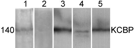
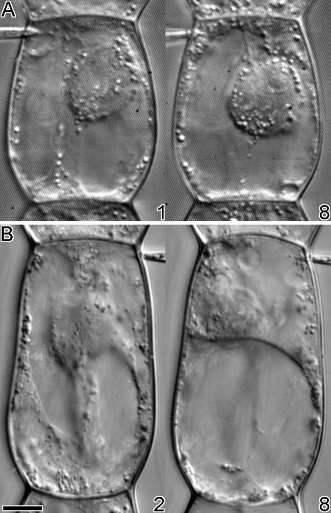
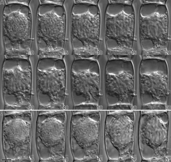
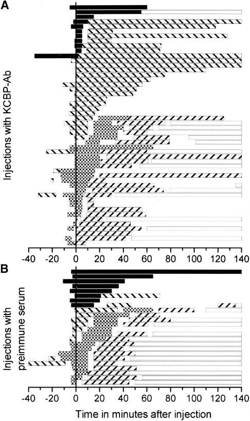
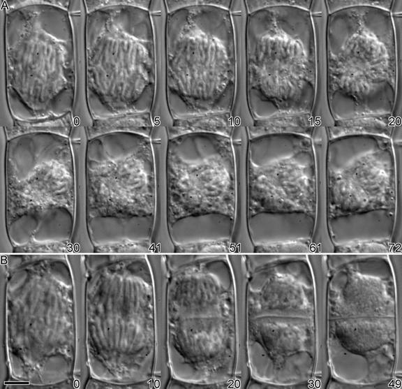
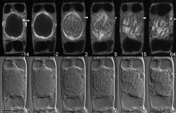
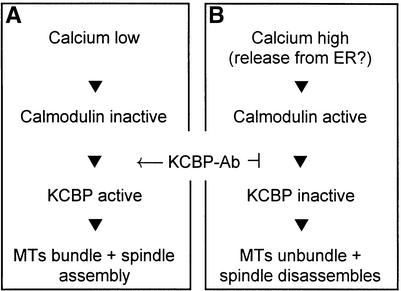
The kinesin-like calmodulin (CaM) binding protein (KCBP), a minus end-directed microtubule motor protein unique to plants, has been implicated in cell division. KCBP is negatively regulated by Ca(2)+ and CaM, and antibodies raised against the CaM binding region inhibit CaM binding to KCBP in vitro; therefore, these antibodies can be used to activate KCBP constitutively. Injection of these antibodies into Tradescantia virginiana stamen hair cells during late prophase induces breakdown of the nuclear envelope within 2 to 10 min and leads the cell into prometaphase. However, mitosis is arrested, and the cell does not progress into anaphase. Injection of antibodies later during cell division has no effect on anaphase transition but causes aberrant phragmoplast formation and delays the completion of cytokinesis by approximately 15 min. These effects are achieved without any apparent degradation of the microtubule cytoskeleton. We propose that during nuclear envelope breakdown and anaphase, activated KCBP promotes the formation of a converging bipolar spindle by sliding and bundling microtubules. During metaphase and telophase, we suggest that its activity is downregulated.


Figures

Figure 1.
Figure 1.
Protein Gel Blot Analysis of KCBP. Total protein preparations of T. virginiana inflorescences (lanes 1 and 2), tobacco BY-2 suspension cell culture (lane 3), Lidium longiflorum anthers (lane 4), and Arabidopsis suspension cell culture (lane 5) were separated on SDS–polyacrylamide gels, blotted, and incubated with 1:500 dilutions of affinity-purified KCBP-Ab or a 1:500 dilution of unpurified preimmune serum (lane 2). In lanes 1 and 3 to 5, a single band at 140 kD was detected, indicating the presence of endogenous KCBP.
Figure 2.
Figure 2.
Injection of Antibodies into T. virginiana Stamen Hair Cells during Interphase. (A) Eight minutes after microinjection with KCBP-Ab, no effects on cytoplasmic streaming or viability were visible. (B) Within 8 min after injection with CaM-Ab, cytoplasmic streaming ceased, cytoplasmic strands collapsed, and most of the cytoplasm was localized to the lower part of the cell. The time in minutes after injection is given at the bottom right of each photograph. formula image.
Figure 3.
Figure 3.
Injection of Antibodies during Late Prophase. (A) In a KCBP-Ab–injected cell, the nuclear envelope broke down between 5 and 10 min after injection. Initially, the chromosomes lined up at the metaphase plate, but at ∼40 min after injection they started losing their orientation. After an hour, the cell was still arrested in prometaphase or metaphase. (B) As the control, a late-prophase cell was injected with affinity-purified preimmune serum. Nuclear envelope breakdown did not occur until ∼20 min after injection, after which the cell completed cell division normally (data not shown). The time in minutes after injection is shown at the bottom right of each photograph. formula image.
Figure 4.
Figure 4.
Mitosis Transition Times and Observation Times. (A) Dividing cells injected with KCBP-Ab. (B) Dividing cells injected with preimmune serum. In both (A) and (B), the microinjections are ordered according to the length of time spent in a mitotic phase after injection at time 0. Observations >140 min were cut off. Each bar represents the observation time (not necessarily the beginning and end of a mitotic phase), except for the bars representing interphase, which were continued to show the observed completion of cell division. Clearly visible are the differences in the lengths of prophase and metaphase and the variation in the length of telophase between KCBP-Ab–injected cells and their controls. Prophase is indicated with black bars; metaphase with striped (\) bars; anaphase with stippled bars; telophase with striped (//) bars; and interphase with open bars.
Figure 5.
Figure 5.
Injection of Antibody during Anaphase. (A) Injection with KCBP-Ab did not affect anaphase; the chromatids separated normally (0 to 15 min after injection). At the end of anaphase, no clear zone or initiation of the cell plate was visible (20 min). The whole spindle apparatus tilted 45° (30 min), and the cell was arrested in telophase for >2 hr. (B) The cell was injected with preimmune serum and progressed through anaphase and telophase normally. At the end of anaphase (20 min), a phragmoplast formed and vesicles coalesced to form a cell plate. The new cell wall was formed in ∼30 min (49 min after injection). The time in minutes after injection is shown at the bottom right of each photograph. formula image.
Figure 6.
Figure 6.
Microtubule Distribution after Injection with KCBP-Ab. A cell in mid-prophase was injected with rhodamine-labeled bovine tubulin; after 32 min, the same cell was injected with KCBP-Ab. The bovine tubulin was incorporated into the endogenous microtubule arrays of the preprophase band and the perinuclear basket. Within 2 min after injection of KCBP-Ab, the nuclear envelope broke down, the microtubules entered the nuclear area, and a bipolar spindle started to form. Although chromosomes lined up at the metaphase plate and kinetochore microtubule bundles were visible (15 and 17 min after injection), after 1 hr the cell was still arrested. (Top) Tubulin fluorescence. (Bottom) Differential interference contrast images of the same cell. The time in minutes after KCBP-Ab injection is shown at the bottom right of each photograph. formula image.
Figure 7.
Figure 7.
Simplified Functional Model of KCBP during Cell Division. (A) Diagram of a cell at nuclear envelope breakdown or anaphase. (B) Representation of a cell in prometaphase, metaphase, or telophase. KCBP-Ab inhibits the deactivation of KCBP and bypasses regulation by Ca2+-CaM. ER, endoplasmic reticulum; MTs, microtubules.

Comment in

  • Cytokinesis: the art of partitioning.
    Jürgens G. Jürgens G. Plant Cell. 2000 Jun;12(6):827-8. doi: 10.1105/tpc.12.6.827. Plant Cell. 2000. PMID: 10970140 Free PMC article. No abstract available.

Similar articles

Cited by

References

    1. Asada, T., and Collings, D. (1997). Molecular motors in higher plants. Trends Plant Sci. 2, 29–37.
    1. Asada, T., and Shibaoka, H. (1994). Isolation of polypeptides with microtubule-translocating activity from phragmoplasts of tobacco BY-2 cells. J. Cell Sci. 107, 2249–2257. - PubMed
    1. Asada, T., Kuriyama, R., and Shibaoka, H. (1997). TKRP125, a kinesin-related protein involved in the centrosome-independent organization of the cytokinetic apparatus in tobacco BY-2 cells. J. Cell Sci. 110, 179–189. - PubMed
    1. Bajer, A.S., and Molè-Bajer, J. (1986). Reorganization of microtubules in endosperm cells and cell fragments of the higher plant Haemanthus in vivo. J. Cell Biol. 102, 263–281. - PMC - PubMed
    1. Bowser, J., and Reddy, A.S.N. (1997). Localization of a kinesin-like calmodulin-binding protein in dividing cells of Arabidopsis and tobacco. Plant J. 12, 1429–1437. - PubMed

Publication types

LinkOut - more resources