. 2000 Jun;12(6):991-1002.
doi: 10.1105/tpc.12.6.991.

Arabidopsis MutS homologs-AtMSH2, AtMSH3, AtMSH6, and a novel AtMSH7-form three distinct protein heterodimers with different specificities for mismatched DNA

Affiliations

Arabidopsis MutS homologs-AtMSH2, AtMSH3, AtMSH6, and a novel AtMSH7-form three distinct protein heterodimers with different specificities for mismatched DNA

K M Culligan et al. Plant Cell. 2000 Jun.

Abstract

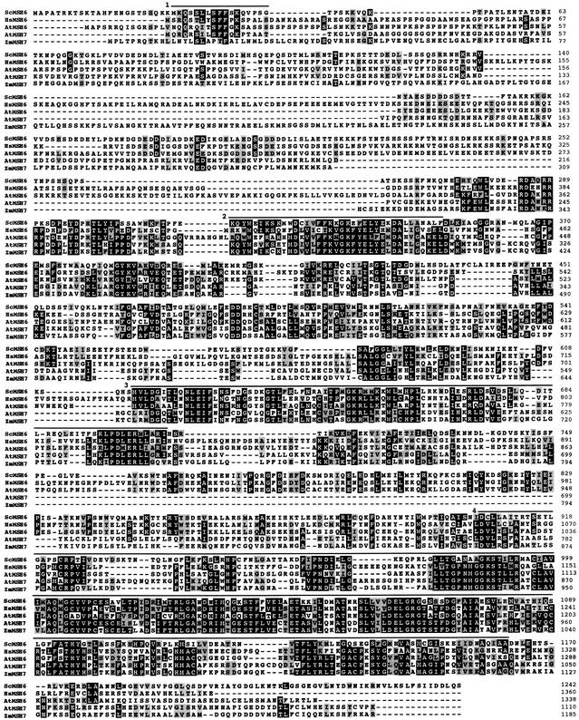
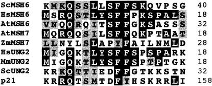
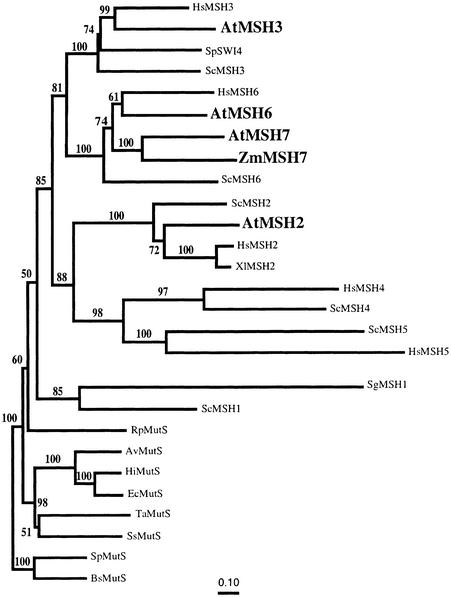
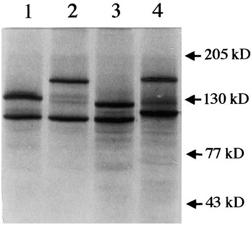
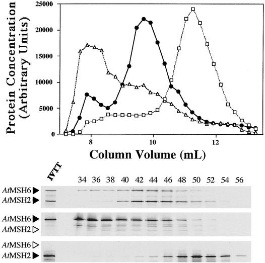
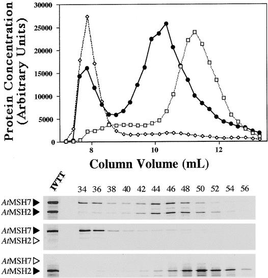
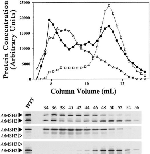
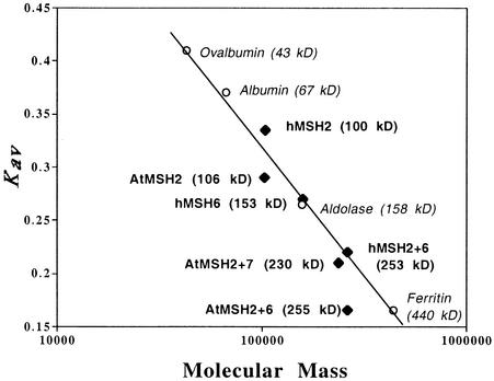
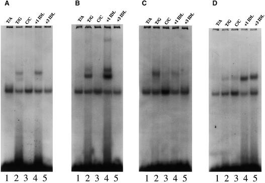
Arabidopsis mismatch repair genes predict MutS-like proteins remarkably similar to eukaryotic MutS homologs-MSH2, MSH3, and MSH6. A novel feature in Arabidopsis is the presence of two MSH6-like proteins, designated AtMSH6 and AtMSH7. Combinations of Arabidopsis AtMSH2 with AtMSH3, AtMSH6, or AtMSH7 proteins-products of in vitro transcription and translation-were analyzed for interactions by analytical gel filtration chromatography. The AtMSH2 protein formed heterodimers with AtMSH3, AtMSH6, and AtMSH7, but no single proteins formed homodimers. The abilities of the various heterodimers to bind to mismatched 51-mer duplexes were measured by electrophoretic mobility-shift assays. Similar to the behavior of the corresponding human proteins, AtMSH2*AtMSH3 heterodimers bound "insertion-deletion" DNA with three nucleotides (+AAG) or one nucleotide (+T) looped out much better than they bound DNA with a base/base mispair (T/G), whereas AtMSH2*AtMSH6 bound the (+T) substrate strongly, (T/G) well, and (+AAG) no better than it did a (T/A) homoduplex. However, AtMSH2*AtMSH7 showed a different specificity: moderate affinity for a (T/G) substrate and weak binding of (+T). Thus, AtMSH2*AtMSH7 may be specialized for lesions/base mispairs not tested or for (T/G) mispairs in special contexts.


Figures

Figure 1.
Figure 1.
Alignment of MSH6-like Protein Sequences. Black boxes highlight identical amino acids and gray boxes highlight similar amino acids in at least three of the sequences, based on Dayhoff's PAM250 matrix. Dashes denote gaps. The sequence prefixes At-, Hs-, Sc-, and Zm- denote Arabidopsis thaliana, Homo sapiens, Saccharomyces cerevisiae, and Zea mays, respectively. Amino acid positions are shown at right. The lines above the alignment denote conserved regions found in MSH proteins: line 1, the putative N-terminal PCNA/RPA interaction domain; line 2, the N-terminal mismatch recognition domain; line 3, the middle-conserved domain; and line 4, the highly conserved C-terminal domain.
Figure 2.
Figure 2.
Alignment of the N-Terminal PCNA/RPA Interaction Domain of MSH6, MSH7, UNG2, and Human p21 Proteins as in Figure 1. Mm, Mus musculus.
Figure 3.
Figure 3.
Neighbor-Joining Tree for Dayhoff PAM Distances among a Representative Set of Complete MutS/MSH Protein Sequences. Gaps and regions of ambiguous alignment were excluded from the analysis. Numbers above each branch represent the number of times the branch was found in 100 bootstrap replicas. The Gram-positive Bacillus subtilis and Streptococcus pneumoniae MutS protein sequences were used as an outgroup. All eukaryotic MutS homologs are encoded by the nuclear genome except for the mitochondrially encoded SgMSH1. At, A. thaliana; Av, Azotobacter vinelandii; Bs, B. subtilis; Ec, E. coli; Hi, Haemophilus influenzae; Hs, H. sapiens; Rp, Rickettsia prowazekii; Sc, S. cerevisiae; Sg, Sarcophytom glaucum; Sp (SW14), Saccharomyces pombe; Sp (MutS), S. pneumoniae; Ss, Synechocystis sp; Ta, Thermus aquaticus; Xl, Xenopus leavis; Zm, Z. mays.
Figure 4.
Figure 4.
SDS-PAGE Analysis of Human and Arabidopsis Cosynthesis Reaction Mixtures Used. Lane 1 contains AtMSH2 and AtMSH3; lane 2, AtMSH2 and AtMSH6; lane 3, AtMSH2 and AtMSH7; and lane 4, hMSH2 and hMSH6. Numbers at the right denote molecular mass.
Figure 5.
Figure 5.
Gel Filtration Chromatography Analysis of Arabidopsis 35S-Labeled Proteins. Fifty-microliter synthesis mixture samples were layered onto the gel filtration column, fractionated, and analyzed by liquid scintillation and SDS-PAGE (see Methods). (Top) Elution profiles for AtMSH2 (open squares), AtMSH6 (open triangles), and AtMSH2•AtMSH6 (closed circles) synthesis mixtures. Fractions 32 to 59 for each of the three mixtures are shown (elution profile). (Bottom) Corresponding SDS-PAGE autoradiographs of the eluted fractions (even numbers 34 to 56). A small amount of the original transcription–translation synthesis reaction mixture is shown in the leftmost lane at bottom (IVTT lane). Arrowheads to the left of the gel panels denote the position expected for each polypeptide. Arrowheads in outline designate theoretical positions of polypeptides not actually present.
Figure 6.
Figure 6.
Gel Filtration Chromatography Analysis of Arabidopsis 35S-Labeled Proteins. Shown are the elution profiles and the corresponding SDS-PAGE autoradiographs of the eluted fractions for AtMSH2 (open squares), AtMSH7 (open diamonds), and AtMSH2•AtMSH7 (closed circles) synthesis mixtures. See Figure 5 for details.
Figure 7.
Figure 7.
Gel Filtration Chromatography Analysis of Arabidopsis 35S-Labeled Proteins. Shown are the elution profiles and the corresponding SDS-PAGE autoradiographs of the eluted fractions for AtMSH2 (open squares), AtMSH3 (open triangles), and AtMSH2•AtMSH3 (closed circles) synthesis mixtures. See Figure 5 for details.
Figure 8.
Figure 8.
Kav versus Log Molecular Mass Plot of Standards and MSH Polypeptides Analyzed by Gel Filtration Chromatography. Open circles denote globular standards used to calibrate the gel filtration column. Black diamonds denote MSH polypeptides used in this study. The predicted (theoretical) molecular mass is shown in parentheses to the right of each monomeric or heterodimeric polypeptide.
Figure 9.
Figure 9.
Representative Mobility Shift Assays of Cosynthesis Reaction Mixtures of Human and Arabidopsis Polypeptides. Cosynthesis mixtures were incubated with 32P-labeled homoduplex DNA (T/A, lane 1, all panels), or heteroduplex DNA (T/G, C/C, +1 IDL [insertion–deletion DNA with one T nucleotide looped out], and +3 IDL [insertion–deletion DNA with AAG nucleotides looped out], lanes 2 to 5, respectively, all panels) and analyzed on nondenaturing PAGE (see Methods). (A) Representative mobility shift assay of hMSH2•hMSH6 cosynthesis mixtures. (B) Representative mobility shift assay of AtMSH2•AtMSH6 cosynthesis mixtures. (C) Representative mobility shift assay of AtMSH2•AtMSH7 cosynthesis mixtures. (D) Representative mobility shift assay of AtMSH2•AtMSH3 cosynthesis mixtures.

References

    1. Acharya, S., Wilson, T., Gradia, S., Kane, M.F., Guerrette, S., Marsischky, G.T., Kolodner, R., and Fishel, R. (1996). hMSH2 forms specific mispair-binding complexes with hMSH3 and hMSH6. Proc. Natl. Acad. Sci. USA 93, 13629–13634. - PMC - PubMed
    1. Ade, J., Belzile, F., Philippe, H., and Doutriaux, M.P. (1999). Four mismatch repair paralogues coexist in Arabidopsis thaliana: AtMSH2, AtMSH3, AtMSH6–1 and AtMSH6–2. Mol. Gen. Genet. 262, 239–249. - PubMed
    1. Allen, D.J., Makhov, A., Grilley, M., Taylor, J., Thresher, R., Modrich, P., and Griffith, J.D. (1997). MutS mediates heteroduplex loop formation by a translocation mechanism. EMBO J. 16, 4467–4476. - PMC - PubMed
    1. Bevan, M., et al. (1998). Analysis of 1.9 Mb of contiguous sequence from chromosome 4 of Arabidopsis thaliana. Nature 391, 485–488. - PubMed
    1. Chen, C., Merrill, B.J., Lau, P.J., Holm, C., and Kolodner, R.D. (1999). Saccharomyces cerevisiae pol30 (proliferating cell nuclear antigen) mutations impair replication fidelity and mismatch repair. Mol. Cell. Biol. 19, 7801–7815. - PMC - PubMed

Publication types

MeSH terms

LinkOut - more resources