. 2000 Jun 20;97(13):7178-83.
doi: 10.1073/pnas.120074297.

A role of transcriptional activators as antirepressors for the autoinhibitory activity of TATA box binding of transcription factor IID

Affiliations

A role of transcriptional activators as antirepressors for the autoinhibitory activity of TATA box binding of transcription factor IID

T Kotani et al. Proc Natl Acad Sci U S A. .

Abstract

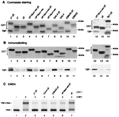
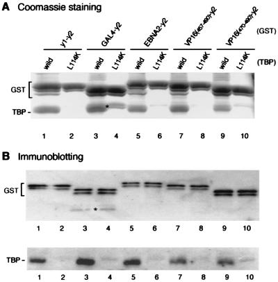
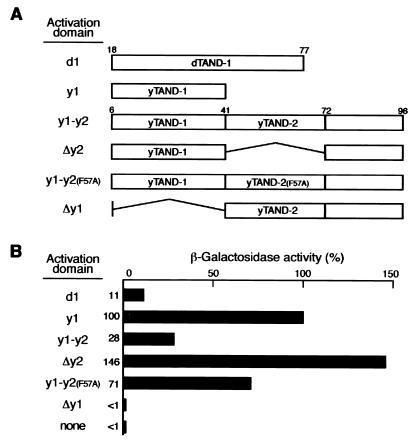
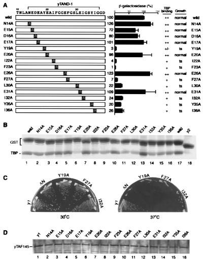
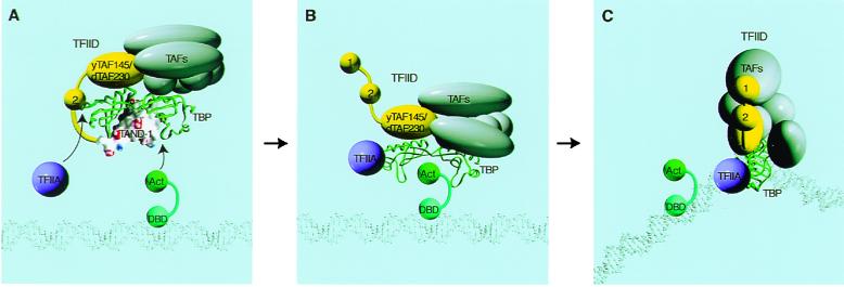
The TATA box-binding activity of transcription factor IID (TFIID) is autoinhibited by the N-terminal domain of the Drosophila TATA box-binding protein- (TBP) associated factor 230/yeast TBP-associated factor 145 subunit, which binds to the TATA box-binding domain of TBP by mimicking the TATA box structure. Here, we propose a mechanism of transcriptional activation that involves antirepression of this autoinhibitory activity by transcriptional activators. Like the autoinhibitory domain of TFIID, various acidic activators interact with the TATA box-binding domain of TBP. Moreover, the autoinhibitory domain of TFIID, which is known to interact with only the TATA box-binding domain of TBP, acts as an activation domain when fused to the GAL4 DNA-binding domain, indicating that interaction with the TATA-binding domain of TBP is crucial for activation of transcription. In a reciprocal fashion, the acidic activation domains can function as the autoinhibitory domain when the latter is replaced by the former within TFIID. These results indicate that activation domains and the autoinhibitory domain of TFIID are interchangeable, supporting a role for transcriptional activators as antirepressors of the autoinhibitory activity of the TATA box binding of TFIID.


Figures

Figure 1
Figure 1
Acidic activation domains form stable complexes with TBP and inhibit TBP binding to the TATA box when fused to TAND-2. (A and B) VP16 (residues 457–490 or 470–490; lanes 4, 6, and 12), GAL4 (lane 8), EBNA2 (lane 10), and Sp1 (lane 14) activation domains as well as yTAND-1 (lanes 1) were fused to yTAND-2 (y2) and expressed in E. coli as GST-tagged proteins. These activation domains (lanes 5, 7, 9, and 11), yTAND-1 (lane 2), and yTAND-2 (lanes 3 and 13) also were expressed as GST-tagged proteins. GST fusion proteins were incubated with an equimolar amount of yTBP and purified by glutathione Sepharose. The purified materials were analyzed by SDS-PAGE followed by Coomassie blue staining (A) or immunoblotting with anti-GST and anti-TBP antibodies (B). The positions of GST fusions and copurified TBP are indicated. GST fusions appeared as multiple bands due to the protease-hypersensitive sites near the C terminus. Although the bands marked by asterisks in lanes 9 and 14 migrated near TBP, these bands derived from GST fusion proteins according to immunoblotting shown in B. (C) Inhibition of TBP binding to the TATA box by GST-activation domain-TAND-2 chimeras. Gel retardation assays were performed with equimolar amounts of TBP and GST-fusion derivatives that were affinity purified. The positions of TBP-DNA complex and free probe were indicated at the left. Much larger amounts of proteins were required to see the inhibitory effects for activation domains alone (data not shown).
Figure 2
Figure 2
Acidic activation domains interact with the concave surface of TBP. yTAND-1 (lanes 1 and 2) and various activation domains including GAL4 (lanes 3 and 4), EBNA2 (lanes 5 and 6), VP16 (residues 457–490 or 470–490; lanes 7–10) were fused to yTAND-2 (y2), and expressed as GST fusion proteins. These GST proteins were incubated with equimolar amounts of wild-type (odd lanes) or L114K mutant TBP (even lanes). After GST precipitation, the purified materials were analyzed by Coomassie blue staining (A) or immunoblotting with anti-GST and anti-TBP antibodies (B) as described in the Fig. 1 legend. Degraded GST proteins that closely migrated with TBP are indicated by asterisks.
Figure 3
Figure 3
TAND-1 functions as an activator when fused to a DNA-binding domain. (A) Overall structures of GAL4 fusions tested in this experiment. Fragments shown here were fused with GAL4 DNA-binding domain (residues 1–147) for expression in yeast. Numbers on the bars indicate the amino acid positions in the respective factors. (B) GAL4-dependent transcriptional activation in yeast. Expression vectors described in A were transformed into yeast, and transcription activity was determined by measuring the lacZ-reporter activity. The relative β-galactosidase activity of authentic activation domains measured under the same conditions was as follows: VP16(457–490), 430%; VP16(470–490), 220%; GAL4, 300%; EBNA2, 270%; and Sp1, <1%.
Figure 4
Figure 4
The interaction of yTAND-1 with the concave surface of TBP is essential for its functions. (A) Alanine-scanning substitution mutants of yTAND-1 used in this study are schematically presented. These mutants were used for TBP-binding, growth, and transcription assays, and the results are summarized. For transcription analysis, indicated fragments were expressed in yeast as fusions with the GAL4 DNA-binding domain and tested for GAL4-dependent transcription, as described in the Fig. 3 legend. Note that although the point mutations within the yTAF145 gene caused only subtle effects, more obvious, but correlated, effects were observed when the point mutations were introduced into the yTAF145 gene in which TAND-2 (43–71 aa) had been internally deleted. The results with the gene background lacking TAND-2 are represented in this figure. (B) TBP-binding activity of the yTAND-1 mutants. The yTAND-1 mutants were fused to yTAND-2 and expressed in E. coli as GST-tagged proteins. Interaction of these fusions with TBP was determined as described in the Fig. 1 legend, and a Coomassie blue-stained gel is represented. (C) Growth phenotype of yeast carrying a defined mutation in yTAF145 gene. The yTAF145 genes harboring alanine-scanning mutations were introduced into yeast, replacing the wild-type gene by plasmid shuffling. After plasmid shuffling, the resulting strains were grown on yeast extract/peptone/dextrose plates at 30°C (Left) and 37°C (Right) for 3 days. Results are summarized in A, and photographs of yeast plates represent the selected mutants. Strains ΔN and y1 express yTAF145 proteins lacking the whole TAND (6–96 aa) and TAND-2 (43–71 aa), respectively (D). Expression level of the yTAF145 mutants in yeast. Yeast strains harboring yTAF145 mutants were grown at 25°C and then shifted to 37°C. Cells were harvested 24 h after the temperature shift and tested for expression level of yTAF145 mutants by immunoblotting.
Figure 5
Figure 5
Acidic activation domains function as yTAND-1 when integrated into TFIID. (A) Suppression analysis of growth defect by deleting yTAND-1 from the yTAF145 gene. yTAND-1 was deleted from the yTAF145 gene (Δy1) or replaced with various activation domains. Activation domains used are as follows: Sp1, residues 82–263; EBNA2, residues 426–462; GAL4, residues 842–874; VP16, residues 457–490 or 470–490, mutant VP16 (referred to as mVP16); triple mutant (F475S, M478T, F479S) in residues 470–490. The yTAF145 genes with these substitutions were transformed into yeast, replacing the wild-type gene by plasmid shuffling. After plasmid shuffling, the resulting strains were grown on yeast extract/peptone/dextrose plates at 30°C (Left) and 37°C (Right) for 3 days. (B) TBP-binding activity of the VP16 mutant. Wild-type (lane 2) and the triple mutant (F475S, M478T, F479S) (lane 1) of VP16 activation domain (residues 470–490) were fused to yTAND-2 and expressed in E. coli as GST fusions. TBP-binding activity was determined as described in the Fig. 1 legend, and a Coomassie blue stained gel was represented. (C) Expression level of the VP16-yTAF145 fusions. Expression level of the VP16-yTAF145 chimeric proteins was detected as described in the legend to Fig. 4. Growth of these strains is represented in A.
Figure 6
Figure 6
A two-step “hand off” model for reversal of the autoinhibitory effect of TFIID. When activators are absent, TAND-1 and -2 bind to the concave and convex surface of TBP, respectively, and keep TFIID in a latent state as shown in A. Acidic activators and TAND-1 competitively bind to the concave surface of TBP, whereas TFIIA and TAND-2 competitively bind to the convex surface of TBP. Competitive binding by activation domains and TFIIA could cause a conformational alteration of TFIID that may result in formation of an intermediate state as illustrated in B. Importantly, several lines of evidence shown in this paper indicate that this intermediate stage is essential for activation of transcription. The activation domain still prevents TBP from binding to the TATA box in the intermediate stage. However, activators and TFIIA may induce conformational alteration of TFIID, allowing stabilized interaction of TFIID with sequences near and downstream from the transcriptional initiation site. Through these interactions, TBP is handed from activation domains to TATA box as shown in C.

Similar articles

Cited by

References

    1. Orphanides G, Lagrange T, Reinberg D. Genes Dev. 1996;10:2657–2683. - PubMed
    1. Myer V E, Young R A. J Biol Chem. 1998;273:27757–27760. - PubMed
    1. Ptashne M, Gann A. Nature (London) 1997;386:569–577. - PubMed
    1. Ranish J A, Yudkovsky N, Hahn S. Genes Dev. 1999;13:49–63. - PMC - PubMed
    1. Burley S K, Roeder R G. Annu Rev Biochem. 1996;65:769–799. - PubMed

Publication types

Substances

LinkOut - more resources