. 2000 Jun 20;97(13):7290-5.
doi: 10.1073/pnas.140210297.

v-Crk activates the phosphoinositide 3-kinase/AKT pathway in transformation

Affiliations

v-Crk activates the phosphoinositide 3-kinase/AKT pathway in transformation

T Akagi et al. Proc Natl Acad Sci U S A. .

Abstract

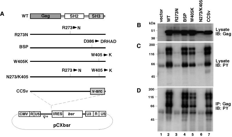
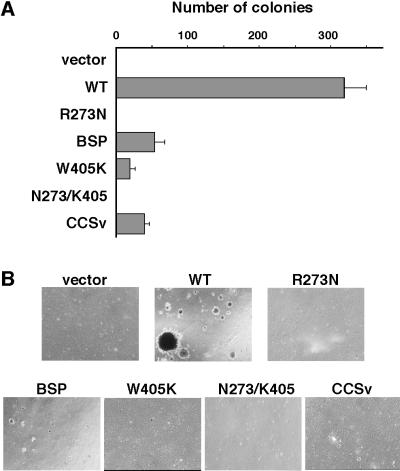
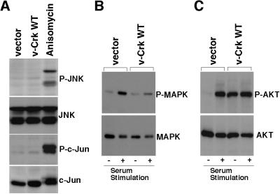
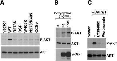
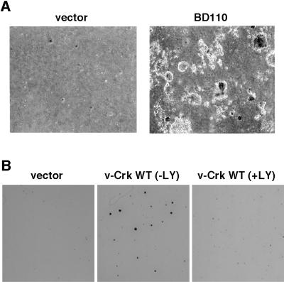
v-Crk induces cellular tyrosine phosphorylation and transformation of chicken embryo fibroblasts (CEF). We studied the molecular mechanism of the v-Crk-induced transformation. Experiments with Src homology (SH)2 and SH3 domain mutants revealed that the induction of tyrosine phosphorylation of cellular proteins requires only the SH2 domain, but both the SH2 and SH3 domains are required for complete transformation. Analysis of three well defined signaling pathways, the mitogen-activated protein kinase (MAPK) pathway, the Jun N-terminal kinase (JNK) pathway, and the phosphoinositide 3-kinase (PI3K)/AKT pathway, demonstrated that only the PI3K/AKT pathway is constitutively activated in v-Crk-transformed CEF. Both the SH2 and SH3 domains are required for this activation of the PI3K/AKT pathway in CEF. We also found that the colony formation of CEF is strongly induced by a constitutively active PI3K mutant, and that a PI3K inhibitor, LY294002, suppresses the v-Crk-induced transformation. These results strongly suggest that constitutive activation of the PI3K/AKT pathway plays an essential role in v-Crk-induced transformation of CEF.


Figures

Figure 1
Figure 1
Characterization of CEF expressing v-Crk mutants. (A) Schematic structure of the v-Crk mutants used in this study. Amino acid changes in each mutant are indicated in the single-letter amino acid code. Detailed descriptions about each mutant are given in Materials and Methods. pCXbsr is a Moloney MuLV-based retroviral vector. CMV, human cytomegalovirus; ψ+, extended retroviral packaging signal; IRES, internal ribose entry site; bsr, Blasticidin S resistance gene. (B) Expression of v-Crk mutants. v-Crk proteins were detected by immunoblotting with anti-Gag antibody 3C2 by using total cell lysates from CEF expressing v-Crk mutants as indicated above each lane. (C) Protein tyrosine phosphorylation in CEF expressing v-Crk mutants. Tyrosine phosphorylated proteins were detected by immunoblotting with anti-phosphotyrosine mouse monoclonal antibody 4G10 by using total cell lysates from CEF expressing v-Crk mutants as indicated above each lane. (D) Association of tyrosine-phosphorylated proteins with v-Crk mutants. Tyrosine phosphorylated proteins associated with v-Crk mutants were detected by immunoprecipitation with the anti-Gag antibody 3C2 followed by immunoblotting with the anti-phosphotyrosine rabbit polyclonal antibody.
Figure 2
Figure 2
Soft-agar colony formation of CEF expressing v-Crk mutants. (A) The numbers of colonies in soft-agar were counted 3 weeks after plating. Bars represent the averages (±SD) of three independent experiments. (B) Photomicrographs of colonies were taken 3 weeks after plating. (×40.)
Figure 3
Figure 3
Investigation of the downstream signals of v-Crk. (A) Analysis of the JNK pathway. Total cell lysates were prepared from CEF transduced with control vector, CEF transduced with v-crk WT, and CEF treated with anisomycin (10 μg/ml for 20 min). Cell lysates then were subjected to immunoblot analysis with antibodies specific for the phosphorylated forms of either JNK (P-JNK) (first panel) or c-Jun (P-c-Jun) (third panel). The same blots also were probed with antibodies to JNK (second panel) or to c-Jun (fourth panel) that react with these proteins irrespective of phosphorylation state to confirm that equal amounts of each protein were present in each lane. (B) Analysis of the MAPK pathway. CEF transduced with vector or v-crk WT were serum-starved for 24 h and then stimulated with 20% calf serum for 20 min (+), or left unstimulated (−). Total cell lysates from these cells were subjected to immunoblot analysis with antibody specific for the phosphorylated form of MAPK (P-MAPK) (Upper). The same blots were also probed with antibody to MAPK that reacts with MAPK irrespective of phosphorylation state to confirm that equal amounts of this protein were present in each lane (Lower). (C) Analysis of the PI3K/AKT pathway. CEF transduced with vector and CEF transduced with v-crk WT were serum-starved for 24 h and then stimulated with 20% calf serum for 20 min (+), or left unstimulated (−). Total cell lysates from these cells were subjected to immunoblot analysis with antibody specific for the phosphorylated form of AKT (P-AKT) (Upper). The same blots also were probed with antibody to AKT that reacts with AKT irrespective of phosphorylation state to confirm that equal amounts of this protein were present in each lane (Lower).
Figure 4
Figure 4
Analysis of v-Crk-induced AKT phosphorylation. (A) AKT phosphorylation in CEF expressing v-Crk mutants. Total cell lysates from CEF expressing v-Crk mutants (indicated above each lane) were subjected to immunoblot analysis with antibody specific for the phosphorylated form of AKT (P-AKT) (Upper) or with antibody to AKT that reacts with AKT irrespective of phosphorylation state (Lower). (B) AKT phosphorylation in an inducible v-Crk expression system. CEF dually infected with CXneo/TR-2 and CXbsrR(TO/v-Crk WT) were cultured with the indicated concentrations of doxycycline for 24 h. Total cell lysates from these cells then were subjected to immunoblot analysis with antibody specific for the phosphorylated form of AKT (P-AKT) (Top), with antibody to AKT that reacts with AKT irrespective of phosphorylation state (Middle), or with anti-Gag antibody (Bottom). (C) Effects of PI3K inhibitors. CEF expressing WT v-Crk were treated for 2 h with LY294002 (Calbiochem) at 10 μM, wortmannin (Calbiochem) at 200 nM, or with the solvent DMSO (−). Then, total cell lysates from these cells as well as from vector-transduced cells were subjected to immunoblot analysis with antibody specific for the phosphorylated form of AKT (P-AKT) (Upper) or with antibody to AKT that reacts with AKT irrespective of phosphorylation state (Lower)
Figure 5
Figure 5
Transformation by a constitutively active mutant of PI3K and suppression of v-Crk-induced transformation by a PI3K inhibitor. (A) Soft-agar colony formation by the PI3K constitutively active mutant BD110. CEF transduced with BD110 or with vector were subjected to soft-agar colony formation assay. Photomicrographs of colonies were taken 3 weeks after plating. (×40.) (B) Suppression of v-Crk-induced soft-agar colony formation by a PI3K inhibitor. CEF expressing v-Crk WT were subjected to soft-agar colony formation assay with (+LY) or without (−LY) LY294002. LY294002 was included in top agar layer at 10 μM final concentration, and 0.5 ml of medium containing the same final concentration of LY294002 was added onto the top agar every 4 days. We confirmed that long-term treatment with this concentration of LY294002 showed no apparent toxicity to CEF cultured in monolayer (data not shown). CEF transduced with vector without such treatment also were examined. At 3 weeks after plating, colonies were stained with 3-(4,5-dimethylthiazol-2-yl)-2,5-diphenyltetrazolium bromide (MTT) as described (37) and photographs of the stained colonies were taken. (×1.)

Similar articles

Cited by

References

    1. Mayer B J, Hamaguchi M, Hanafusa H. Nature (London) 1988;332:272–275. - PubMed
    1. Matsuda M, Tanaka S, Nagata S, Kojima A, Kurata T, Shibuya M. Mol Cell Biol. 1992;12:3482–3489. - PMC - PubMed
    1. Reichman C T, Mayer B J, Keshav S, Hanafusa H. Cell Growth Differ. 1992;3:451–460. - PubMed
    1. Pawson T, Gish G D. Cell. 1992;71:359–362. - PubMed
    1. Birge R B, Knudsen B S, Besser D, Hanafusa H. Genes Cells. 1996;1:595–613. - PubMed

Publication types

MeSH terms

Substances

Associated data