. 2000 May 25;257(1):111-26.
doi: 10.1006/excr.2000.4882.

Induction of apoptosis in rat cardiocytes by A3 adenosine receptor activation and its suppression by isoproterenol

Affiliations

Induction of apoptosis in rat cardiocytes by A3 adenosine receptor activation and its suppression by isoproterenol

V Shneyvays et al. Exp Cell Res. .

Abstract

The purpose of the present study was to investigate the mechanisms involved in the induction of apoptosis in newborn cultured cardiomyocytes by activation of adenosine (ADO) A3 receptors and to examine the protective effects of beta-adrenoceptors. The selective agonist for A3 ADO receptors Cl-IB-MECA (2-chloro-N6-iodobenzyl-5-N-methylcarboxamidoadenosine) and the antagonist MRS1523 (5-propyl-2-ethyl-4-propyl-3-(ethylsulfanylcarbonyl)-6-phenylpy rid ine-5-carboxylate) were used. High concentrations of the Cl-IB-MECA (> or = 10 microM) agonist induced morphological modifications of myogenic cells, such as rounding and retraction of cell body and dissolution of contractile filaments, followed by apoptotic death. In addition, Cl-IB-MECA caused a sustained and reversible increase in [Ca2+]i, which was prevented by the selective antagonist MRS1523. Furthermore, MRS1523 protected the cardiocytes if briefly exposed to Cl-IB-MECA and partially protected from prolonged (48 h) agonist exposure. Apoptosis induced by Cl-IB-MECA was not redox-dependent, since the mitochondrial membrane potential remained constant until the terminal stage of cell death. Cl-IB-MECA activated caspase-3 protease in a concentration-dependent manner after 7 h of treatment and more effectively after 18 h of exposure. Bcl-2 protein was readily detected in control cells, and its expression was significantly decreased after 24 and 48 h of treatment with Cl-IB-MECA. Beta-adrenergic stimulation antagonized the pro-apoptotic effects of Cl-IB-MECA, probably through a cAMP/protein kinase A-independent mechanism, since addition of dibutyryl-cAMP did not abolish the apoptosis induced by Cl-IB-MECA. Incubation of cultured myocytes with isoproterenol (5 microM) for 3 or 24 h almost completely abolished the increase in [Ca2+]i. Prolonged incubation of cardiomyocytes with isoproterenol and Cl-IB-MECA did not induce apoptosis. Our data suggest that the apoptosis-inducing signal from activation of adenosine A3 receptors (or counteracting beta-adrenergic signal) leads to the activation of the G-protein-coupled enzymes and downstream pathways to a self-amplifying cascade. Expression of different genes within this cascade is responsible for orchestrating either cardiomyocyte apoptosis or its protection.


Figures
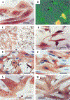
FIG. 1.
FIG. 1.
Light micrographs of cultured cardiac cells after exposure to Cl-IB-MECA. (A) Control cells cultured in serum-free medium. Cross-striated myofibrils arranged in perinuclear area of the cell. (B) Effect of 20 µM Cl-IB-MECA on cardiocytes after 24 h of exposure. Shortening and shrinkage of viable cells (exclude propidium iodide, black arrows). Propidium iodide-stained cells were scored as necrotic (white arrows). Combined visualization by phase-contrast and fluorescence microscopy. (C) Effect of 30 µM Cl-IB-MECA after 48 h of treatment. (Ca) Control, untreated cardiocytes. (Cb) Complete destruction of cardiomyocytes; only very shortened cells or particles (apoptotic bodies) were observed. (D) Effect of 20 µM Cl-IB-MECA after 48 h of treatment. (E) Effect of 20 µM Cl-IB-MECA after 2 h of treatment (arrows show apoptotic cells). (F) Cardiocytes treated with the antagonist MRS1523 before A3 agonist. The antagonist MRS1523 prevents the toxic effect of Cl-IB-MECA (20 µM for 2 h). (G) Morphology of cardiomyocytes 2 days incubated with 5 µM isoproterenol. (H) Reduction of apoptosis by isoproterenol in Cl-IB-MECA-stimulated cells. A and C–H, immunohistochemical staining of α-sarcomeric actin and counterstaining with hematoxylin. No counterstain in Ca and Cb. Bars, 10 µm. Ca and Cb, bars, 30 µm.
FIG. 2.
FIG. 2.
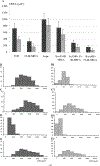
Histograms of cardiomyocytes area and myofibrils area after various treatments. (A) Cardiomyocyte area (black bars) and myofibril area (gray bars) following treatment with Cl-IB-MECA (20 µM), isoproterenol (5 µM), dibutyryl-cAMP, I- (1 µM), or II- (200 µM). (B–E) Histograms of cardiac cells distribution in control (B), isoproterenol (C), Cl-IB-MECA (D), Cl-IB-MECA, and isoproterenol (E) cultures. (B1–E1) distributions of intracellular myofibrillar area in the cultured cardiomyocytes. Y axis, cell number.
FIG. 3.
FIG. 3.
Histograms of DNA content by image analysis of Feulgen stained nuclei. ( x) Arbitrary units of IOD; ( y) frequency of cells; 2C, diploid content of DNA (arrow); crossed bars, hypodiploid nuclei. (A) Control. (B) 48-h incubation with 5 µM isoproterenol; slight increase in hypodiploid DNA content. (C) 48-h treatment with 20 µM Cl-IB-MECA; considerable increase in hypodiploid DNA transformation. (D) Protective effect of isoproterenol on Cl-IB-MECA-induced damage in cardiocytes DNA content.
FIG. 4.
FIG. 4.
Mitochondrial fluorescence of DASPMI in Cl-IB-MECA-treated cardiocytes. (A) control. (B, C) Cells treated for 48 h with 20 µM Cl-IB-MECA. Intensity of DASPMI fluorescence did not change until the terminal state of cell death. (D–F) Histograms of gray-level distribution show the presentation fluorescence intensity of A–C, respectively. Bar, 10 µm.
FIG. 5.
FIG. 5.
Photographs of Western blots of Bcl-2 proteins, following Cl-IB-MECA treatment. Significant differences among control and treated cells after 24 and 48 h of exposure to A3 RC agonist.
FIG. 6.
FIG. 6.
Caspase-3 (CPP32) protease activity in cardiocytes following Cl-IB-MECA. Seven and 18 h with Cl-IB-MECA (10 and 20 µM) induced two- to threefold increase in the protease activity. The CPP32 inhibitor DEVO-FMK (50 µM) decreases the caspase-3 activity below the control level.
FIG. 7.
FIG. 7.
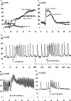
Effect of Cl-IB-MECA on intracellular Ca2+ concentration in cultured cardiomyocytes. (A) Effects of Cl-IB-MECA (20 and 30 µM) and A2A-selective agonist CGS-21680 (C-141, 100 µM) on intracellular calcium level. The adenosine analogues were added at the indicated time (arrows). (B) Effect of the selective A3 adenosine receptor antagonist MRS1523. Ca elevation by Cl-IB-MECA was prevented. (C) Elevation of intracellular calcium levels by Cl-IB-MECA in cardiac cells in calcium-free medium. (D) Incubation with isoproterenol (5 µM) for 24 h causes positive chronotropic effect on cardiomyocyte contractility and significantly reduced calcium elevation after Cl-IB-MECA addition (arrow). (E) Response to Cl-IB-MECA (arrow) after a 3-h incubation with 5 µM isoproterenol.
FIG. 8.
FIG. 8.
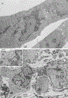
Electron micrographs of a cardiac cells after exposure to 20 µM Cl-IB-MECA. (A) Ultrastructure of control cardiomyocyte, cultured in serum-free medium. (B) Loss of typical cardiomyocytes ultrastructure after 48-h treatment with the agonist, constriction, and fragmentation of nuclei. (B1) High magnification of B micrographs (inset) shows residual myofilaments of damaged cardiocyte. (C) Preapoptotic shrinking cardiomyocyte with reduced myofibrils and maintain of intact mitochondria. (D, E) Condensation of chromatin characteristic for apoptosis. Condensed chromatin occupies considerable part of cross-sectional nucleus area. (F) Nuclear fragments of varying size, surrounded by double nuclear membranes. (G, H) “Secondary necrosis” of apoptotic bodies with signs of extensive chromatinolysis, microvacuolation of sarcoplasm and cell remnants formation.
FIG. 8.
FIG. 8.
Electron micrographs of a cardiac cells after exposure to 20 µM Cl-IB-MECA. (A) Ultrastructure of control cardiomyocyte, cultured in serum-free medium. (B) Loss of typical cardiomyocytes ultrastructure after 48-h treatment with the agonist, constriction, and fragmentation of nuclei. (B1) High magnification of B micrographs (inset) shows residual myofilaments of damaged cardiocyte. (C) Preapoptotic shrinking cardiomyocyte with reduced myofibrils and maintain of intact mitochondria. (D, E) Condensation of chromatin characteristic for apoptosis. Condensed chromatin occupies considerable part of cross-sectional nucleus area. (F) Nuclear fragments of varying size, surrounded by double nuclear membranes. (G, H) “Secondary necrosis” of apoptotic bodies with signs of extensive chromatinolysis, microvacuolation of sarcoplasm and cell remnants formation.
FIG. 9.
FIG. 9.
Effect of Cl-IB-MECA and isoproterenol on contractility of cardiac cells after 48-h exposure to the drugs. (A) Control, contracting cells incubated in serum-free medium. (B) Positive chronotropic effect of isoproterenol (5 µM). (C) After application of Cl-IB-MECA (20 µM) cells showed irregular and delayed contractions. (D) Cells treated with Cl-IB-MECA (20 µM) and maintained in 5 µM of isoproterenol.

Similar articles

Cited by

References

    1. Zhou Q, Li C, Olah ME, Jonson RA, Stiles GL, and Civelli O (1992). Molecular cloning and characterization of an adenosine receptor: The A3 adenosine receptor. Proc. Natl. Acad. Sci. USA 89, 7432–7436. - PMC - PubMed
    1. Giannella E, Mochmann HC, and Levi R (1997). Ischemic preconditioning prevents the impairment of hypoxic coronary vasodilatation caused by ischemia/reperfusion: Role of adenosine A1/A3 and bradykinin B2 receptor activation. Circ. Res 81, 415–422. - PubMed
    1. Fozard JR, Pfannkuche HJ, and Schuurman HJ (1996). Mast cell degranulation following adenosine A3 receptor activation in rats. Eur. J. Pharmacol 298, 293–297. - PubMed
    1. Liang BT, and Jacobson KA (1998). A physiological role of the adenosine A3 receptor: Sustained cardioprotection. Proc. Natl. Acad. Sci. USA 95, 6995–6999. - PMC - PubMed
    1. Jacobson KA (1998). Adenosine A3 receptors: Novel ligands and paradoxical effects. Trends Pharmacol. Sci 19, 184–191. - PMC - PubMed

Publication types

Substances