. 2000 Jun;156(6):1865-73.
doi: 10.1016/S0002-9440(10)65060-2.

Disruption of the plasminogen gene in mice abolishes wound healing after myocardial infarction

Affiliations

Disruption of the plasminogen gene in mice abolishes wound healing after myocardial infarction

E Creemers et al. Am J Pathol. 2000 Jun.

Abstract

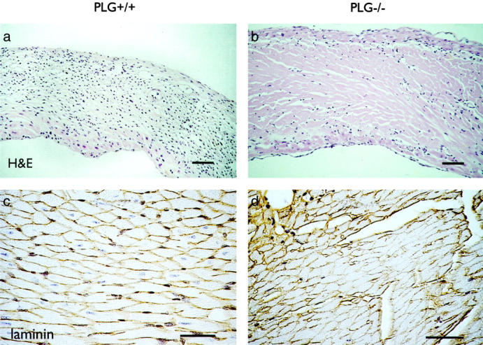
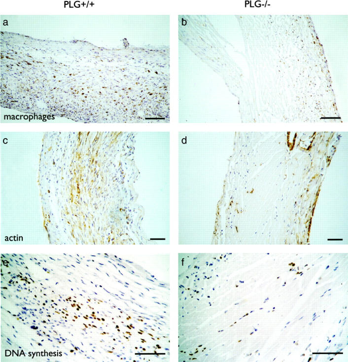
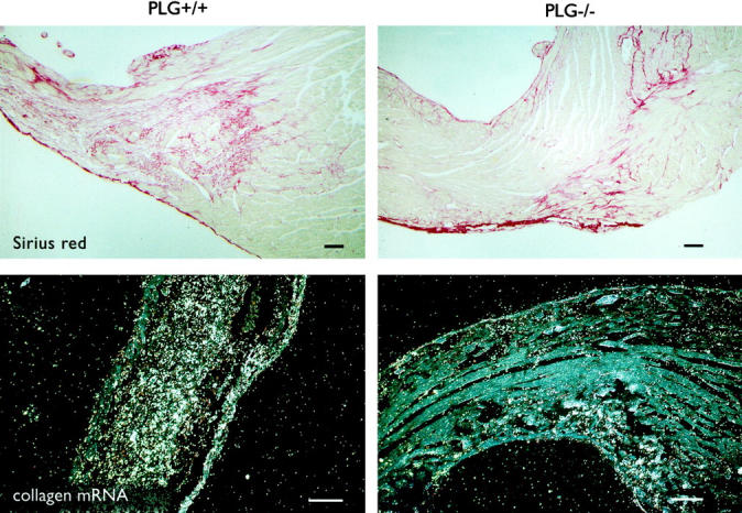
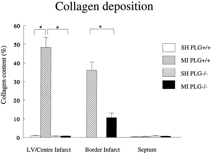
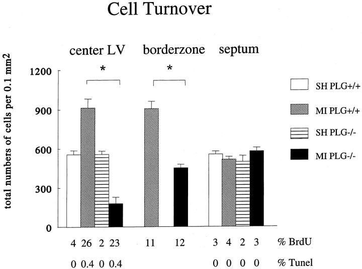
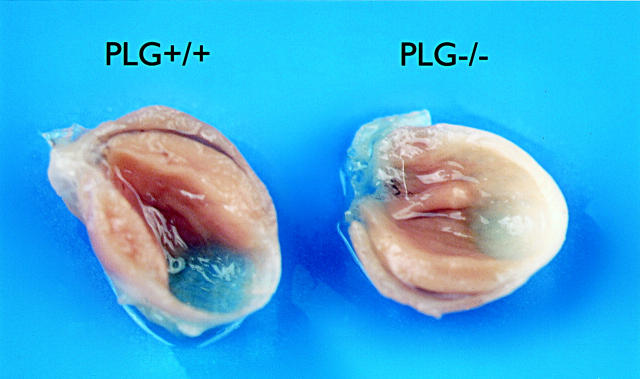
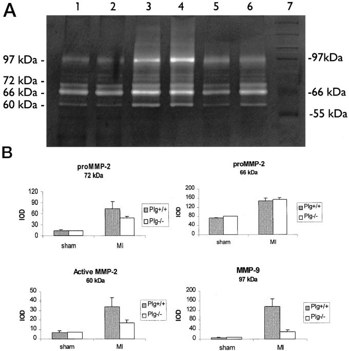
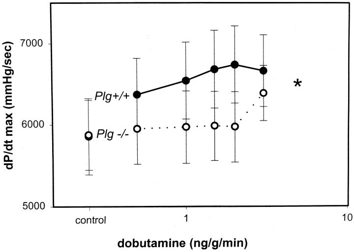
The plasminogen system plays an important role in the proteolytic degradation of extracellular matrices during wound healing. In the present study we investigated the impact of the plasminogen system on cardiac wound healing and function after myocardial infarction. Myocardial infarction was induced in plasminogen-deficient mice (Plg-/-) and in wild-type controls (Plg+/+). Structural analysis 1, 2, and 5 weeks after infarction revealed that infarct healing was virtually abolished in Plg-/- mice, indicating that the plasminogen system is required for the repair process of the heart after infarction. In the absence of plasminogen, inflammatory cells did not migrate into the infarcted myocardium. Necrotic cardiomyocytes were not removed and the formation of granulation tissue and fibrous tissue did not occur. In these non-healing infarcted hearts, LV dilatation was not altered. In addition, gelatinolytic activity of MMP-2 and MMP-9 was depressed in the Plg-/- infarcted hearts, suggesting that the plasmin effect on infarct healing may be mediated by MMPs. Surprisingly, cardiac function was only attenuated to a rather small extent in the Plg-/- infarcted mice when compared to the wild-types. This study provides direct prove that plasmin-mediated proteolysis plays a central role in cardiac wound healing after myocardial infarction in mice.


Figures

Figure 1.
Figure 1.
Structural composition of the infarcts. a and b: Hematoxylin-and-eosin-stained sections of Plg+/+ and Plg−/− infarcts 2 weeks after MI. In Plg+/+ infarcts, the necrotic cardiomyocytes are replaced by granulation tissue, whereas in Plg−/− infarcts wound healing is absent. c and d: laminin, a basement membrane component, encages normal cardiomyocytes in the non-infarcted myocardium (Plg+/+). In the center of 2-week-old Plg−/− infarcts, these basement membranes are still present. The intensity of the staining, however, decreases over time. Bars represent 50 μm.
Figure 2.
Figure 2.
Cellular composition of the infarcts. a and b: Large numbers of macrophages infiltrate into 1-week-old Plg+/+ infarcts in contrast to the Plg−/− infarcts. c and d: α-Smooth muscle actin immunohistochemistry reveals myofibroblasts and smooth muscle cells in the center of the Plg+/+ infarcts. In the center of the Plg−/− infarcts, the only α-sma-positive cells were vascular smooth muscle cells. e and f: BrdU immunohistochemistry in the infarcted myocardium. The absolute number of BrdU-positive cells is higher in the Plg+/+, but the percentage of cells that are BrdU-positive is very similar in the Plg+/+ and Plg−/−. Bars represent 50 μm.
Figure 3.
Figure 3.
Collagen deposition and synthesis. a and b: Sirius red staining 2 weeks after MI shows extensive collagen deposition in the Plg+/+ infarcts and only a little in the Plg−/− infarcts. c and d: In situ hybridization shows that type I collagen mRNA expression is more pronounced in 2-week-old wild-type infarcts than in 2-week-old Plg−/− infarcts. Bars represent 50 μm.
Figure 4.
Figure 4.
Quantification of the Sirius red staining demonstrates completely abolished collagen deposition in the center of the Plg−/− infarcts. MI, myocardial infarction; SH, sham operation.
Figure 5.
Figure 5.
Cell turnover in Plg+/+ and Plg−/− hearts, 2 weeks after MI. Despite reduced absolute cell numbers (bars) in the Plg−/− infarcted hearts, the percentage of BrdU-positive cells is not significantly different from the Plg+/+ infarcts (23% and 26%, respectively; P = 0.33 by Mann-Whitney test). This was also seen for levels of apoptosis, as measured by the TUNEL staining technique. MI, myocardial infarction; SH, sham operation.
Figure 6.
Figure 6.
Infarcted hearts from Plg+/+ and Plg−/− mice, 5 weeks after surgery. Note the extensive infarct thinning in the wild-types, where the infarcted wall has almost become transparent.
Figure 7.
Figure 7.
MMP-2 and MMP-9 activity in Plg−/− and Plg+/+ hearts. A: Gelatin zymography, demonstrating MMP-2 and MMP-9 activity in cardiac extracts, 2 weeks after surgery. Lanes 1 and 2, Plg−/− infarcts; lanes 3 and 4, Plg+/+ infarcts; lane 5, Plg+/+ sham; lane 6, Plg−/− sham; lane 7, marker. The 97-kd band corresponds to MMP-9. Three bands with molecular weights of ∼72, ∼66, and ∼60 kd represent, respectively, two proenzyme forms and one activated form of MMP-2. B: Quantification of the scanned gelatinolytic bands.
Figure 8.
Figure 8.
The cardiac contractile response to dobutamine infusion is blunted in the 2-week Plg−/− infarcts (n = 5–6 per group, P = 0.014 by two-way analysis of variance).

Similar articles

Cited by

References

    1. Willems IE, Havenith MG, De Mey JG, Daemen MJAP: The alpha-smooth muscle actin-positive cells in healing human myocardial scars. Am J Pathol 1994, 145:868-875 - PMC - PubMed
    1. Heymans S, Luttun A, Nuyens D, Theilmeier G, Creemers E, Moons L, Dyspersin GD, Cleutjens JPM, Shipley M, Angellilo A, Levi M, Nube O, Baker A, Keshet E, Lupu F, Herbert JM, Smits JF, Shapiro SD, Baes M, Borgers M, Collen D, Daemen MJ, Carmeliet P: Inhibition of plasminogen activators or matrix metalloproteinases prevent cardiac rupture but impairs therapeutic angiogenesis and causes cardiac failure. Nat Med 1999, 10:1135-1142 - PubMed
    1. Matrisian LM: Metalloproteinases and their inhibitors in matrix remodeling. Trends Genet 1990, 6:121-125 - PubMed
    1. Carmeliet P, Moons L, Lijnen R, Baes M, Lemaitre V, Tipping P, Drew A, Eeckhout Y, Shapiro S, Lupu F, Collen D: Urokinase-generated plasmin activates matrix metalloproteinases during aneurysm formation. Nat Genet 1997, 17:439-444 - PubMed
    1. Carmeliet P, Schoonjans L, Kieckens L, Ream B, Degen J, Bronson R, De Vos R, Van den Oord JJ, Collen D, Mulligan RC: Physiological consequences of loss of plasminogen activator gene function in mice. Nature 1994, 368:419-424 - PubMed

Publication types