. 2000 Jun;156(6):1951-9.
doi: 10.1016/S0002-9440(10)65068-7.

Contrasting effects of CCR5 and CCR2 deficiency in the pulmonary inflammatory response to influenza A virus

Affiliations

Contrasting effects of CCR5 and CCR2 deficiency in the pulmonary inflammatory response to influenza A virus

T C Dawson et al. Am J Pathol. 2000 Jun.

Abstract

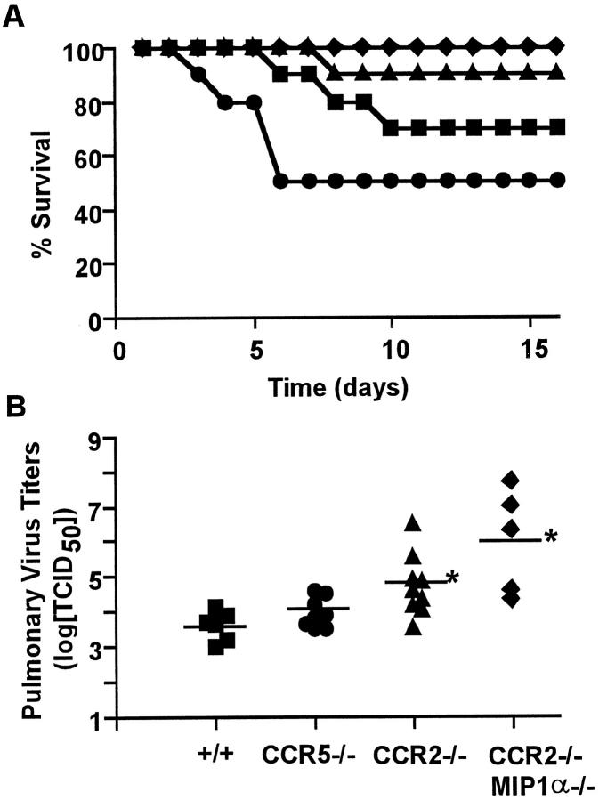
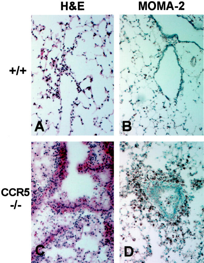
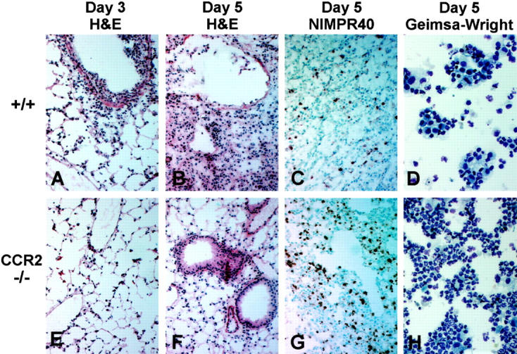
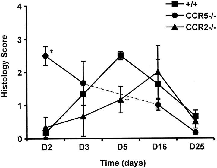
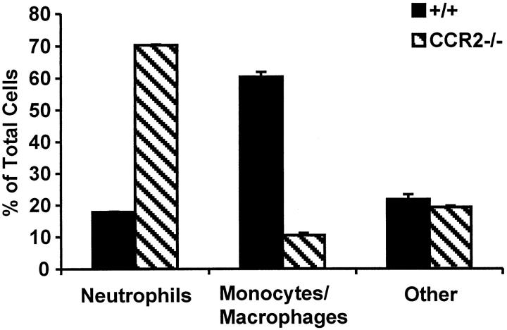
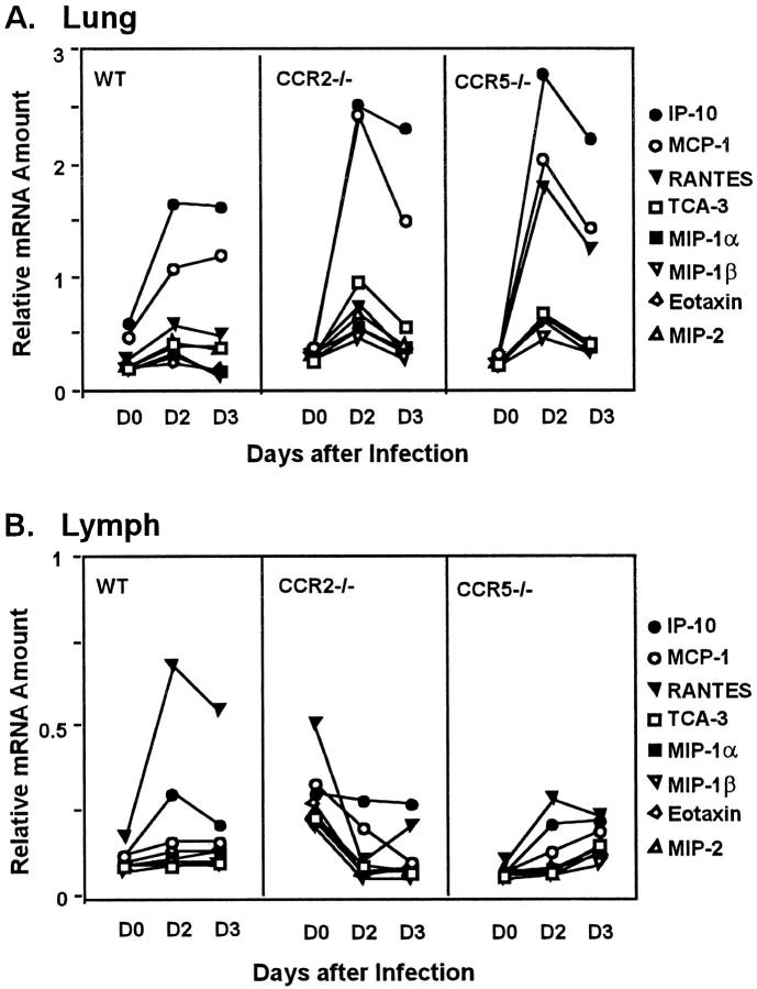
The immune response to influenza A virus is characterized by an influx of both macrophages and T lymphocytes into the lungs of the infected host, accompanied by induced expression of a number of CC chemokines. CC chemokine receptors CCR5 and CCR2 are both expressed on activated macrophages and T cells. We examined how the absence of these chemokine receptors would affect pulmonary chemokine expression and induced leukocyte recruitment by infecting CCR5-deficient mice and CCR2-deficient mice with a mouse-adapted strain of influenza A virus. CCR5(-/-) mice displayed increased mortality rates associated with acute, severe pneumonitis, whereas CCR2(-/-) mice were protected from the early pathological manifestations of influenza because of defective macrophage recruitment. This delay in macrophage accumulation in CCR2(-/-) mice caused a subsequent delay in T cell migration, which correlated with high pulmonary viral titers at early time points. Infected CCR5(-/-) mice and CCR2(-/-) mice both exhibited increased expression of the gene for MCP-1, the major ligand for CCR2(-/-) and a key regulator of induced macrophage migration. These studies illustrate the very different roles that CCR5 and CCR2 play in the macrophage response to influenza infection and demonstrate how defects in macrophage recruitment affect the normal development of the cell-mediated immune response.


Figures

Figure 1.
Figure 1.
The effect of CCR5 and CCR2 deletion on influenza associated mortality and viral titers. A: Mortality of +/+ (▪), CCR5−/− (•), CCR2−/− (▴), or CCR2−/−/MIP-1α−/− (♦ mice (n = 19 per group) infected intranasally with 5 HAU of influenza A (strain A/PR/8/34) and monitored for 16 days. Data represent two independent experiments combined. B: Influenza A titers in +/+, CCR5−/−, CCR2−/−, and CCR2−/−/MIP-1α−/− mice measured at day 5 after infection with 10 HAU of virus. Data are expressed as log[TCID50] per gram of tissue. *P < 0.01 versus +/+ group by Student’s t-test.
Figure 2.
Figure 2.
Inflammatory response to influenza A in lungs of control and CCR5-deficient mice. All histology sections are from lung tissues taken at day 2 postinfection with 5 HAU of influenza A; original magnification, ×100. A and B: Sections from +/+ lungs show H&E staining of only a few inflammatory cells characteristic for this early time point (A) and MOMA-2 staining of a few, sparse resident alveolar macrophages (B). C and D: Sections from CCR5−/− lungs show H&E staining of massive inflammatory cell infiltration and interstitial pneumonitis (C) and intense MOMA-2 staining of the infiltrating macrophages (D).
Figure 3.
Figure 3.
Inflammatory response to influenza A in lungs of control and CCR2-deficient mice. All histology sections are from lung tissue taken at days 3 and 5 postinfection with 5 HAU of influenza A; original magnification, ×100. Cytological preparations are BAL samples taken at day 5 postinfection; original magnification, ×400. A–C: Sections from +/+ lungs show H&E staining of the acute inflammatory cell infiltrate typically seen 3 days postinfection (A), the areas of chronic inflammation and tissue consolidation, thickening of the septal walls, and disorganization of the bronchial epithelial cells typically seen 5 days postinfection (B), and NIMPR40 staining of the relatively few neutrophils seen at this time point in +/+ mice (C). D: Giemsa-Wright staining of the BAL cells obtained from the +/+ mice 5 days postinfection. E–G: Sections from CCR2−/− lungs show H&E staining of less cellular infiltration 3 days postinfection (E), absence of the epithelial cell damage and consolidating pneumonitis that is seen in control mice at day 5 postinfection (F), and relatively intense NIMPR40 staining of the accumulating neutrophils, which is the dominant leukocyte population present in CCR2−/− lungs 5 days postinfection (G) . H: Giemsa-Wright staining of CCR2−/− BAL cells obtained at day 5 postinfection.
Figure 4.
Figure 4.
Time course of pulmonary pathology scores for surviving +/+, CCR5−/−, and CCR2−/− mice after influenza virus infection. All lung histology sections were scored (blinded) 0+ to 4+ based on the severity of inflammation and tissue damage. For this experiment, CCR5−/− mice were not analyzed at day 5 postinfection. Each data point represents the average of 3 to 5 mice per group for each time point. *, P = 0.002 versus +/+ group, and , P = 0.05 versus +/+ group both by Student’s t-test.
Figure 5.
Figure 5.
Differential leukocyte counts of BAL cells obtained from influenza-infected +/+ and CCR2−/− mice. Data represents the average of 3 counts (of approximately 300 cells per count) from one pooled sample per group (each group = 5 mice pooled).
Figure 6.
Figure 6.
Time course of chemokine mRNA expression levels in the lung (A) and the MLN (B). RNA was obtained from uninfected (day 0) tissues and from postinfection (day 2, day 3) tissues of +/+, CCR5−/−, and CCR2−/− mice. RNA was prepared and chemokine mRNA was detected by a multiprobe RNase protection assay and quantified by image densitometry, as described in Materials and Methods. Each data point represents a sample of pooled RNA from two individual animals and chemokine mRNA amounts were expressed relative to that of GAPDH.

Similar articles

Cited by

References

    1. Cook DN: The role of MIP-1α in inflammation and hematopoiesis. J Leukoc Biol 1996, 59:61-66 - PubMed
    1. Baggiolini M: Chemokines and leukocyte traffic. Nature 1998, 392:565-568 - PubMed
    1. Ward SG, Bacon K, Westwick J: Chemokines and T lymphocytes: more than an attraction. Immunity 1998, 9:1-11 - PubMed
    1. Zlotnik A, Morales J, Hedrick JA: Recent advances in chemokines and chemokine receptors. Crit Rev Immunol 1999, 19:1-47 - PubMed
    1. Devalaraja MN, Richmond A: Multiple chemotactic factors: fine control or redundancy? Trends Pharm Sci 1999, 20:151-156 - PubMed

Publication types

MeSH terms