

Treatment-induced decline of human immunodeficiency virus-1 p24 and HIV-1 RNA in lymphoid tissue of patients with early human immunodeficiency virus-1 infection
- PMID: 10854220
- PMCID: PMC1850084
- DOI: 10.1016/S0002-9440(10)65070-5
Treatment-induced decline of human immunodeficiency virus-1 p24 and HIV-1 RNA in lymphoid tissue of patients with early human immunodeficiency virus-1 infection
Abstract
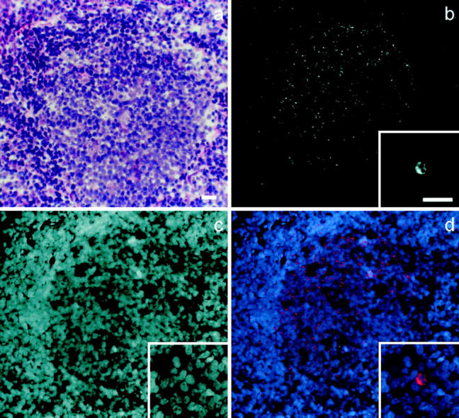
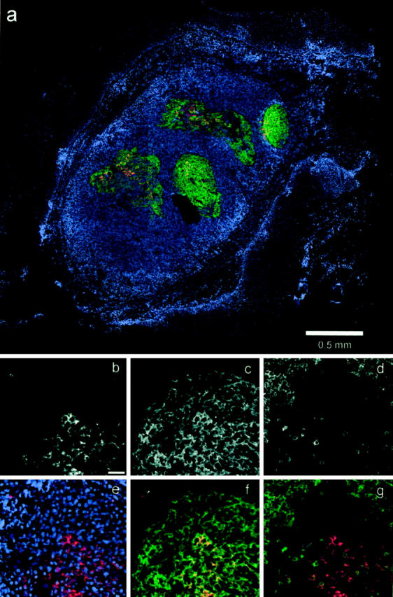
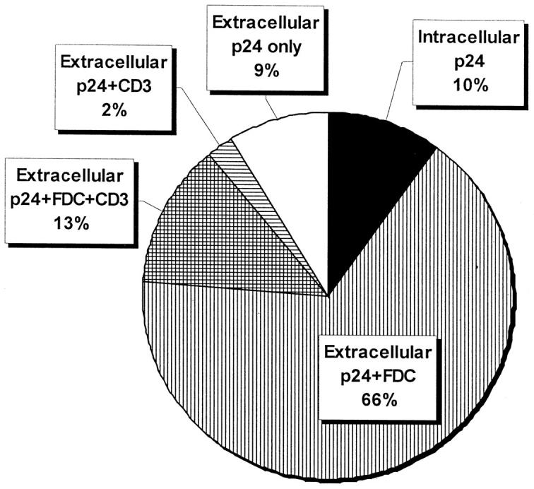
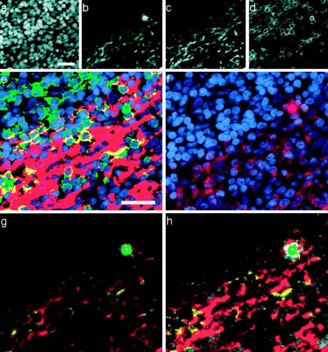
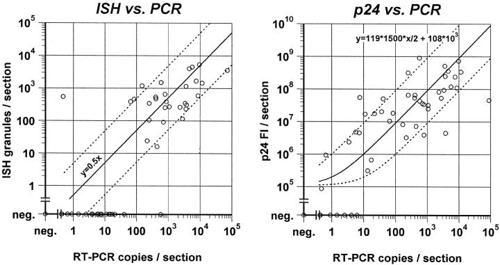
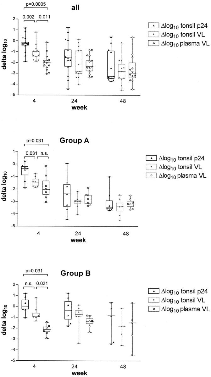
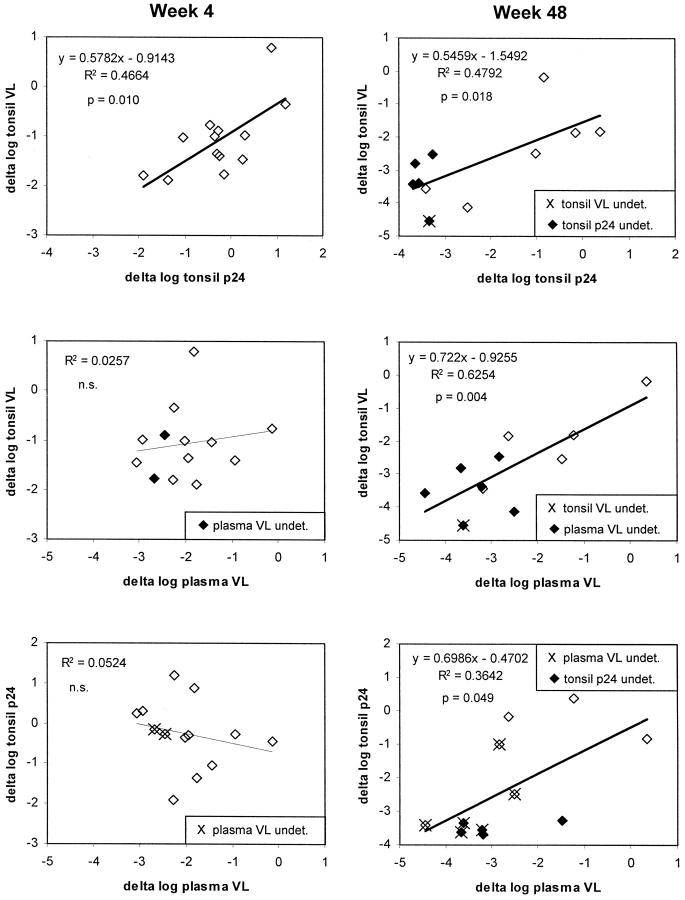
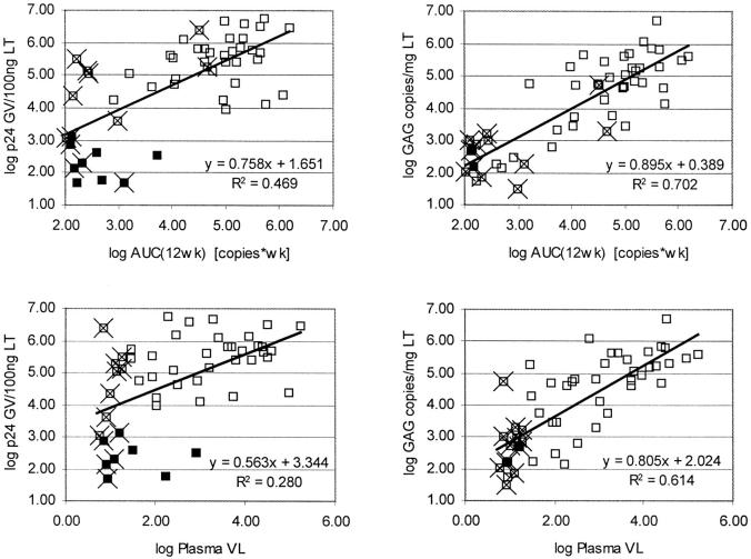
We report detailed quantitative analysis of human immunodeficiency virus-1 (HIV-1) p24 and HIV-1 RNA in tonsil biopsies from 13 patients with early, asymptomatic HIV infection before and during combination antiretroviral therapy. Using fluorescent microscopy in conjunction with reverse transcriptase-polymerase chain reaction of frozen tissue sections, we show that plasma and tissue viral loads decreased by approximately 3 logs during the 1-year treatment period, with good correlation between the HIV-1 p24 and HIV-1 RNA response in tissue. The decrease of tissue viral load was delayed compared to plasma viral load, possibly explained by the observation that the amount of follicular dendritic cell-associated virus correlated best with the area under the curve of plasma HIV-1 RNA throughout the last 12 weeks. Before and during treatment, the relative proportions of HIV-1 on follicular dendritic cells and within mononuclear cells remained constant, suggesting similar decay characteristics in these two lymphoid tissue compartments. However, viral p24 or RNA remained almost always detectable in tissue despite full suppression of HIV-1 RNA in plasma, and increased even after short-term rebounds in plasma viral load. Thus, full and sustained suppression of viral replication was required to efficiently decrease viral load in lymphoid tissue, but complete abolition of residual viral replication was not achieved.
Figures
Similar articles
-
Persistence of HIV-1 transcription in peripheral-blood mononuclear cells in patients receiving potent antiretroviral therapy.N Engl J Med. 1999 May 27;340(21):1614-22. doi: 10.1056/NEJM199905273402102. N Engl J Med. 1999. PMID: 10341273 Clinical Trial.
-
Study of the impact of HIV genotypic drug resistance testing on therapy efficacy.Verh K Acad Geneeskd Belg. 2001;63(5):447-73. Verh K Acad Geneeskd Belg. 2001. PMID: 11813503 Review.
-
Quantifying residual HIV-1 replication in patients receiving combination antiretroviral therapy.N Engl J Med. 1999 May 27;340(21):1605-13. doi: 10.1056/NEJM199905273402101. N Engl J Med. 1999. PMID: 10341272 Clinical Trial.
-
In vivo cytolysis and fusion of human immunodeficiency virus type 1-infected lymphocytes in lymphoid tissue.J Infect Dis. 2000 Jul;182(1):338-42. doi: 10.1086/315640. Epub 2000 Jun 30. J Infect Dis. 2000. PMID: 10882620
-
Molecular biological assessment methods and understanding the course of the HIV infection.APMIS Suppl. 2003;(114):1-37. APMIS Suppl. 2003. PMID: 14626050 Review.
Cited by
-
Detection of HIV gp120 in plasma during early HIV infection is associated with increased proinflammatory and immunoregulatory cytokines.AIDS Res Hum Retroviruses. 2010 Oct;26(10):1139-45. doi: 10.1089/aid.2009.0290. Epub 2010 Aug 19. AIDS Res Hum Retroviruses. 2010. PMID: 20722464 Free PMC article.
-
HIV-1 Structural Proteins or Cell-Signaling Factors? That Is the Question!Curr Issues Mol Biol. 2024 May 22;46(6):5100-5116. doi: 10.3390/cimb46060306. Curr Issues Mol Biol. 2024. PMID: 38920978 Free PMC article. Review.
-
Challenges in detecting HIV persistence during potentially curative interventions: a study of the Berlin patient.PLoS Pathog. 2013;9(5):e1003347. doi: 10.1371/journal.ppat.1003347. Epub 2013 May 9. PLoS Pathog. 2013. PMID: 23671416 Free PMC article. Clinical Trial.
-
Both R5 and X4 human immunodeficiency virus type 1 variants persist during prolonged therapy with five antiretroviral drugs.J Virol. 2002 Mar;76(6):3054-8. doi: 10.1128/jvi.76.6.3054-3058.2002. J Virol. 2002. PMID: 11861873 Free PMC article.
-
Vpx mediated degradation of SAMHD1 has only a very limited effect on lentiviral transduction rate in ex vivo cultured HSPCs.Stem Cell Res. 2015 Sep;15(2):271-80. doi: 10.1016/j.scr.2015.06.012. Epub 2015 Jul 3. Stem Cell Res. 2015. PMID: 26207584 Free PMC article.
References
-
- Pantaleo G, Graziosi C, Demarest JF, Butini L, Montroni M, Fox CH, Orenstein JM, Kotler DP, Fauci AS: HIV infection is active and progressive in lymphoid tissue during the clinically latent stage of disease. Nature 1993, 362:355-358 - PubMed
-
- Haase AT, Henry K, Zupancic M, Sedgewick G, Faust RA, Melroe H, Cavert W, Gebhard K, Staskus K, Zhang ZQ, Dailey PJ, Balfour HHJ, Erice A, Perelson AS: Quantitative image analysis of HIV-1 infection in lymphoid tissue. Science 1996, 274:985-989 - PubMed
-
- Cavert W, Notermans DW, Staskus K, Wietgrefe SW, Zupancic M, Gebhard K, Henry K, Zhang ZQ, Mills R, McDade H, Schuwirth CM, Goudsmit J, Danner SA, Haase AT: Kinetics of response in lymphoid tissues to antiretroviral therapy of HIV-1 infection. Science 1997, 276:960-964 - PubMed
Publication types
MeSH terms
Substances
LinkOut - more resources
Full Text Sources
Medical
